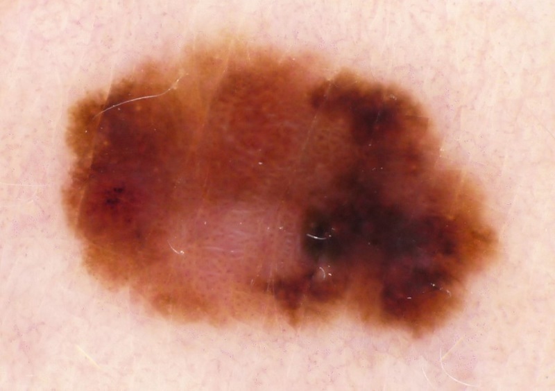
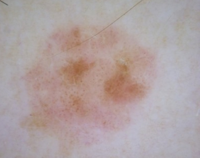
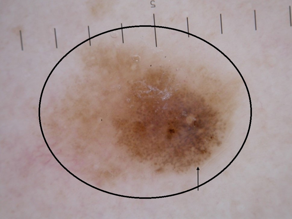
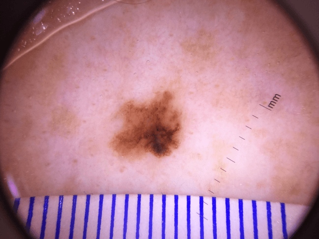
Melanoma (including melanoma in situ, superficial spreading melanoma, nodular melanoma)
LAST UPDATED: Jun 05, 2026
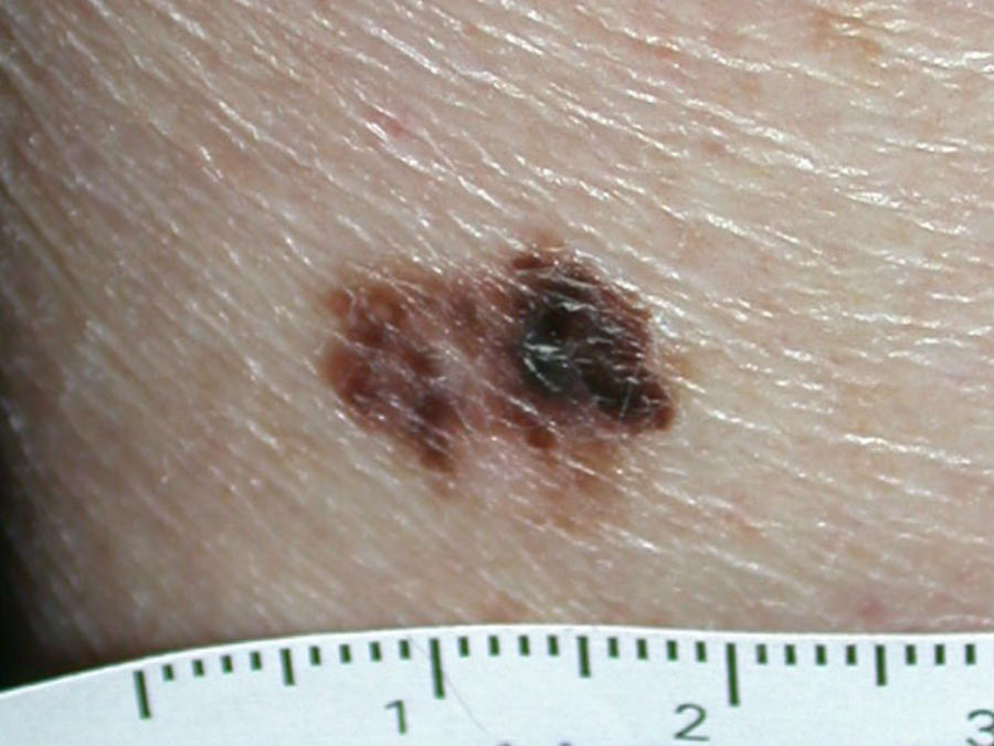
Introduction
Melanoma is a type of skin cancer that arises from melanocytes, the cells that produce melanin, which is the pigment that gives the skin its colour. Melanoma predominantly affects the skin (cutaneous melanoma), less commonly mucosal surfaces, and rarely other organs such as the eyes. According to the Global Cancer Observatory (GLOBOCAN) report, melanoma is the 17th most common cancer, with 324,635 new cases, and the 23rd leading cause of death among all cancers, resulting in 57,043 deaths in 2020. Melanoma has the highest incidence in Europe (46.4%) and North America (32.4%). Melanoma occurs less frequently in persons of colour but is associated with higher rates of morbidity and mortality resulting from later diagnosis.
This chapter focuses on melanoma of the trunk, limbs, and mucosal surfaces. For detailed information on the palms/soles/nails, and the face, refer to the chapters acral-lentiginous melanoma and lentigo maligna melanoma respectively.
This chapter is set out as follows:
Aetiology
Causal factors
Melanoma is thought to begin as an uncontrolled growth of melanocytic stem cells that have undergone a mutation (genetic transformation). The cause of these cell mutations can be acquired or inherited.
- Acquired 'sporadic' mutations are the most common cause of melanoma, accounting for approximately 90% of cases. The mutations result from cell damage as people age. The most common source of damage is overexposure to UV radiation, eg sun exposure and sunbed use. In the case of superficial spreading melanoma there appears to be an association with intermittent sun exposure especially in childhood, for nodular melanoma the picture is less clear. In lentigo maligna melanoma the risk is increased by the total number of lifetime hours of sun exposure
- Germline 'inherited' mutations are passed down from the parent accounting for approximately 10% of melanoma cases. CDKN2A (also called p16INK4A or MTS1) is the gene primarily linked, occuring in up to 20–40% of familial melanomas. In recent years, a number of other genes have been implicated including CDK4 and BAP1. Both CDKN2A and CDK4 are associated with an increased risk for developing pancreatic cancer (28% absolute risk compared to less than 1% in the general population), and possibly other cancers, including upper gastrointestinal cancers, astrocytomas, neurofibromas, and schwannomas. For further information refer to the section on management
Identifying risk factors
- Slightly increased risk (1-3 times that of the general population)
- Skin that burns easily
- Red or blonde hair
- A high density of freckles
- Any family history of melanoma
- Moderately increased risk (6-10 times that of the general population)
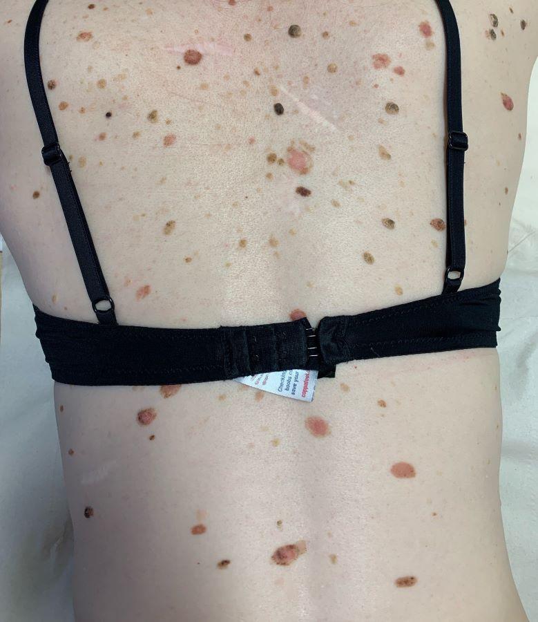
- Large number of naevi - a linear relationship exists with an increased relative risk of cutaneous melanoma as the number of total body common melanocytic naevi increased. Patients with more than 100 common naevi on their body have a relative risk of developing cutaneous melanoma almost sevenfold higher compared to those with only 15 naevi or less
- Atypical mole syndrome - seen in approximately 2% of the population. Defined by an individual with more than 50 naevi of which three or more are atypical
- A personal history of melanoma
- Organ transplant recipients
- High-risk (more than 10 times that of the general population)
- Patients with giant congenital melanocytic naevi
- Those with inherited mutations (such as CDKN2A) carry a 28%–67% lifetime risk of developing melanoma up to the age of 80. Individuals with the Familial Atypical Multiple Mole Melanoma (FAMMM) syndrome (multiple melanocytic nevi, usually more than 50, and a family history of melanoma) are 25 times more likely to develop melanoma compared to unaffected individuals
History
- The majority of melanomas arise de-novo (approximately 80 %), others arise from pre-existing naevi
- Age - the risk of melanoma increases with age; it is extremely rare pre-puberty. Those with an inherited cause are more likely to develop melanoma when younger with a median age of 33–45
- Hormones - the use of use of unopposed oestrogen therapy in HRT might increase the risk of melanoma, there is no evidence for an increased risk in combined HRT. Pregnancy does not appear to increase the risk. Several studies have reported a positive association between oral contraceptive (OC) use and melanoma risk; however, OC users have also been found more likely to use sunscreen and tanning beds - overall, the link does not appear to strong, the findings conflicting, and data from large prospective studies is lacking
- Reliability of history - depending on site and number of moles it can be difficult to ascertain whether or not a particular mole is new, or, if it long-term whether or not it has changed. Additionally, if a pigmented lesions looks suspicious, regardless of the history the patient should be referred
- Although melanoma can itch, so can healthy naevi. Often melanoma is asymptomatic. Nodular melanoma tends to grow more quickly than superficial spreading melanoma, and is more likely to bleed or crust
Clinical findings
Melanoma found at typical skin sites arises as melanoma in situ, superficial spreading melanoma, or nodular melanoma. Lentigo maligna (and lentigo maligna melanoma) is predominantly a skin cancer of the head and neck, occasionally affecting the trunk and limbs.
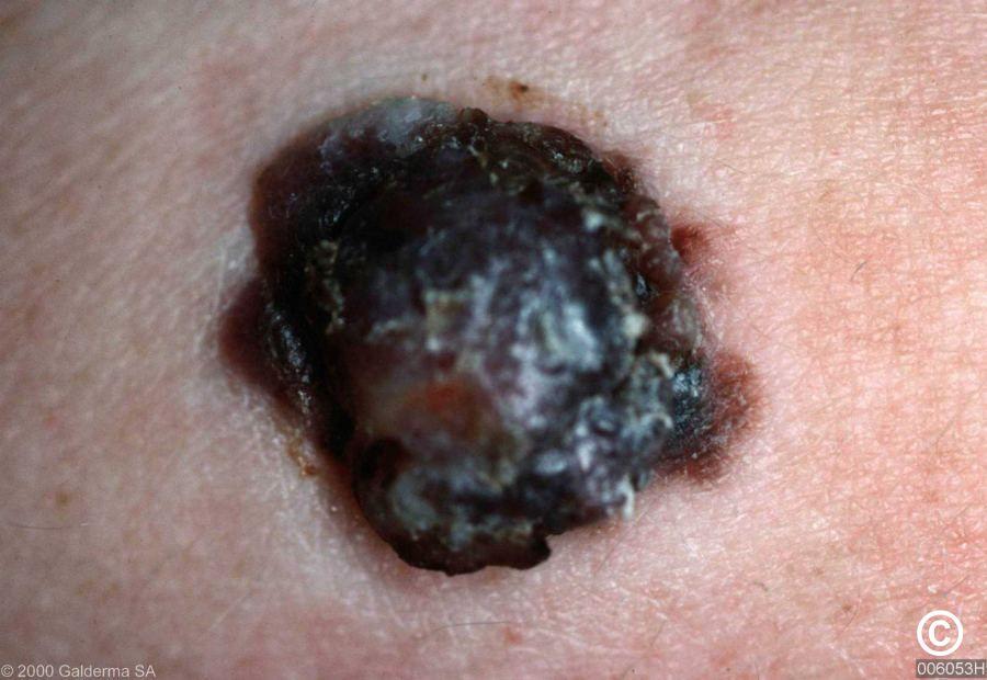
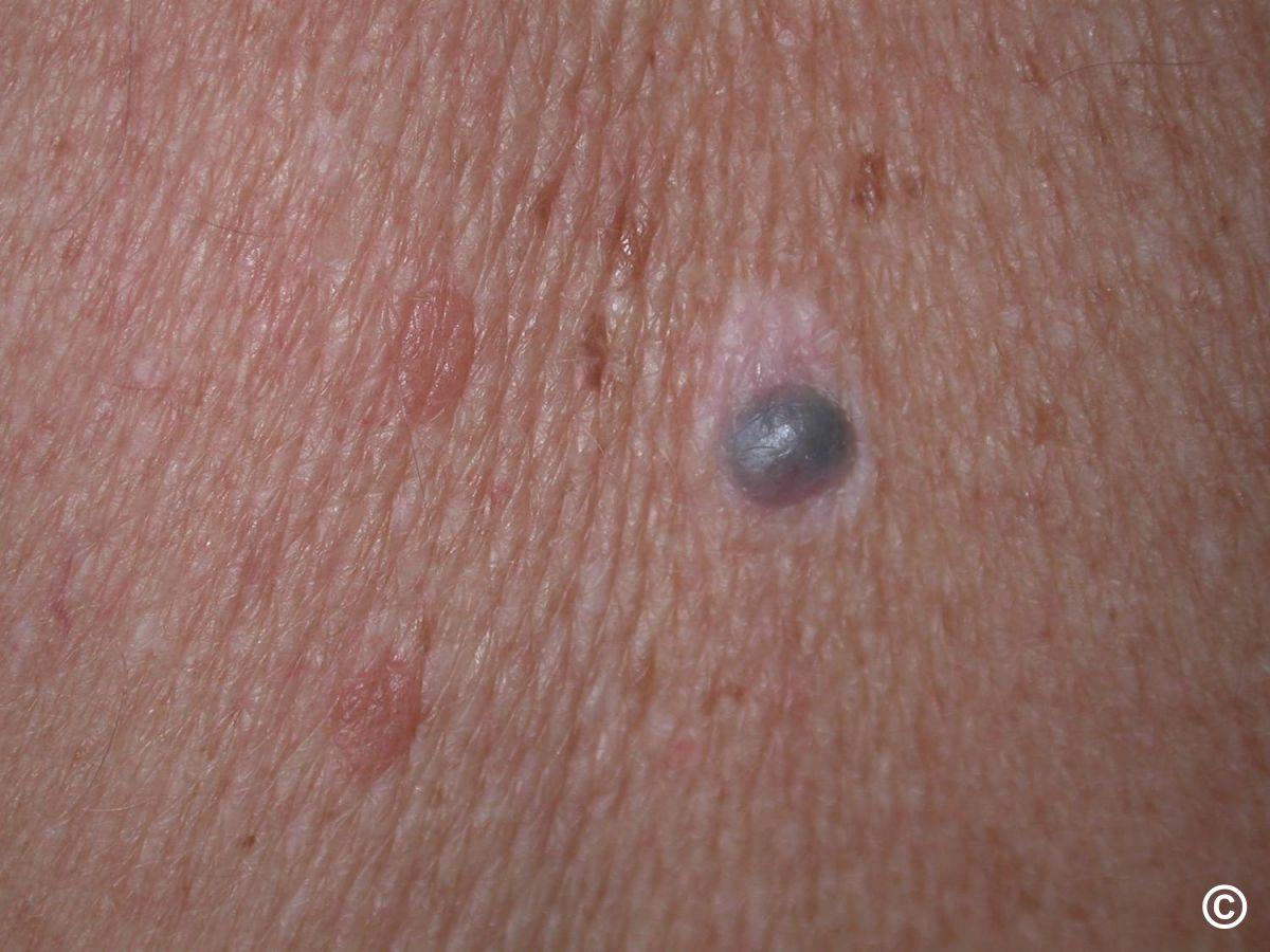
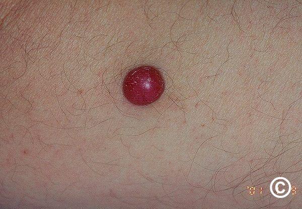
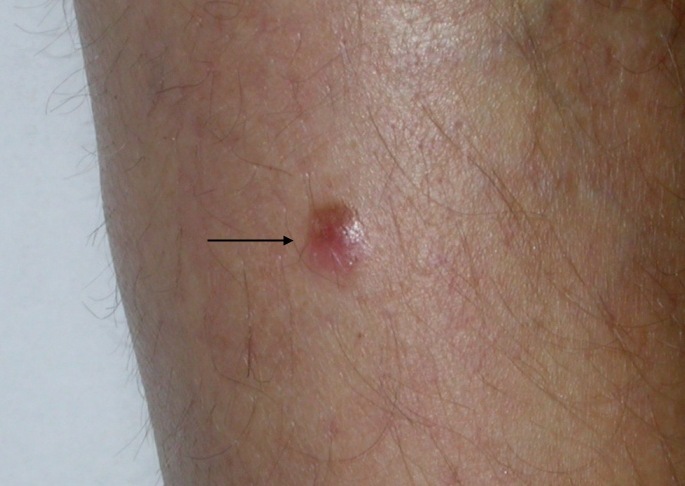
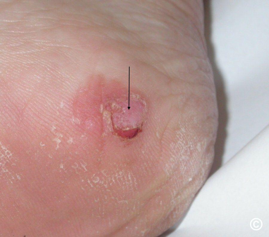
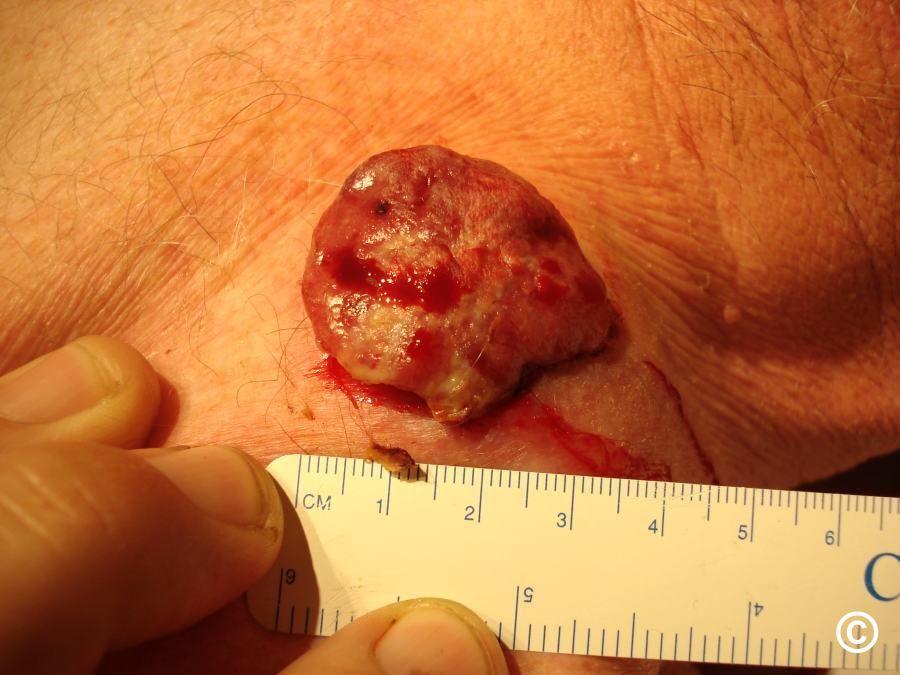
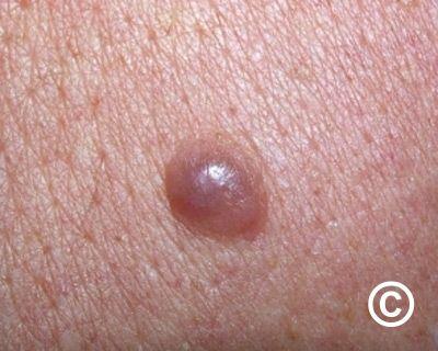
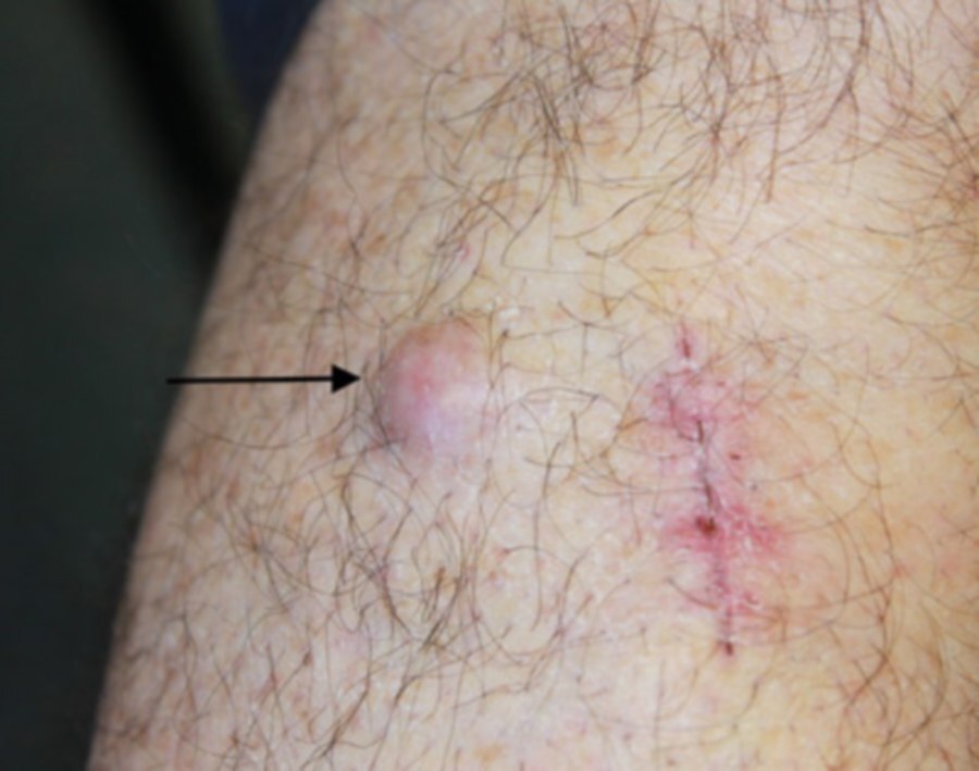
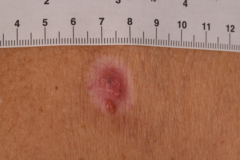
Superficial (thin) forms of melanoma initially spread out within the epidermis. If all the melanoma cells are confined to the epidermis it is termed melanoma in situ. When the cancerous cells have grown through the basement membrane and into the dermis it is known as invasive melanoma and is termed superficial spreading melanoma. Superficial spreading melanoma is the most common type of invasive melanoma and accounts for 50% of all melanoma in the UK. Nodular melanoma, presenting as a firm papule, nodule or plaque accounts for 20-25% of cases of melanoma and is the most aggressive type, it is more common in males and often presents in the fifth or sixth decade, but can occur at any age.
In terms of screening out harmless naevi, the following are more likely to be benign:
- Those growing gradually and symmetrically in younger patients
- Brown naevi that gradually become dome-shaped and soft/wobbly to palpate whilst maintaining a regular symmetrical edge
- Colour - brown naevi that are one colour, or two colours where the colours are similar (eg two shades of brown) AND where the pigment is arranged in a symmetrical fashion eg darker centre and lighter edge
- Itchy naevi that are not changing and otherwise appear normal
- Naevi that change rapidly (over a few days) becoming swollen, inflamed and crusty AND which then settle back down to their original appearance - these naevi are likely to have been traumatised or become acutely infected. Such patients must be followed up after 2-3 weeks to make sure that the symptoms are settling, if not the patient should be referred urgently
- Classical halo naevi in young patients (refer to the chapter Halo naevi)
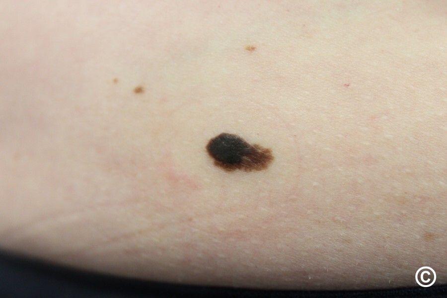
The most important aspect of melanoma diagnosis is the ABCD / EFG rule:
The ABCD rule
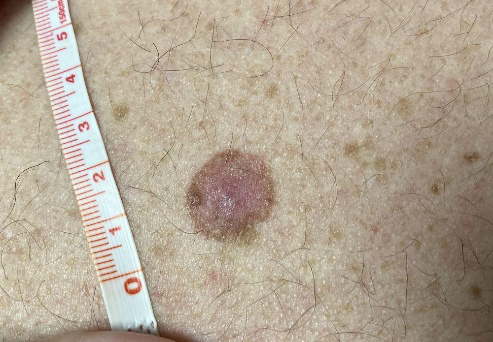
The ABCD rule can be used by health professionals and patients to check for the main warning signs of melanoma:
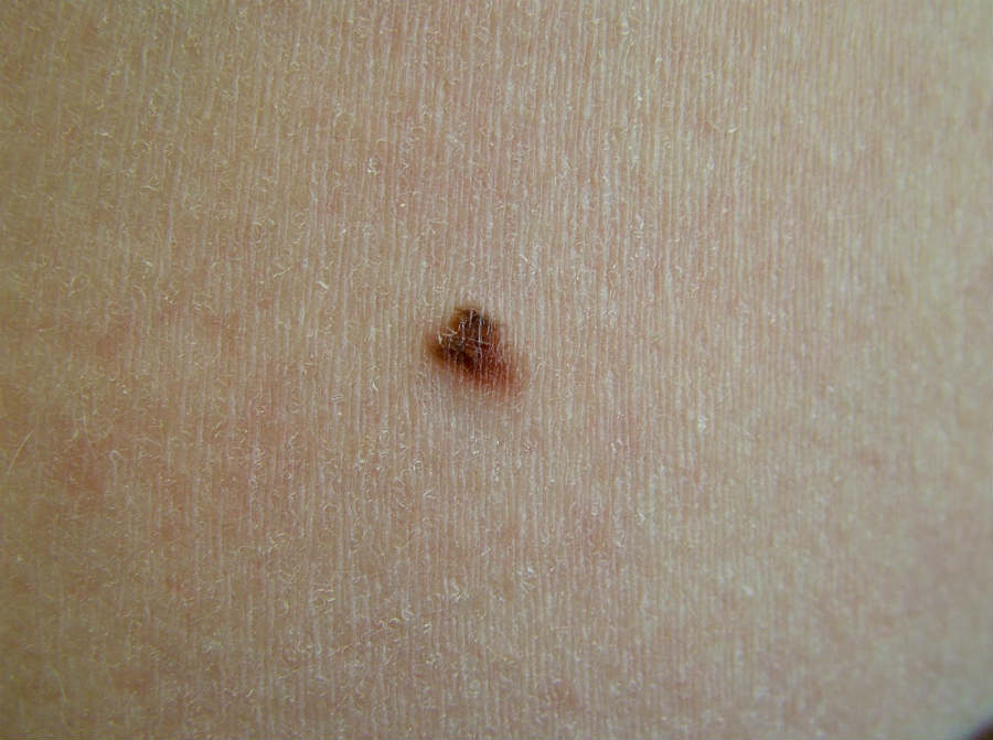
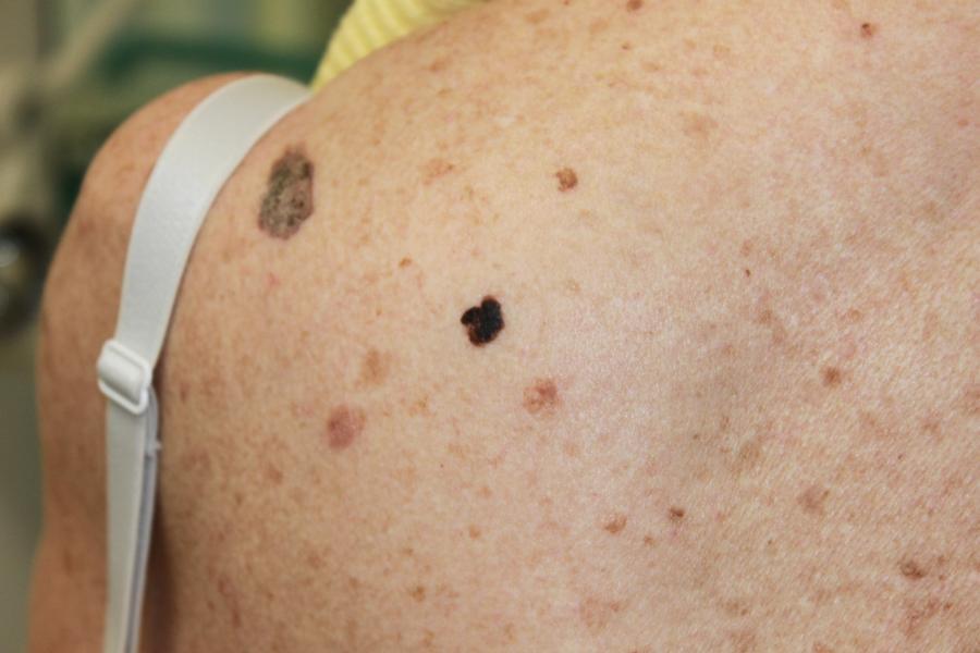
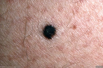
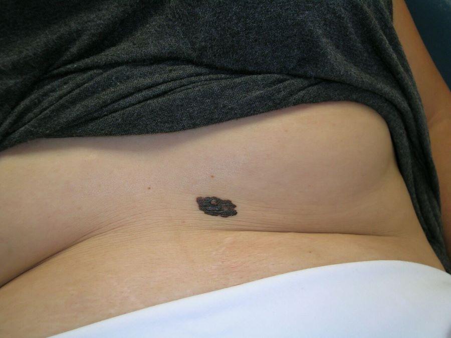
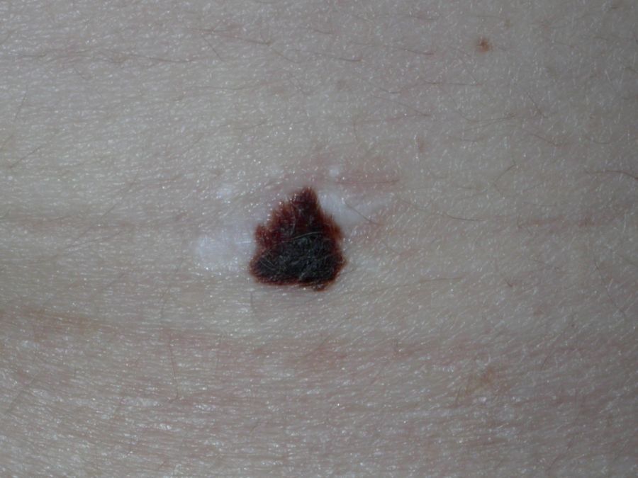
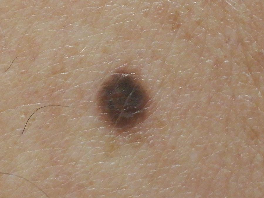
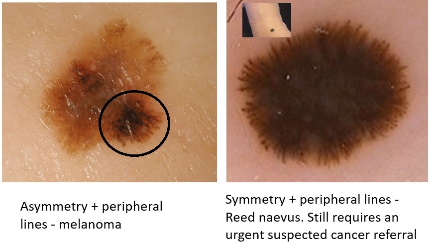
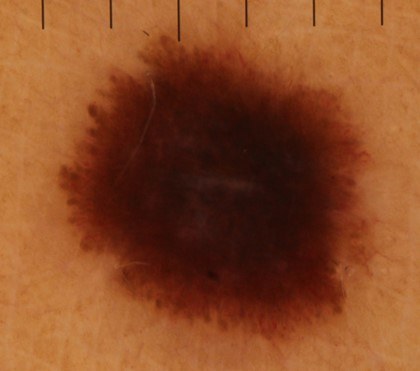
- A= asymmetry - if you draw a line through the middle of the lesion, the two halves don't match
- B = border irregular - the edges of the lesion may be irregular or blurred, and sometimes show notches
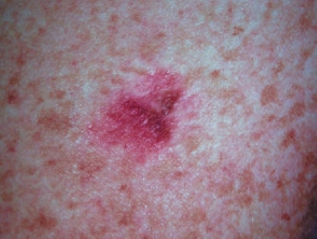
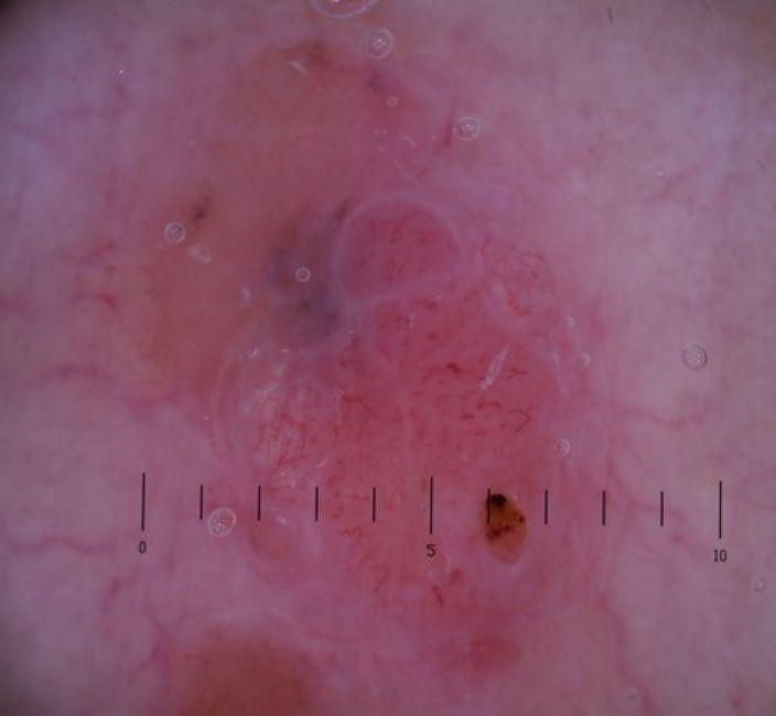
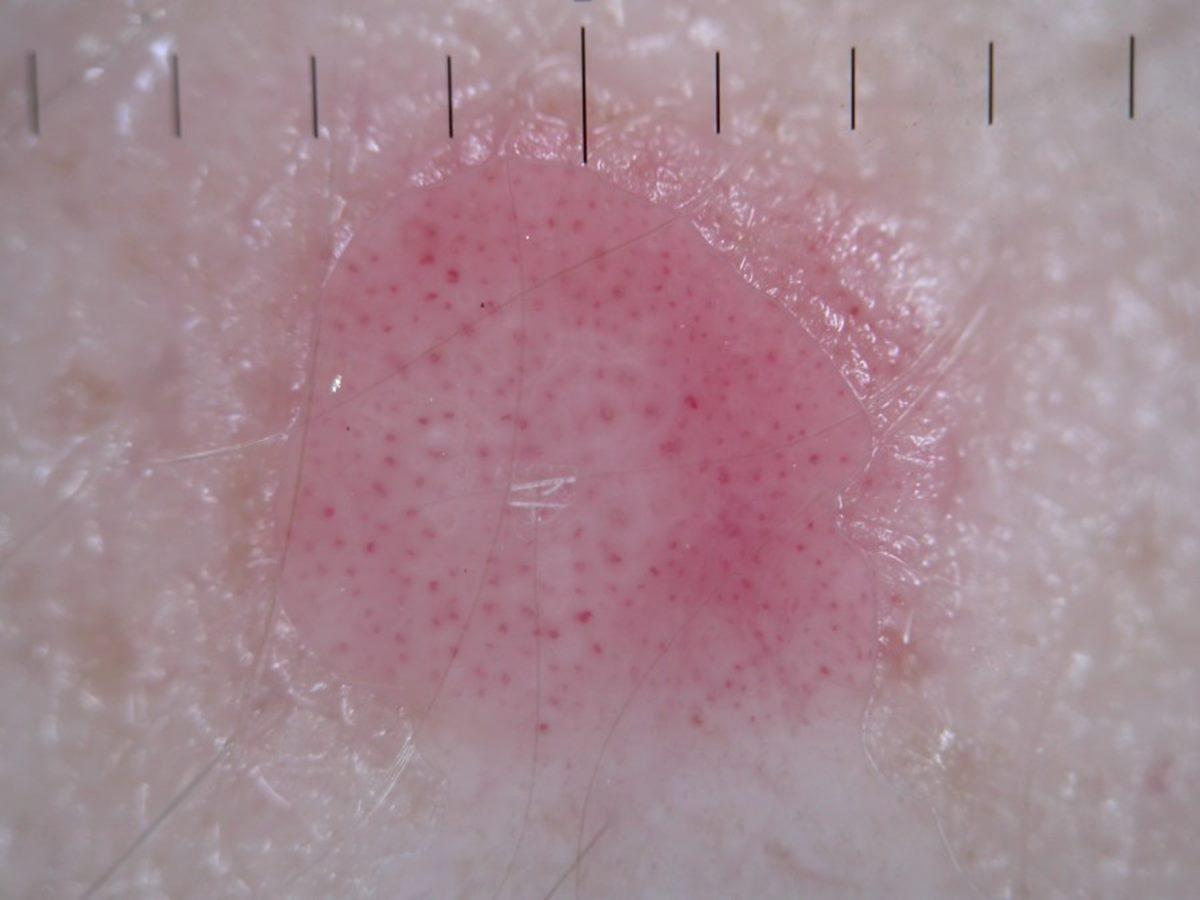
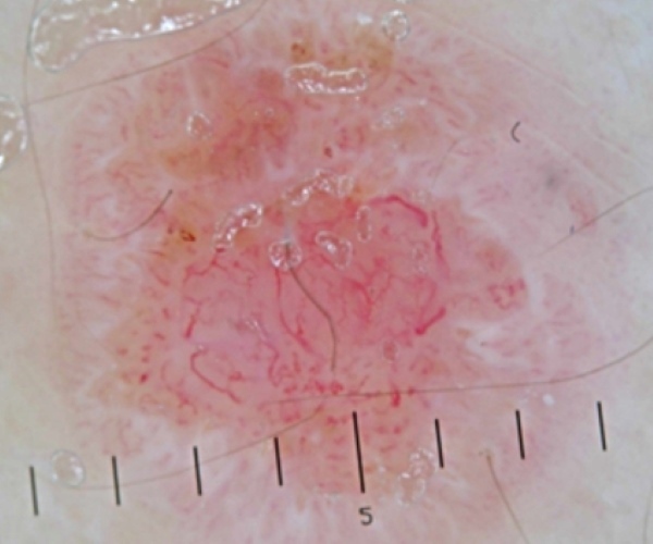
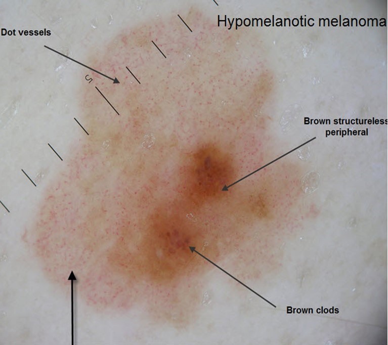
- C = colour - 2 or more colours (or shades of colour) that are asymmetrically arranged; sometimes a single colour that differs to the rest of the patient's moles, eg black or pink (melanoma with no pigment is sometimes referred to as amelanotic melanoma, melanoma with a small amount of pigment is a hypomelanotic melanoma)
- C also stands for comparison ie the ugly-duckling that looks different to the patient's other lesions
- D, as first described, stands for diameter > 6mm, however, many skin cancers start small and therefore can present at any size. Thus, D also stands for changing dimensions. It is also important to note:
- Melanomas grow at different rates - even if the patient states that the lesion is not changing, if it looks suspicious still refer
- Melanoma often has an irregular appearance, however, if a symmetrical lesion continues to grow out of proportion to the patient's other moles, especially if aged > 45, then melanoma must be considered
The EFG rule (raised lesions can lack the ABCD rule)
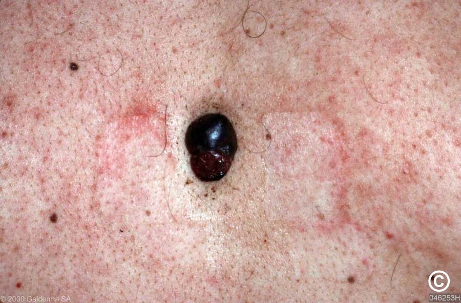
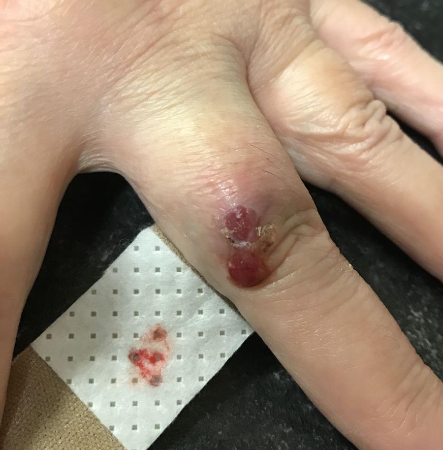
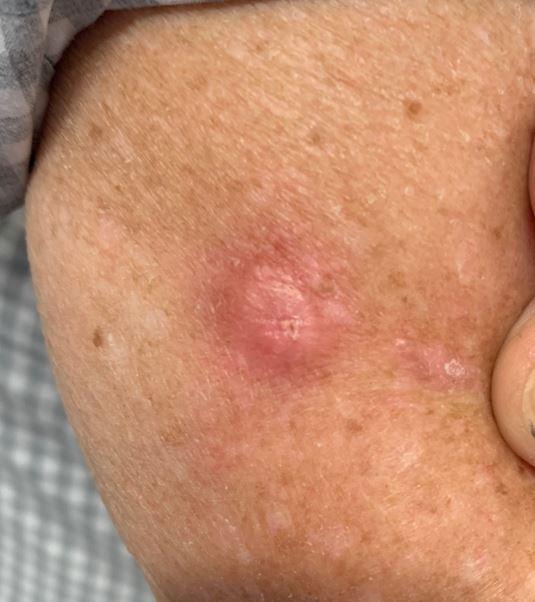
Nodular melanoma can be brown, black, blue-black, and up to 50% are amelanotic (pink-red or skin-coloured). The surface can be smooth, rough, or crusted. Nodular melanoma often lacks dermoscopic clues, and the diagnosis should be considered in any skin lesion demonstrating all of EFG:
- E = Elevation, AND
- F = Firmness to touch, AND
- G = Growth. Sustained growth for 3-4 weeks
Melanoma affecting mucosal surfaces
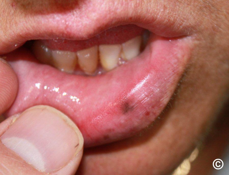
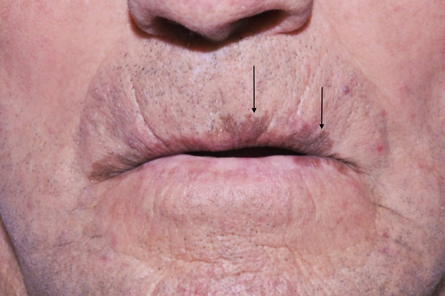
- Stable regular brown macules on the lips are very likely to be mucosal melanocytic macules, which are harmless
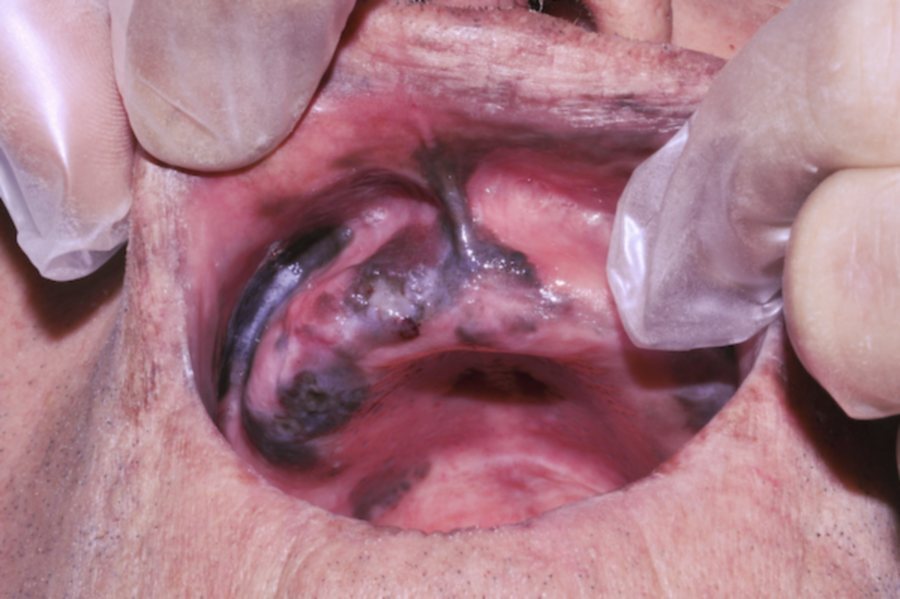
- The following should be considered as an urgent referral for possible melanoma:
- Lips, any of - irregular shape, atypical colour (black, or more than one colour / shade of colour), a firm papule/nodule
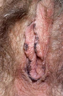
- Oral / genital - any new or changing pigmented lesion. Have a low threshold for referral as an accurate history is usually absent (check local guidelines for where to refer)
Melanoma affecting the palms/soles/nails, or face
Melanoma in skin of colour
- African Americans have deeper tumours at time of diagnosis in addition to increased rates of regionally advanced and distant disease. Lesions are generally located on the lower extremities and have an increased propensity for ulceration. Acral lentiginous melanoma (ALM) is the most common melanoma subtype found in AA patients
- In Hispanics, superficial spreading melanoma is the most common melanoma subtype. Lower extremity lesions are more common relative to Caucasians. Hispanics have the highest rate of oral cavity melanomas across all ethnic groups
- In Asians, acral and subungual sites are most common. Specifically, Pacific Islanders have the highest proportion of mucosal melanomas across all ethnic groups
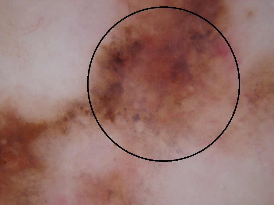
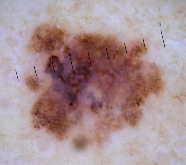
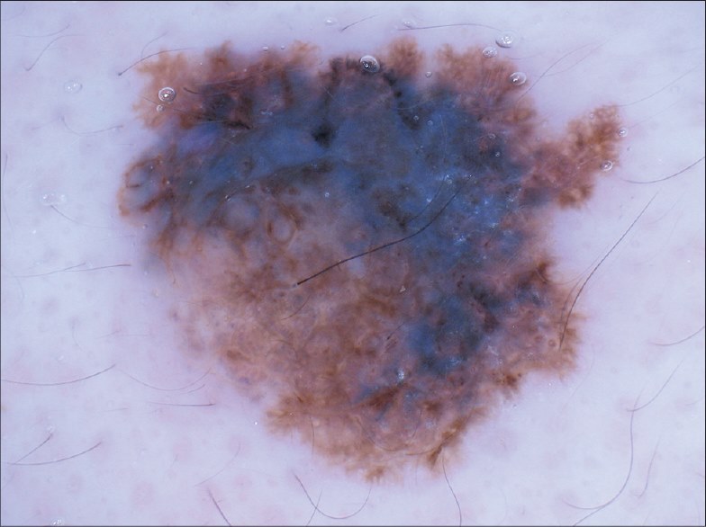
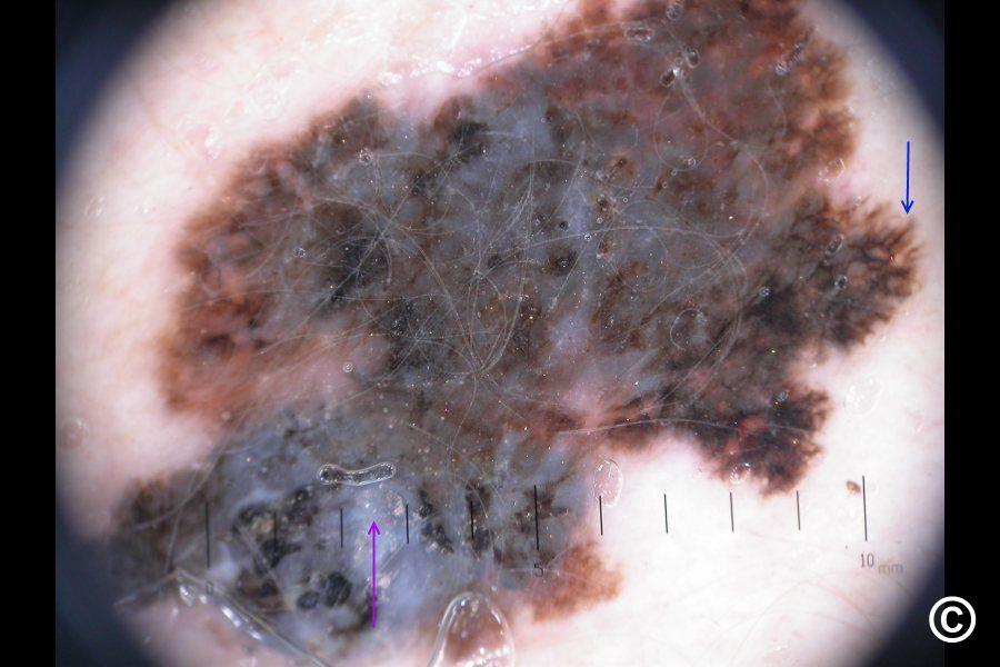
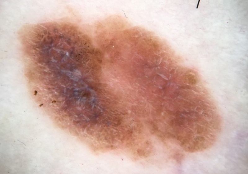
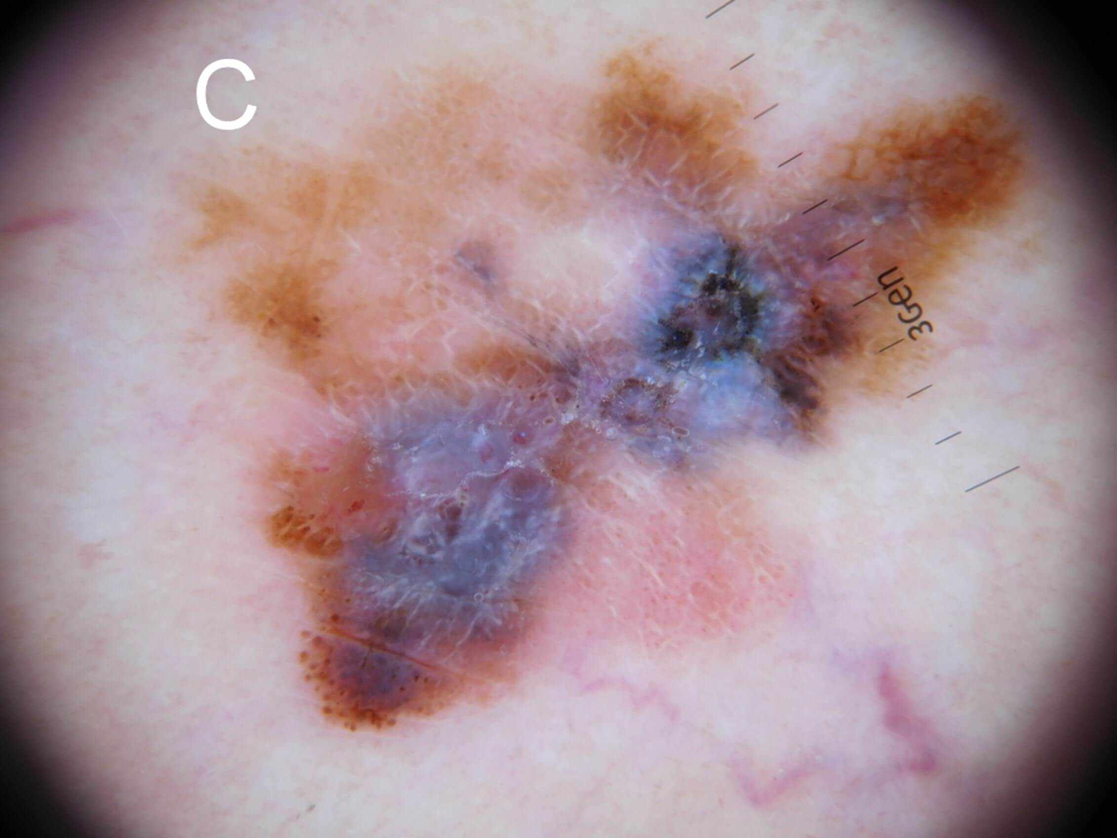
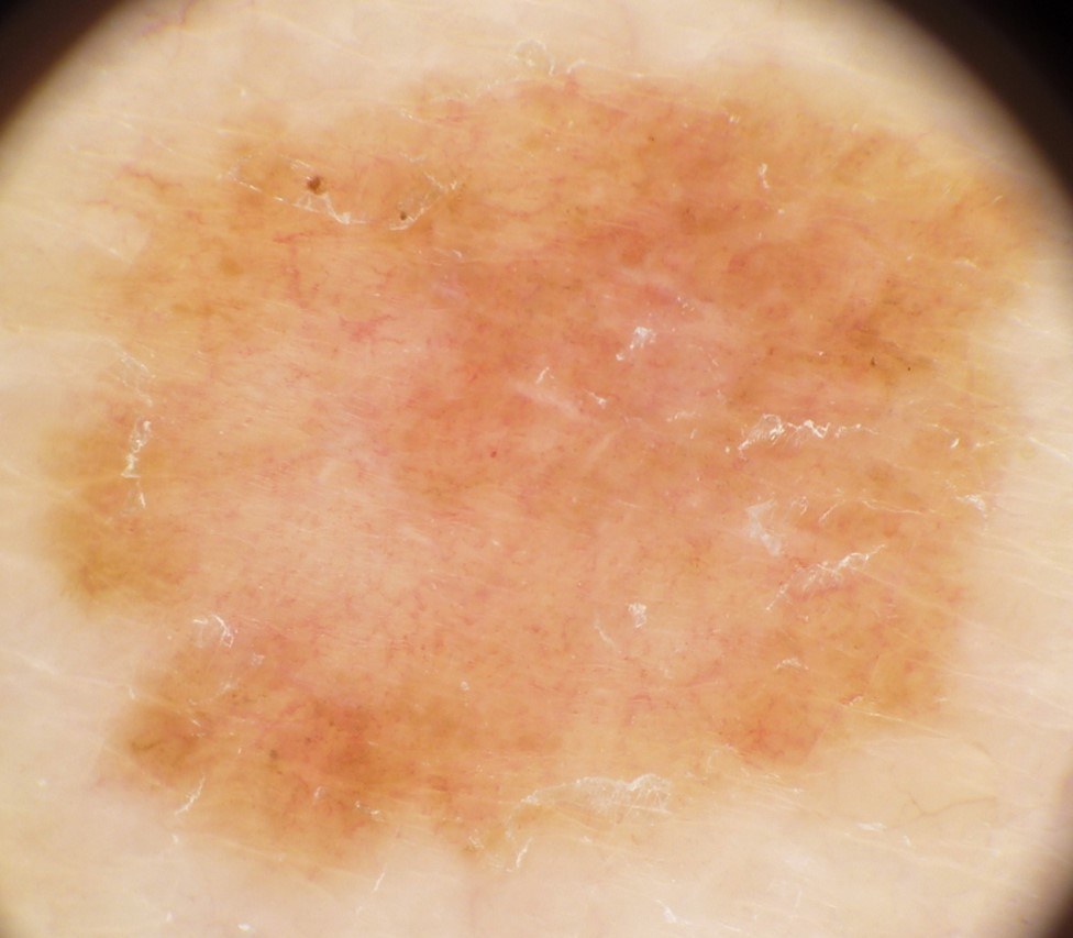
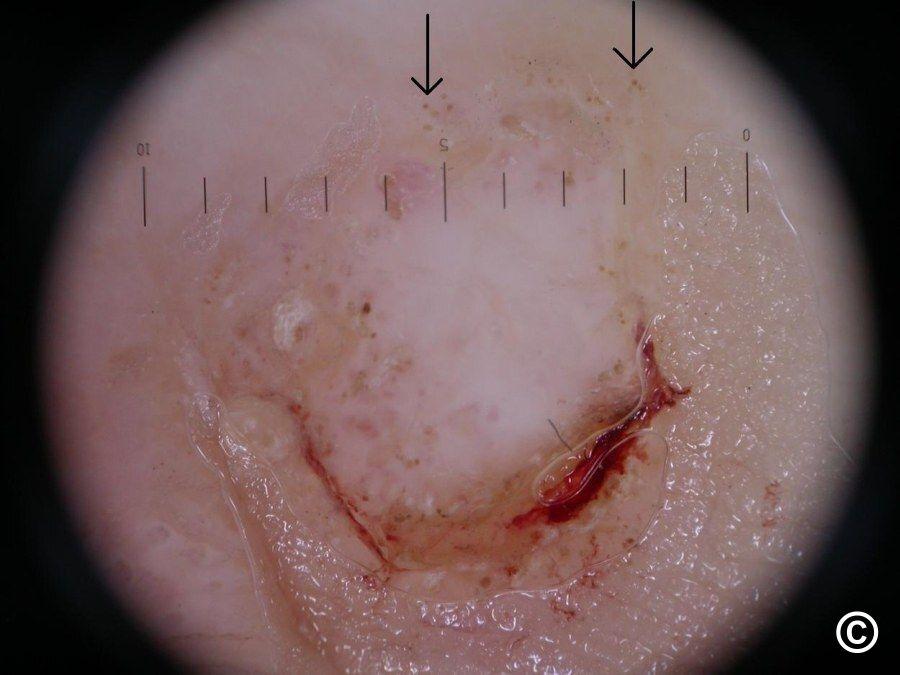
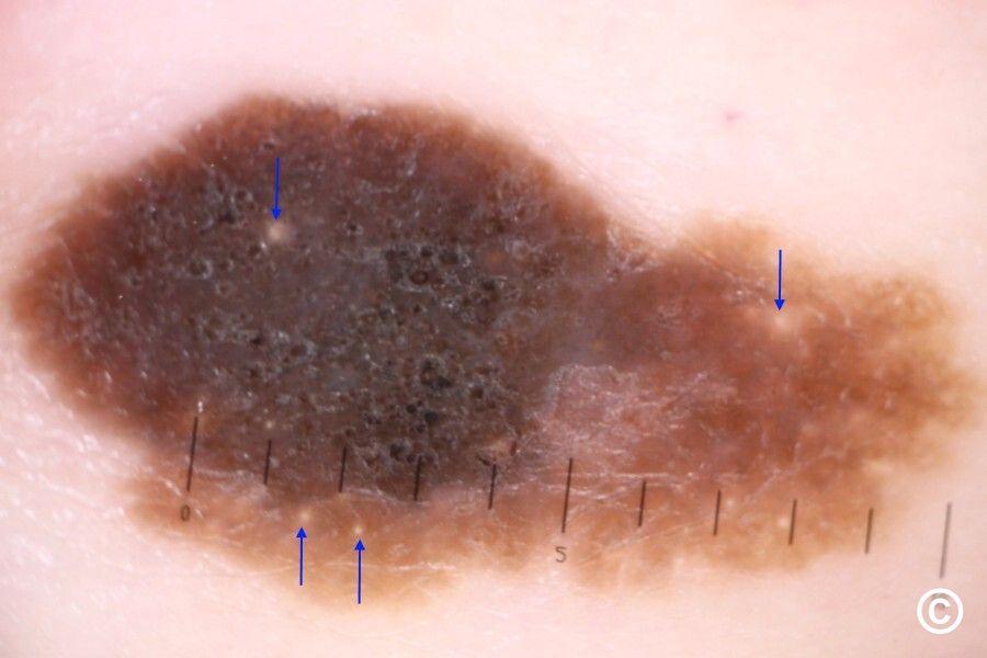
Dermoscopic features of melanoma (hover over terminologies for description)
This section refers to dermoscopic features of melanoma on the trunk and limbs, as opposed to the so called ''special sites'' such as the face, palms and soles, which have different features.
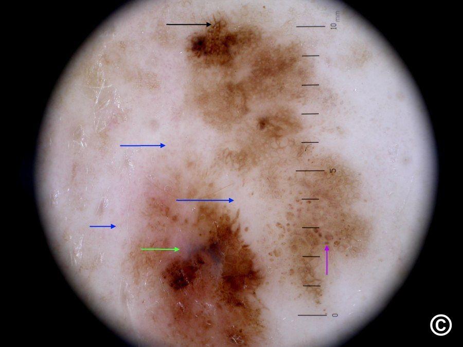
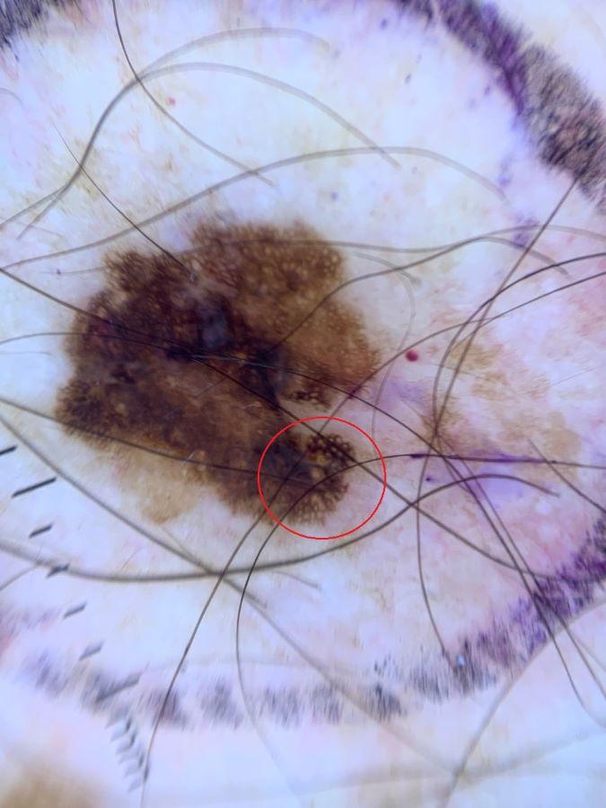
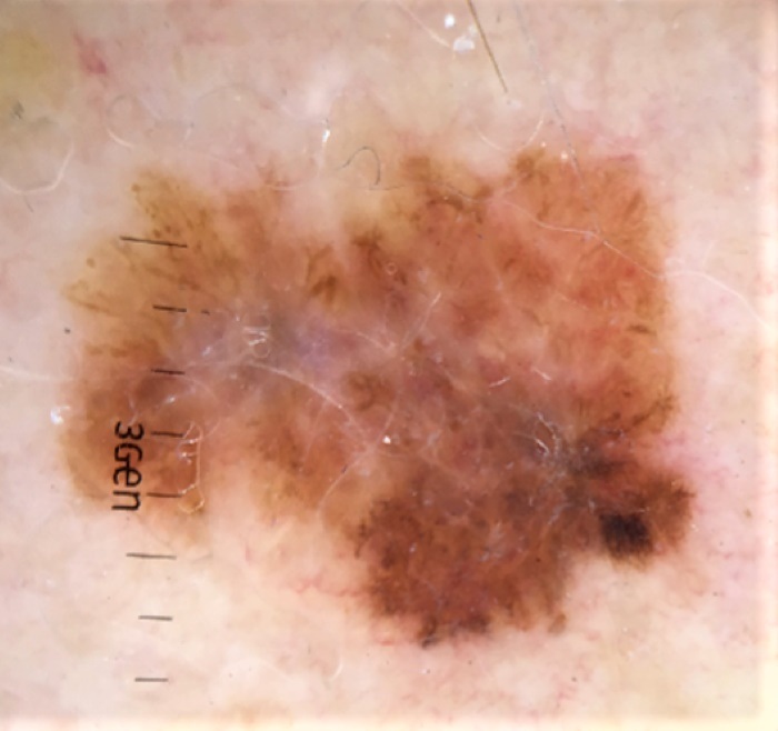
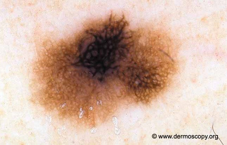
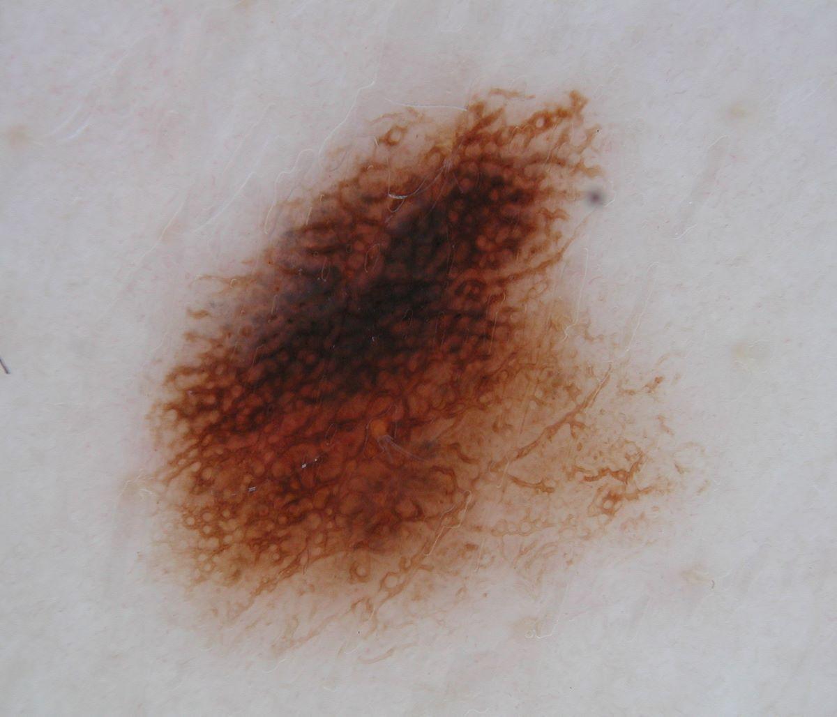
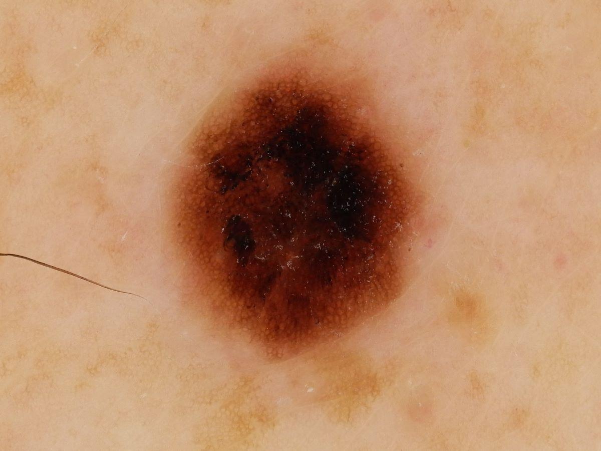
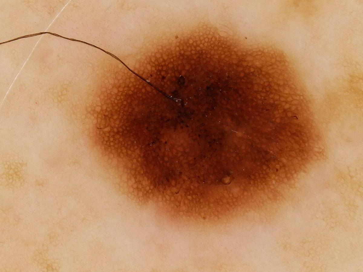
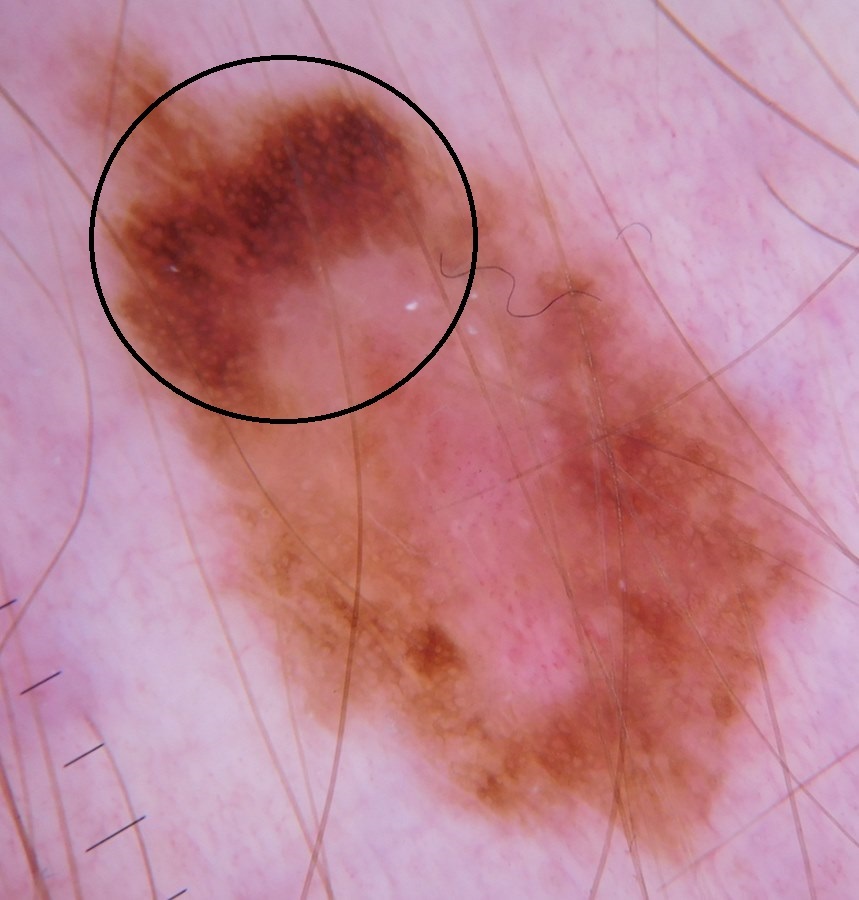
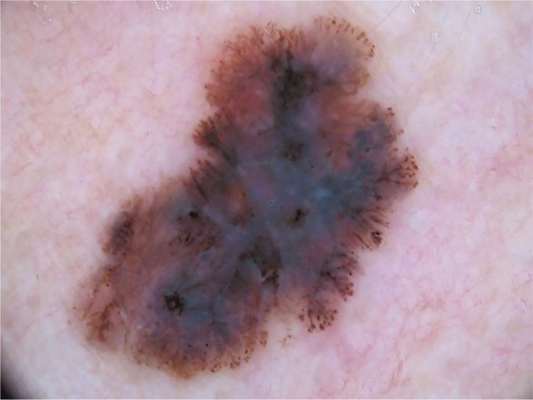
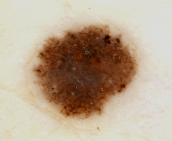
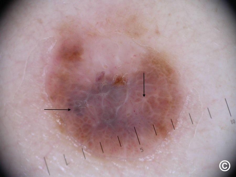
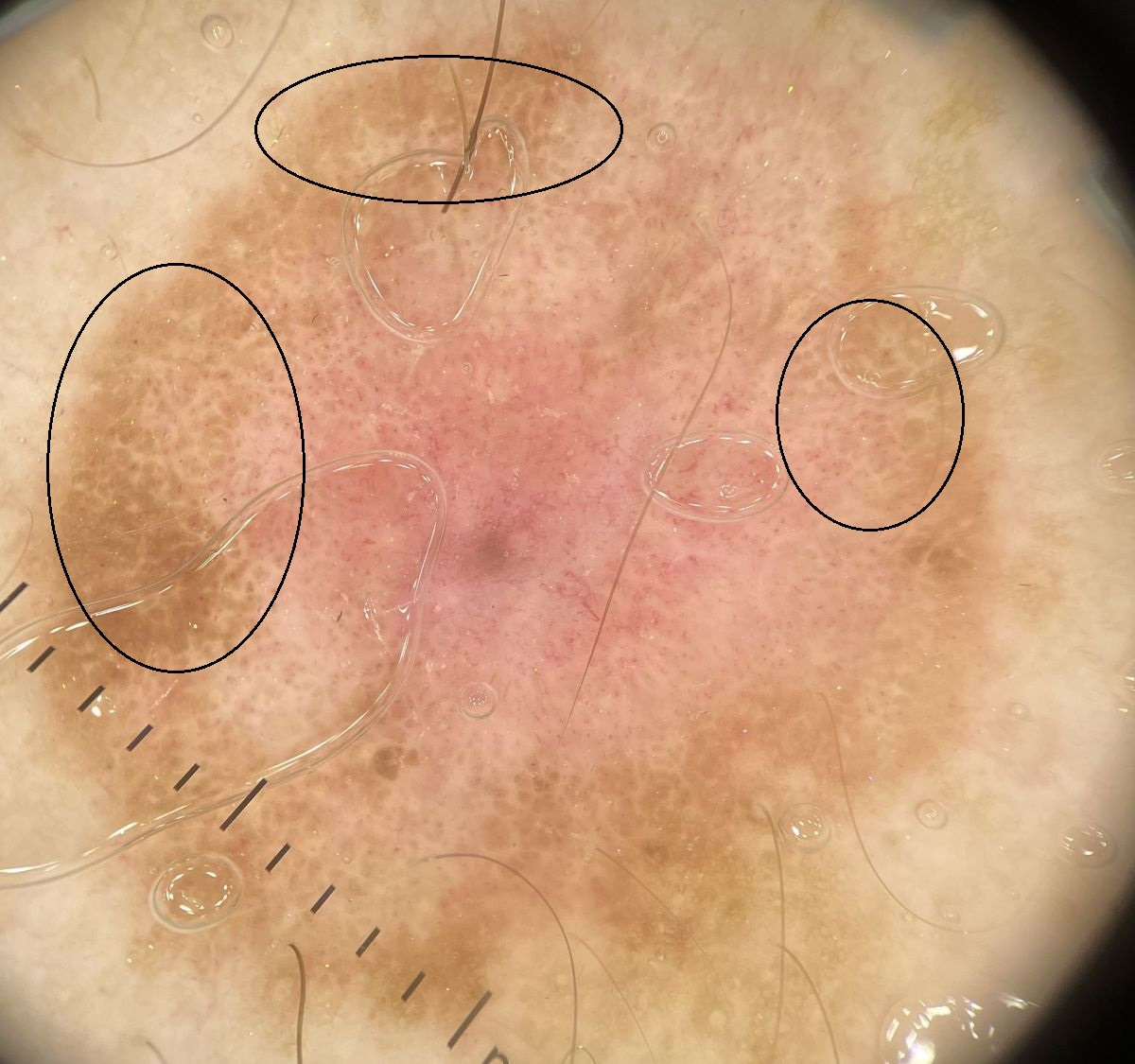
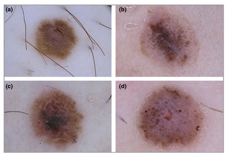
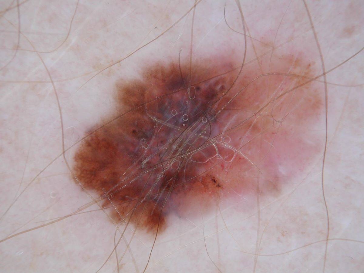
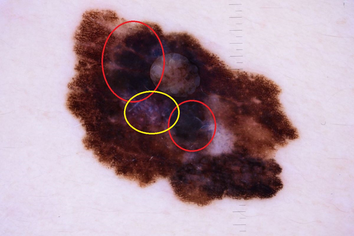
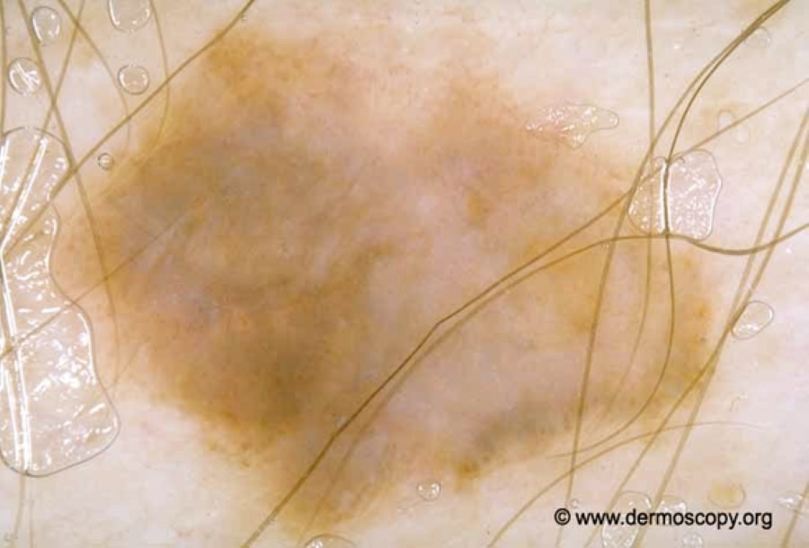
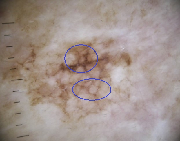
From a dermoscopic perspective melanoma can be chaotic and/or present with one or more clues:
- In terms of chaos we refer to the lesion appearing disorganised in one or more of - structures, colour, or having a variable border
- In terms of clues, melanoma can present with one or more of the following clues:
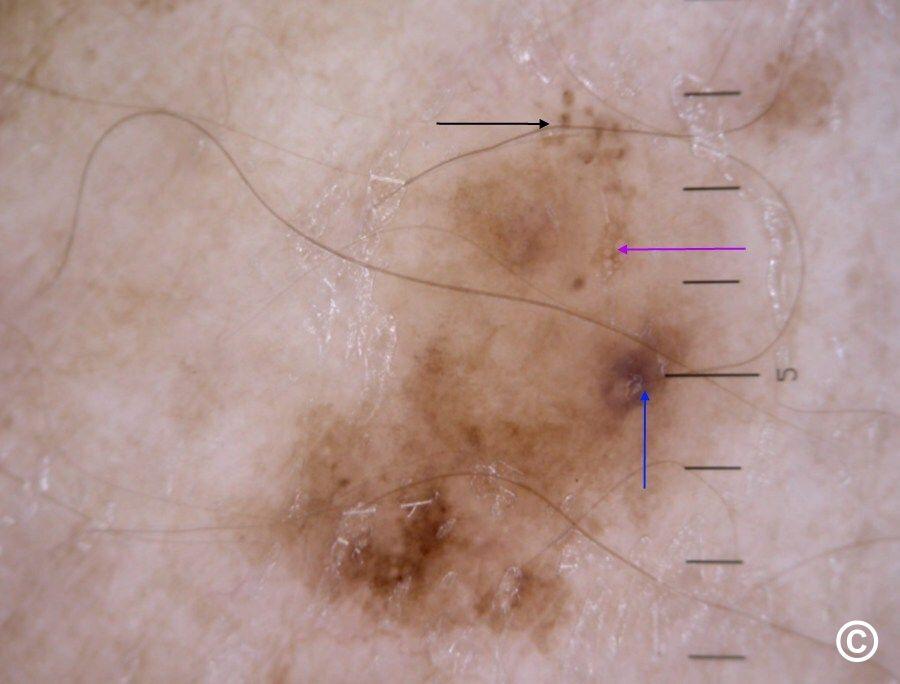
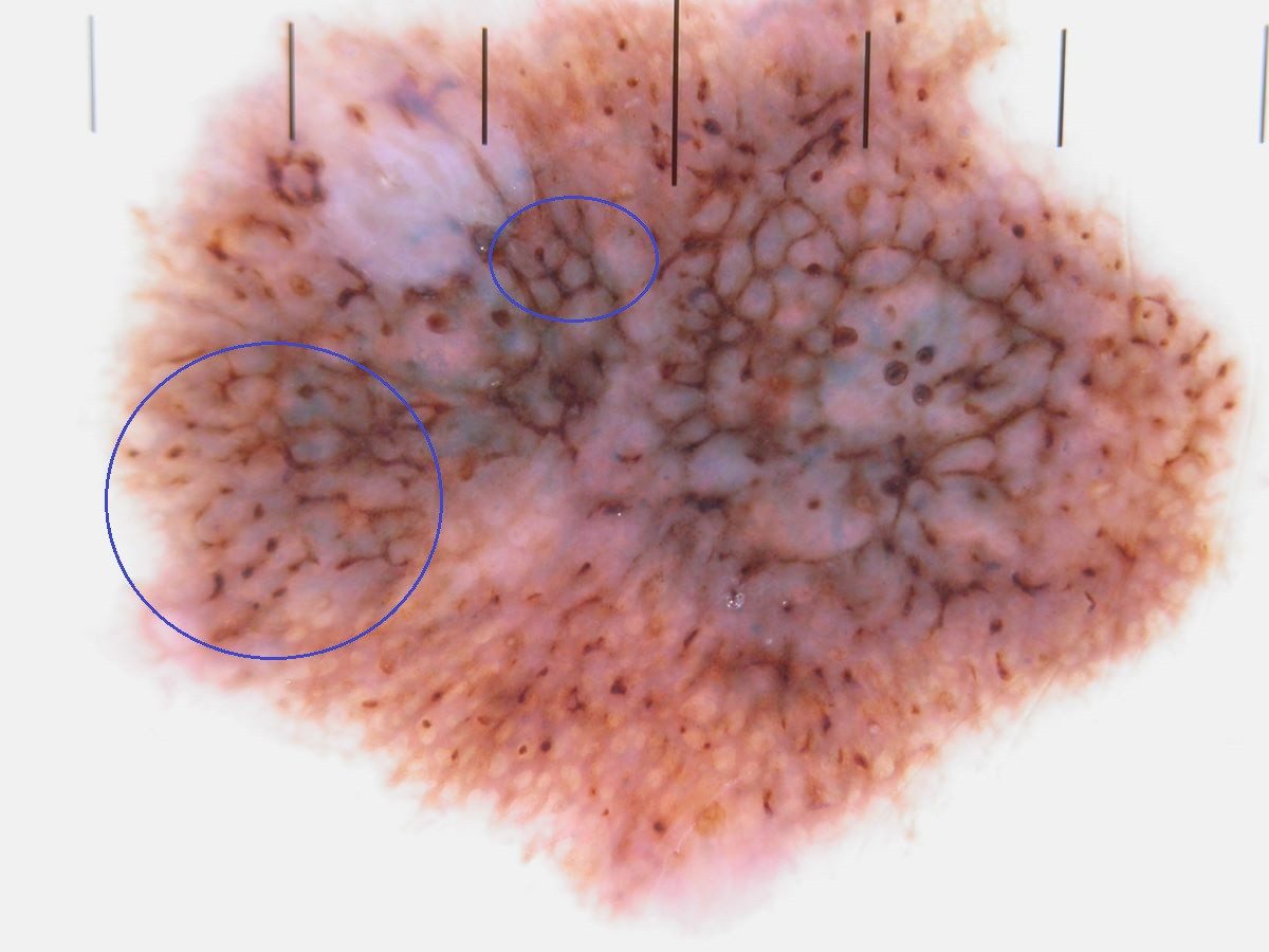
- Atypical pigment network
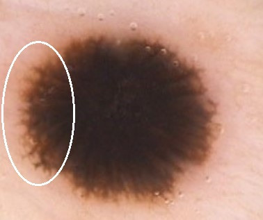
- Streaks or pseudopods, especially if asymmetrical
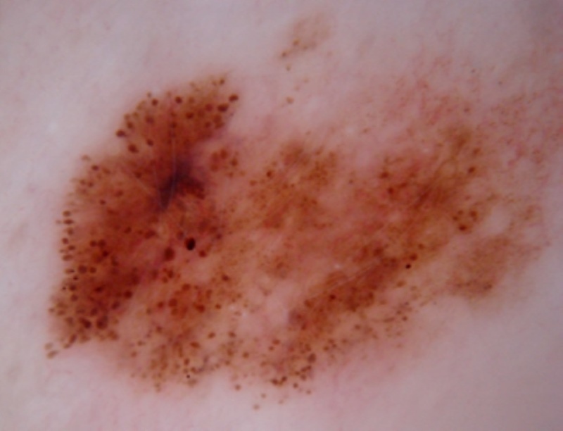
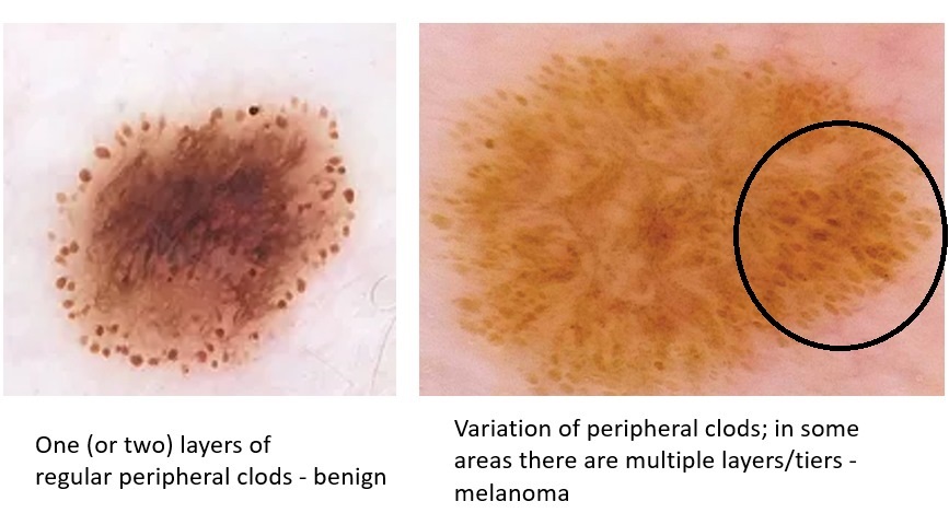
- Irregular dots and/or globules (sometimes referred to as clods) that can be:
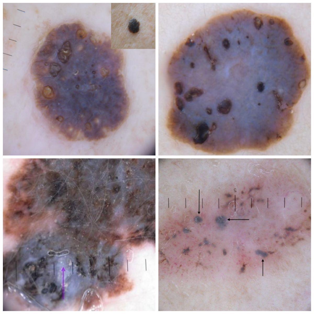
- Within/throughout the lesion - variation of size, shape, or colour. The size can vary from dots to small globules, to larger irregular clods (sometimes referred to as pigment blotches)
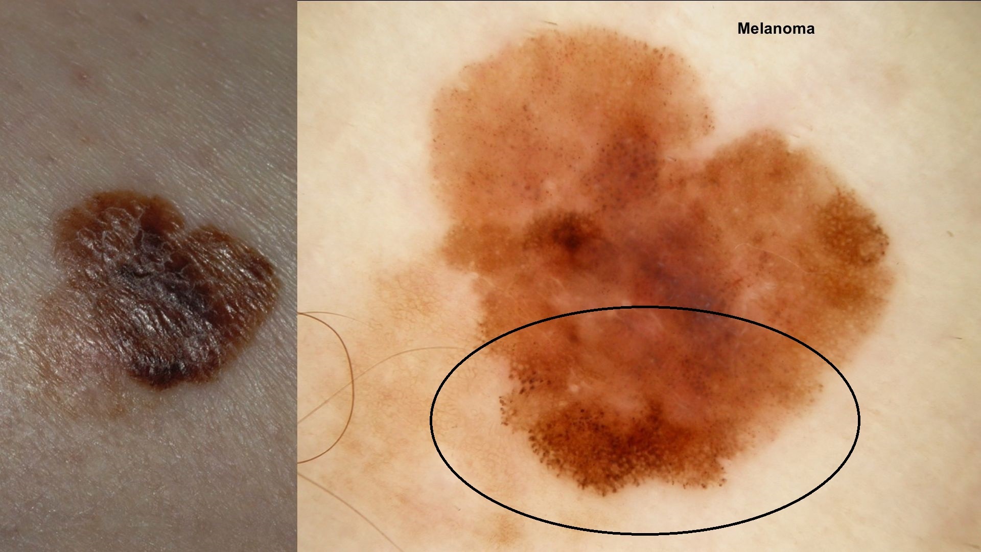
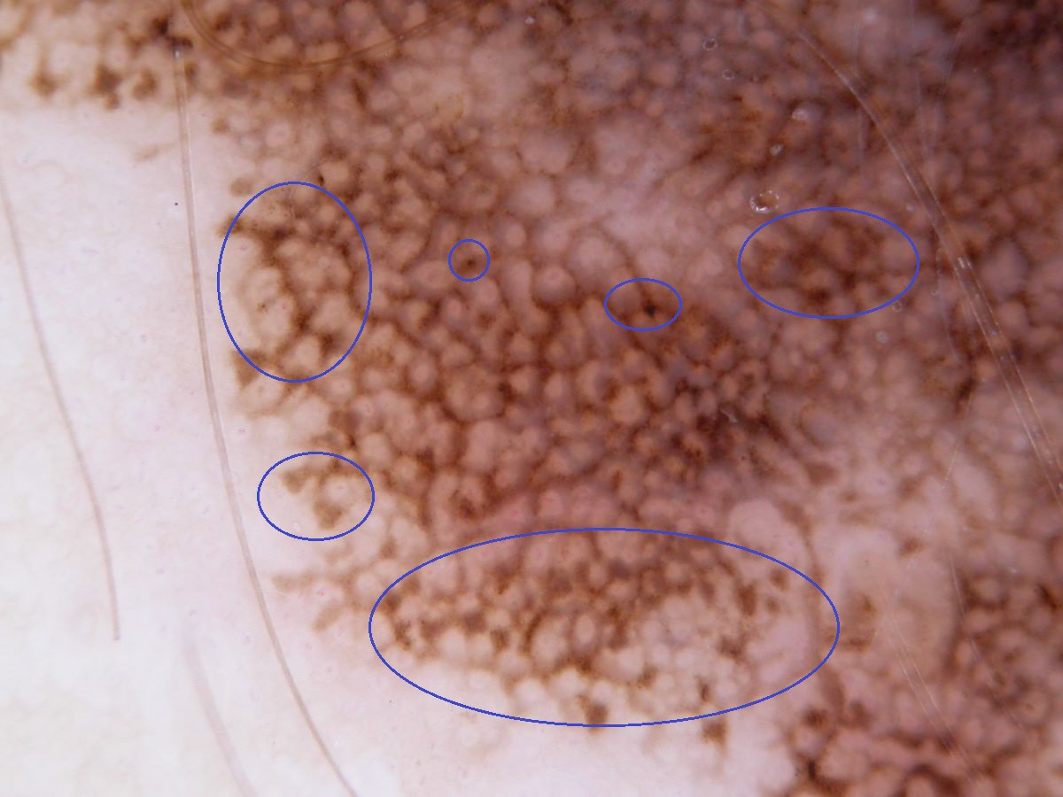
- Peripheral - multiple layers (tiers) of globules can suggest a Spitz naevus or melanoma. The greater the variation and/or asymmetry leans more towards melanoma
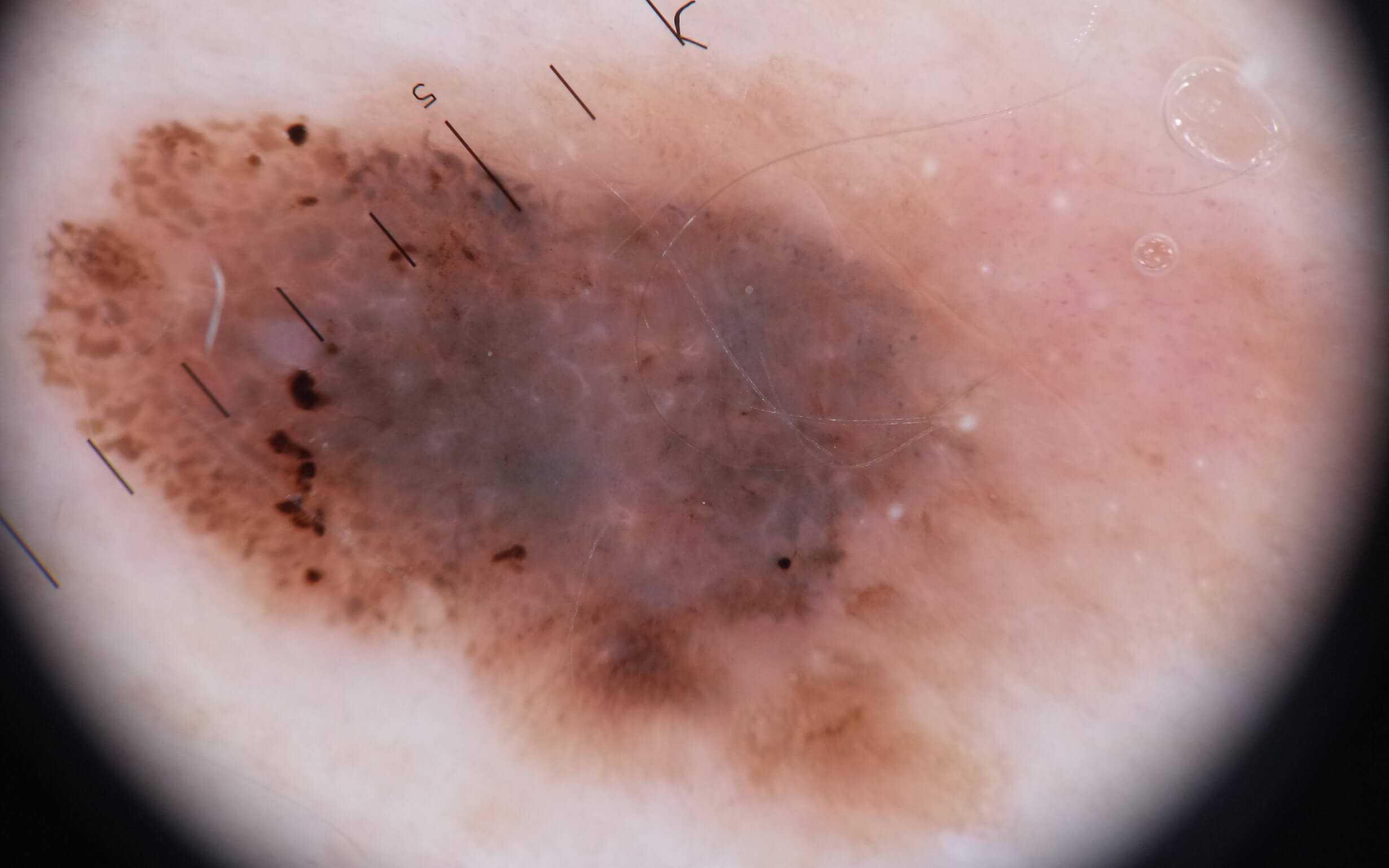
- Peripheral - black dots and globules. Even a single black globule can be of concern
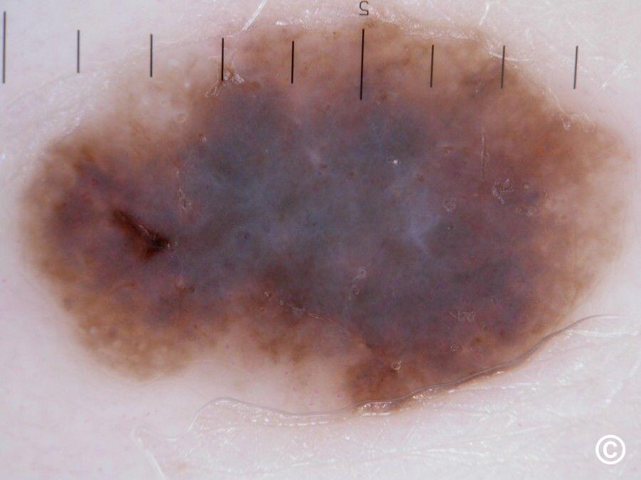
- Irregular blue-grey structures, often with white scar-like areas, usually represent regression
- Blue-white veil
- A reticular-globular network with a beaded appearance - an uncommon features of melanoma in which dots/small globules appear at intersections along the pigment network
- Shiny white lines (seen with polarised light), especially if orthogonal (ie lines perpendicular to each other)
- A negative network (seen with polarised or non-polarised light) - this can be a very difficult sign to spot
- Eccentric structureless areas
- Broad angulated lines seen in lesions not located on the the head and neck is a clue for lentiginous melanoma
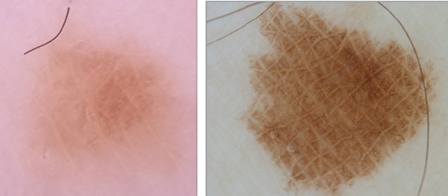
- Accentuated skin surface markings
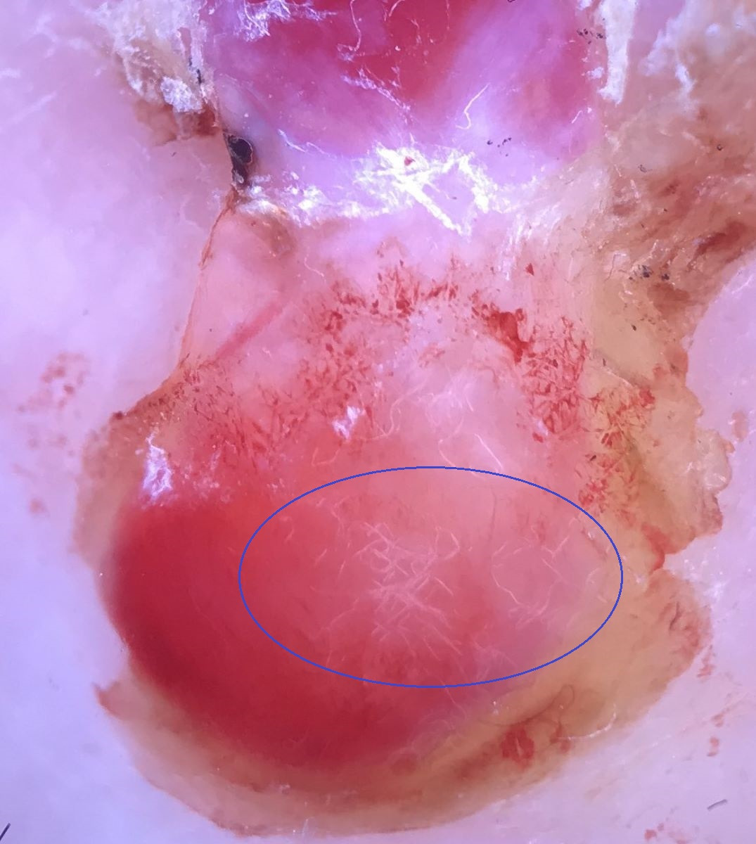
- Atypical vascular pattern - dotted, linear-irregular, helical, polymorphous (ie more than one pattern). Click here for an overview of vessel patterns in dermoscopy (click as opposed to hover)
- A solitary pink lesion for which there is no confident benign diagnosis
- Occasionally, melanoma has no clues and can be almost 'structureless' ie being devoid of structures. Such lesions will tend to still be the ugly duckling when compared to others in terms of either appearance, size, or the fact that they show signs of sustained growth in comparison to the person's other lesions
- Given the complexity of assessing melanocytic lesions dermoscopically, the dermoscopic examination of melanocytic lesions should only be undertaken by those who manage skin lesions on a regular basis and who have had appropriate training. For more information refer to the section on the PCDS Dermoscopy events, dermoscopy and the skin lesion diagnostic tools
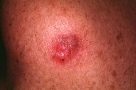
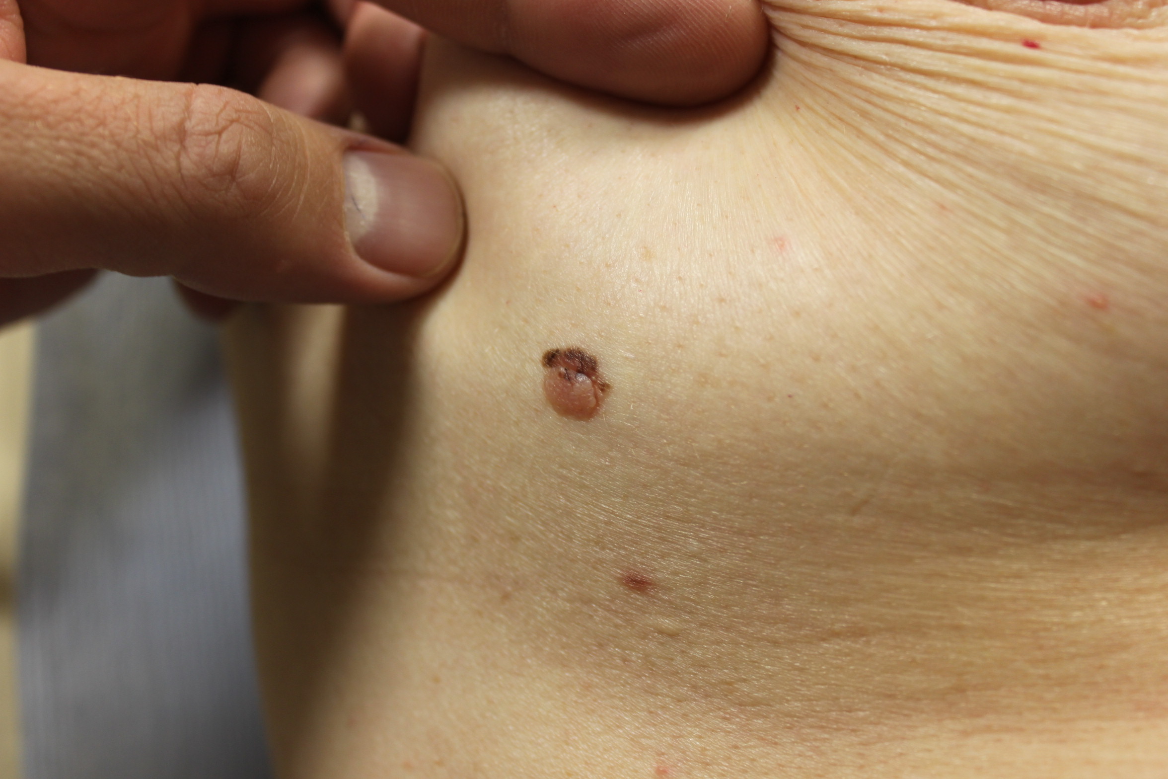
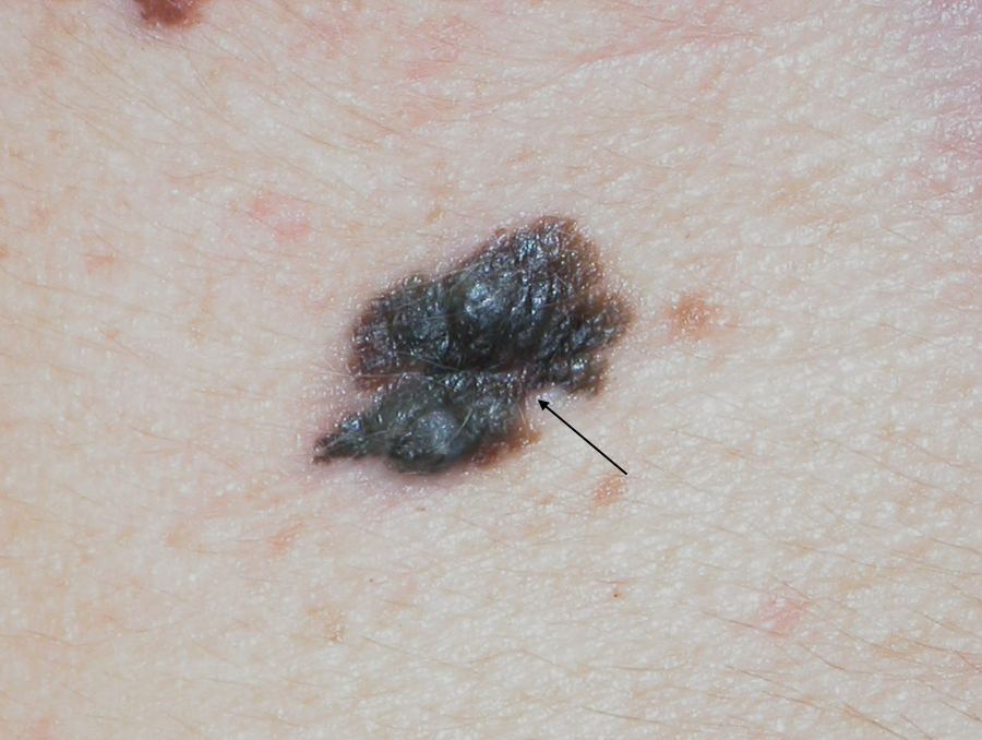
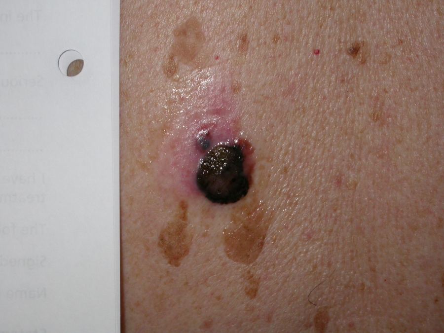
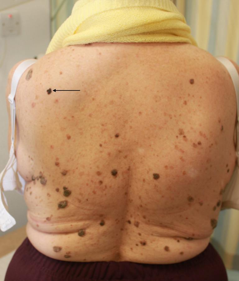
Clinical Images
Please refer to notes on image rights at bottom of the page with regards to individual image
ownership.
1
2
3
4
5
6
7
8
9
10
11
12
13
14
15
16
17
18
19
20
21
22
23
24
25
26
27
28
29
30
31
32
33
34
35
36
37
38
39
40
41
42
43
44
45
46
47
48
49
50
51
52
53
54
55
56
57
58
59
60
61
62
63
64
65
66
67
68
69
70
71
72
73
74
75
76
77
78
79
80
81
82
Investigations
- Lesions clinically suspicious of melanoma (or any incidental histological report of melanoma) should be referred urgently to Secondary Care
- Histological interpretation of melanocytic lesions is not always straightforward:
- It can be difficult to distinguish between high-grade dysplastic naevi and melanoma. To this end all lesions histologically found to be high-grade dysplastic naevi (moderate-severe dysplasia) must be completely excised
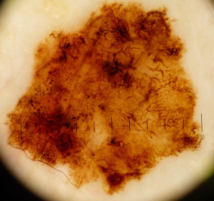
- Melanocytic lesions of unknown malignant potential - this is a term reserved for cases where there is histological uncertainty around whether or not the lesion is a melanoma. Such cases require further discussion at the skin cancer MDT
Management
Self-examination
General notes on management of melanoma / suspected melanoma
- Suspected cases of melanoma must be referred urgently to Secondary Care (2ww/USC pathway)
- Melanoma is primarily managed by surgical excision of the tumour and normal surrounding skin
- Patients with more advanced cases may be offered medical therapy as either:
- Targeted cancer drugs - used in patients who have a BRAF gene mutation
- Immunotherapy - drugs that help the immune system to attack the melanoma
- For more in-depth guidance refer to the NICE guidelines Improving Outcomes for People with Skin Tumours including Melanoma
Genetic testing
- While genetic testing is the role of Secondary Care, family history can be difficult to ascertin and a genetic link may not be picked up; it can be helpful for the referrer to add in any relevant family history on the referral letter
- People who carry a mutation on either CDKN2A or CDK4 (rare) have a higher risk of developing melanoma, cancer of the pancreas, or a tumour in the central nervous system. The following should be tested where the individual +/- family history meets ONE of the following criteria:
- ≥1 melanoma < 18 years, OR
- ≥2 melanomas and/or melanomas in situ age <30 years, OR
- ≥3 melanoma and/or melanomas in situ at any age, OR
- Melanoma and/or melanoma in situ AND ≥2 relatives (first / second / third degree relatives) with melanoma and/or melanoma in situ, OR
- Melanoma and/or melanoma in situ AND ≥1 first degree relative with melanoma and/or melanoma in situ; one individual has multiple melanomas and/or melanomas in situ, OR
- ≥1 melanoma and/or melanoma in situ OR melanoma and/or melanoma in situ and atypical moles AND ≥1 first degree relative with pancreatic cancer aged <60, OR
- Atypical moles AND ≥2 relatives (first / second degree relatives) with melanoma and/or melanoma in situ, OR
- Deceased affected individual (proband) where (i) the individual +/- family history meets one of the above criteria, (ii) appropriate tissue is available (tumour or normal), and (iii) no living affected individual is available for genetic testing
- Another genetic condition that can be tested for is the BAP1 tumour predisposition syndrome, a group of malignant and benign tumours associated with inherited mutations in the BAP1 tumour suppressor gene. The testing criteria is that an individual should have ONE of the following:
- A personal history of two or more core BAP1 associated tumours (mesothelioma, uveal melanoma, cutaneous melanoma, renal cell cancer or BAP1 inactivated melanocytic tumour - BIMT) (excluding two cases of melanoma)
- A personal history of two or more inactivated melanocytic tumours (BIMT); a histological diagnosis (previously known as any of - BAPoma, atypical Spitz naevus, Melanocytic BAP1-associated intradermal tumour (MBAIT) or naevoid melanoma-like melanocytic proliferation)
- Individual with a personal history of a BAP1 associated tumour and a first degree relative with a BAP1 core associated tumour (mesothelioma, uveal melanoma, cutaneous melanoma, renal cell cancer or BAP1 inactivated melanocytic tumour- BIMT) (excluding two cases of melanoma or renal cancer)
- Mesothelioma (less than 60 years) in the absence of asbestos exposure
- Uveal melanoma (<40 years)
- An up-to-date document for genetic testing can be found on the National geonomic test directory - rare and inherited disease eligibility criteria
In terms of further testing, NICE guidelines recommend monitoring for pancreatic cancer in patients with a CDKN2A mutation and one or more first degree relatives with pancreatic cancer.
A first-degree, second-degree, third-degree relative of the individual, or of a dependent of the individual is defined as:
- First-degree relatives include an individual's parents, siblings, and children
- Second-degree relatives include an individual's grandparents, grandchildren, uncles, aunts, nephews, nieces, and half-siblings
- Third-degree relatives include an individual's great-grandparents, great grandchildren, great uncles/aunts, and first cousins
Other resources
Disclaimer - the author PCDS cannot accept responsibility for any misleading or incorrect statements, and the management of individual patients remains the direct responsibility of the individual doctor. We do however hope that visitors to this site can contact us regarding comments that are considered misleading or incorrect so that we can continue to improve the site.
Image Rights - The PCDS would like to thank Dermatoweb, DermQuest (Galderma), and others who have contributed images. All named individuals and organisations maintain copyright for the relevant images.