Melanoma: acral lentiginous melanoma (including subungual melanoma)

LAST UPDATED: Aug 09, 2026

Acknowledgements: Author - Dr Tim Cunliffe.
Introduction

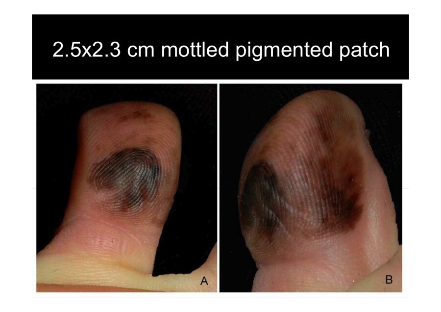
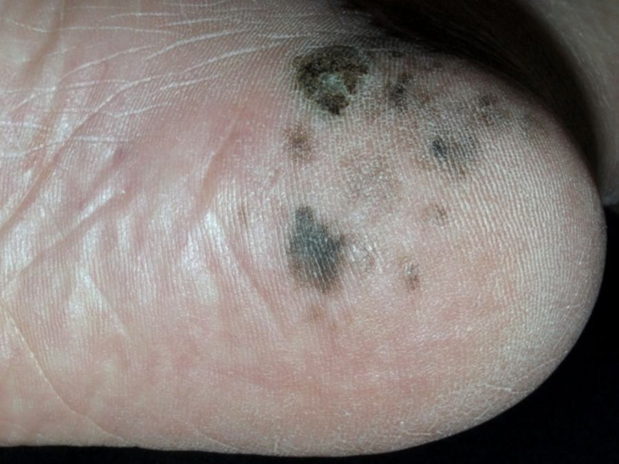
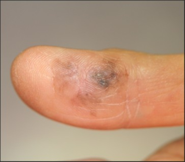
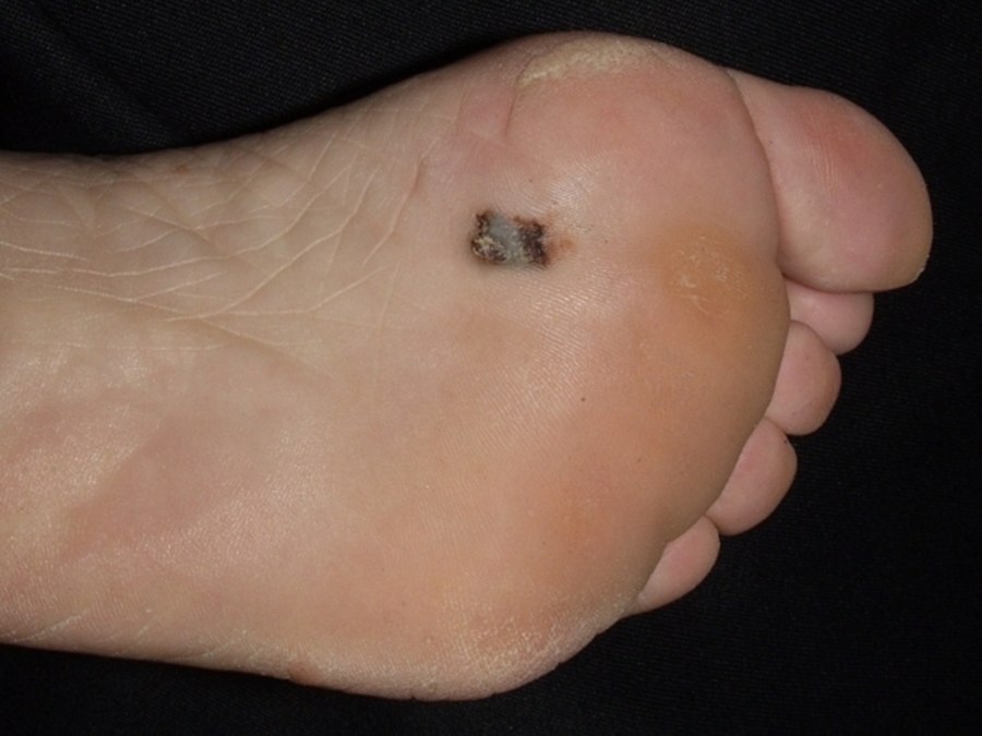
Acral lentiginous melanoma (ALM) is a rare type of skin cancer primarily affecting the palms, soles, and under the nails (subungual melanoma); the soles are the most commonly affected acral site. Unlike other melanomas, ALM is not strongly linked to sun exposure. ALM is often diagnosed at a later stage, which can lead to a higher risk of morbidity and mortality.

This chapter is set out as follows:


Aetiology

  • ALM is the least common subtype of melanoma, accounting for 2–3% of melanoma diagnoses
  • The cause is unknown
  • It is not related to UV exposure
  • It affects men and women equally
  • It affects all skin types, and is the most common type of melanoma in skin of colour

History

  • The majority arise in patients aged over 40
  • Except for nodular ALM, lesions grow relatively slowly, especially in the early stages

Clinical findings

Melanocytic lesions on palms and soles - overview 

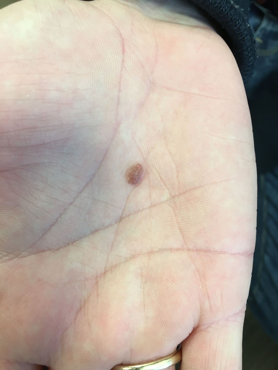
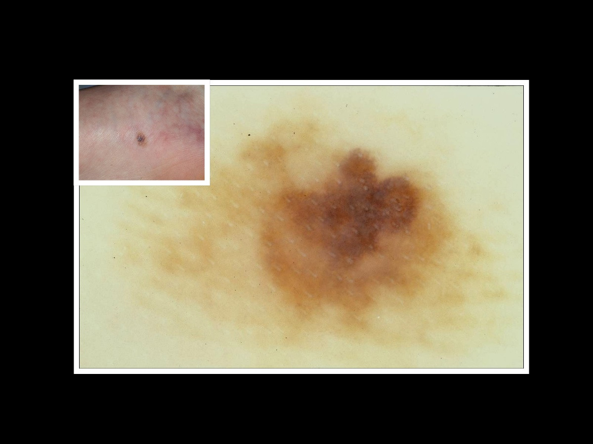
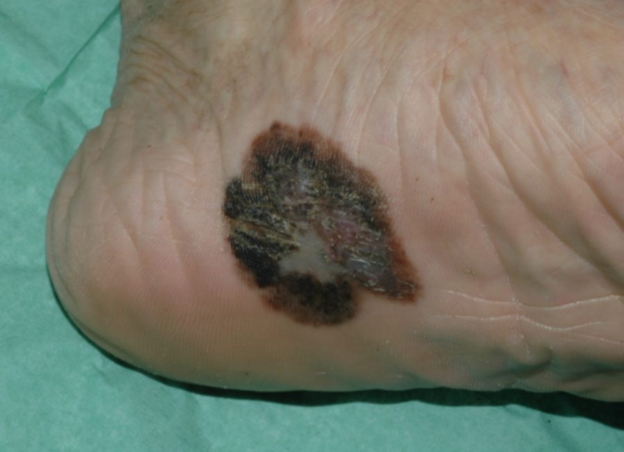
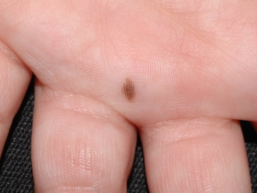
From a clinical perspective, ALM usually presents as a slow growing deceptively banal-looking pigmented macule or patch, hence the term lentiginous. The development of nodular areas within the lesion suggests deeper invasion.

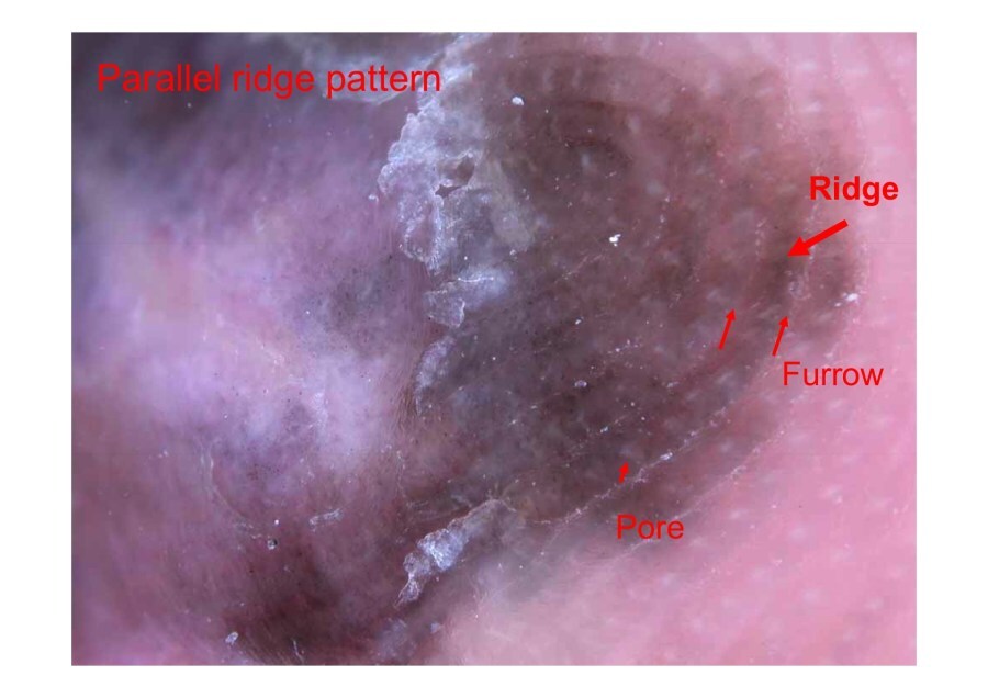
The dermoscopic appearance of melanocytic lesions at acral sites differs to other areas of the body due to the different anatomy. Acral skin consists of a parallel arrangement of the skin surface markings known as the dermatoglyphics, which run straight or in whorls. These surface markings are composed of parallel gyri (ridges) and sulci (furrows). Openings of the eccrine sweat ducts, the acrosyringium, are found in the centre of the ridges. 

Once the observer understands the anatomy of skin at acral sites, we can use similar dermoscopic principes to those used for melanocytic lesions at typical body sites, namely:

  • Benign melanocytic naevi tend to be well-organised
  • Acral melanoma may have chaos or clues
  • Careful dermoscopic follow-up can be used for certain small flat melanocytic lesions (see later); palpable lesions must never be followed up  

As with melanoma at any site, there are rule breakers, which at acral sites include (hover over terminologies for description):

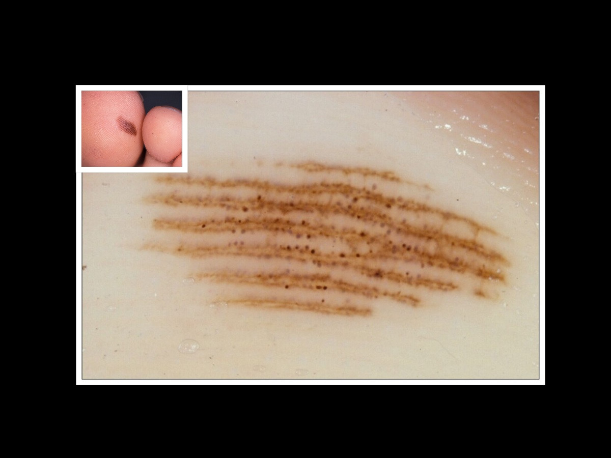
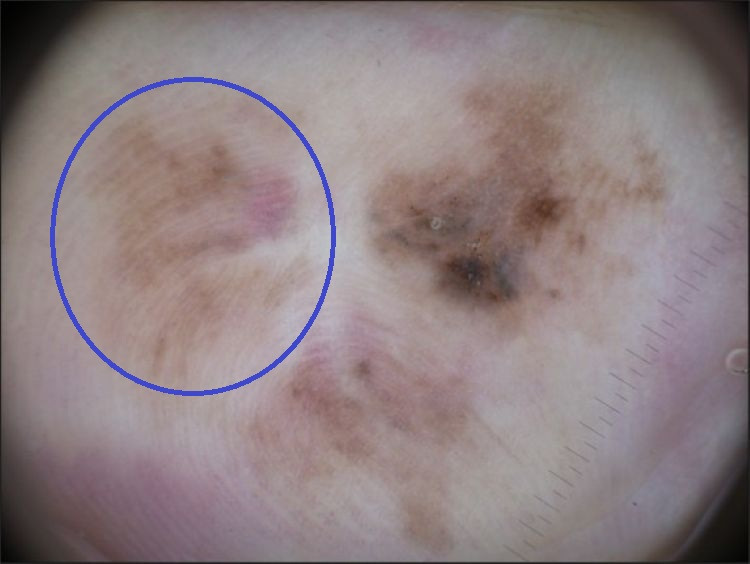
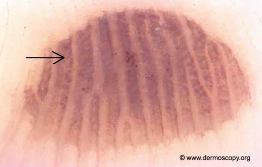
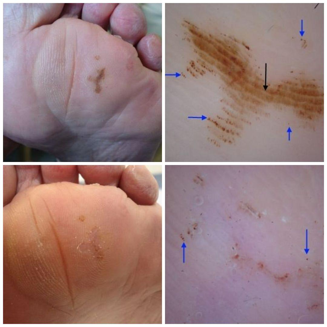
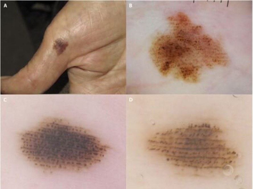
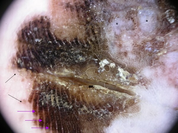
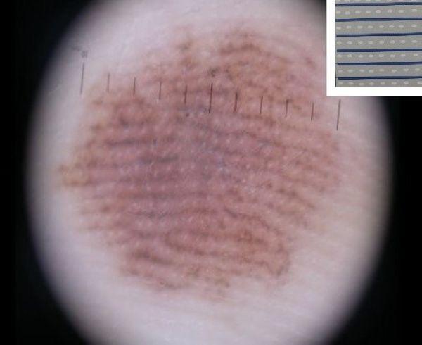
  • The parallel ridge pattern - the most common presentation of acral melanoma, which is not chaotic  
  • The fibrillar pattern - this disguises the real pattern meaning a more detailed clinical assessment is required; most such lesions will turn out to be benign
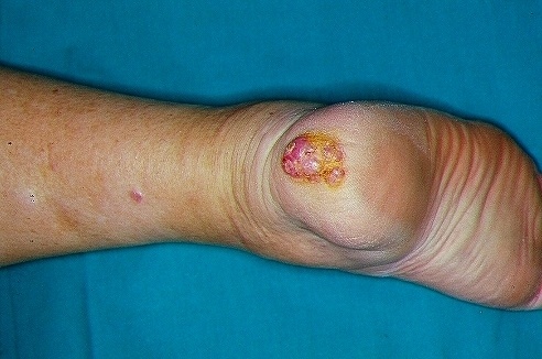
  • Nodular / ulcerative lesions - as at other sites, nodular melanoma may lack typical dermoscopic features of melanoma. As such, all acral EFG lesions (= all of elevated + firm + growing) should be regarded with suspicion (discounting obvious benign lesions such as myxoid cysts), hence why there is no room for following up palpable lesions

Before focusing on ALM, it is important to have a good understand of benign patterns, which are much commoner.


Dermoscopic features of benign acral lesions (hover over terminologies for dermoscopic description)

Benign acral naevi tend to have the following dermoscopic features (hover over terminologies for description):

  • Often < 7 mm in diameter
     
  • One colour relevant to the skin type, occasionally with central hyperpigmentation 
     
  • Tend to be well-organised 
     
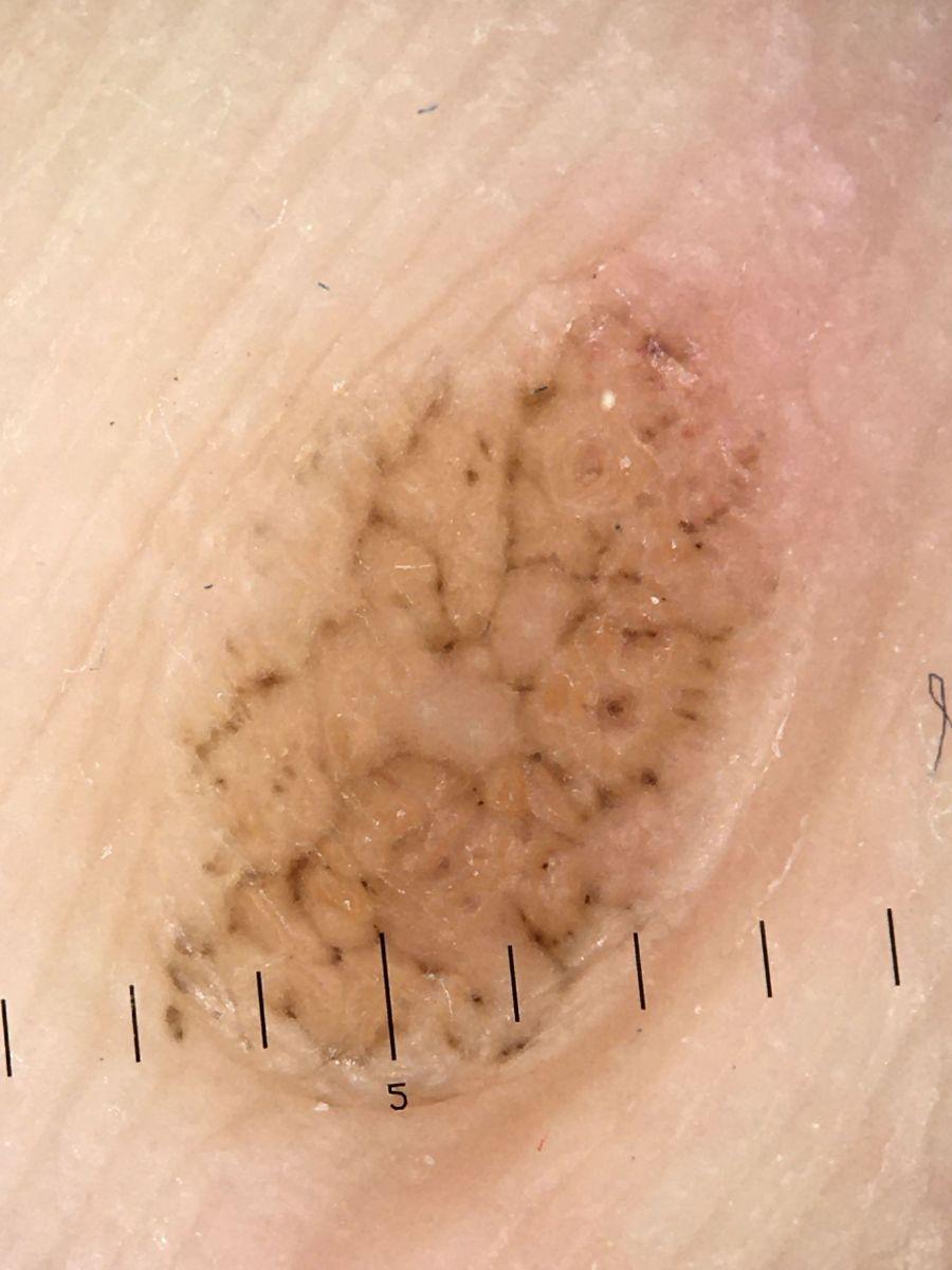
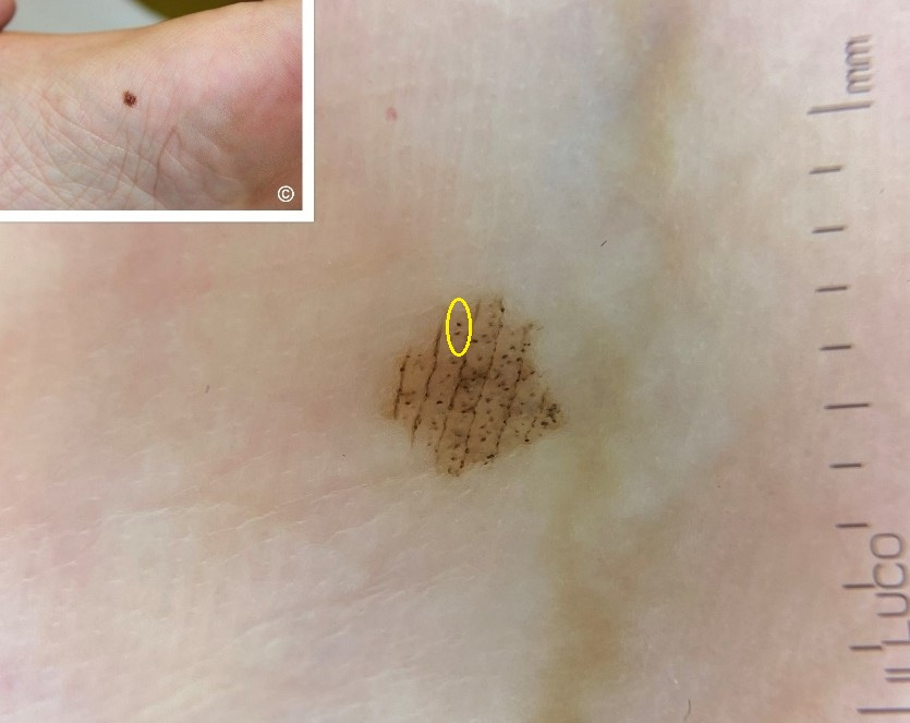
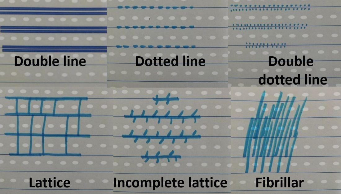
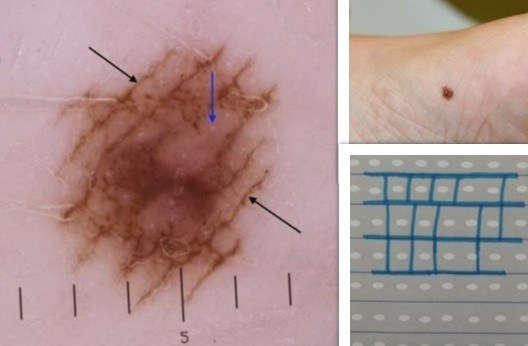
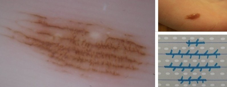
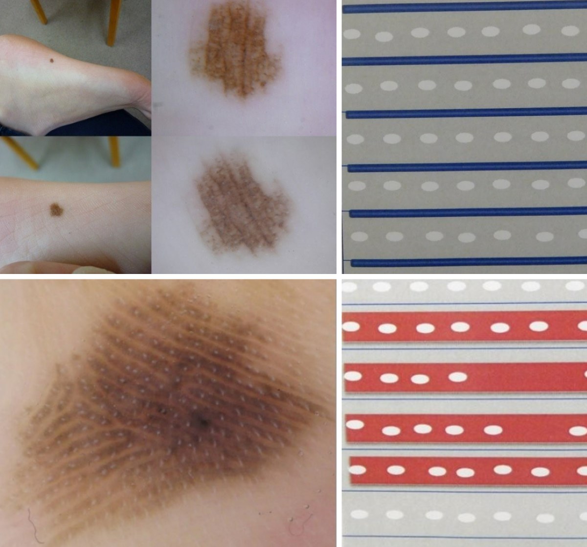
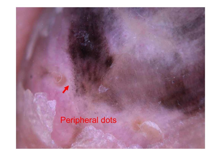
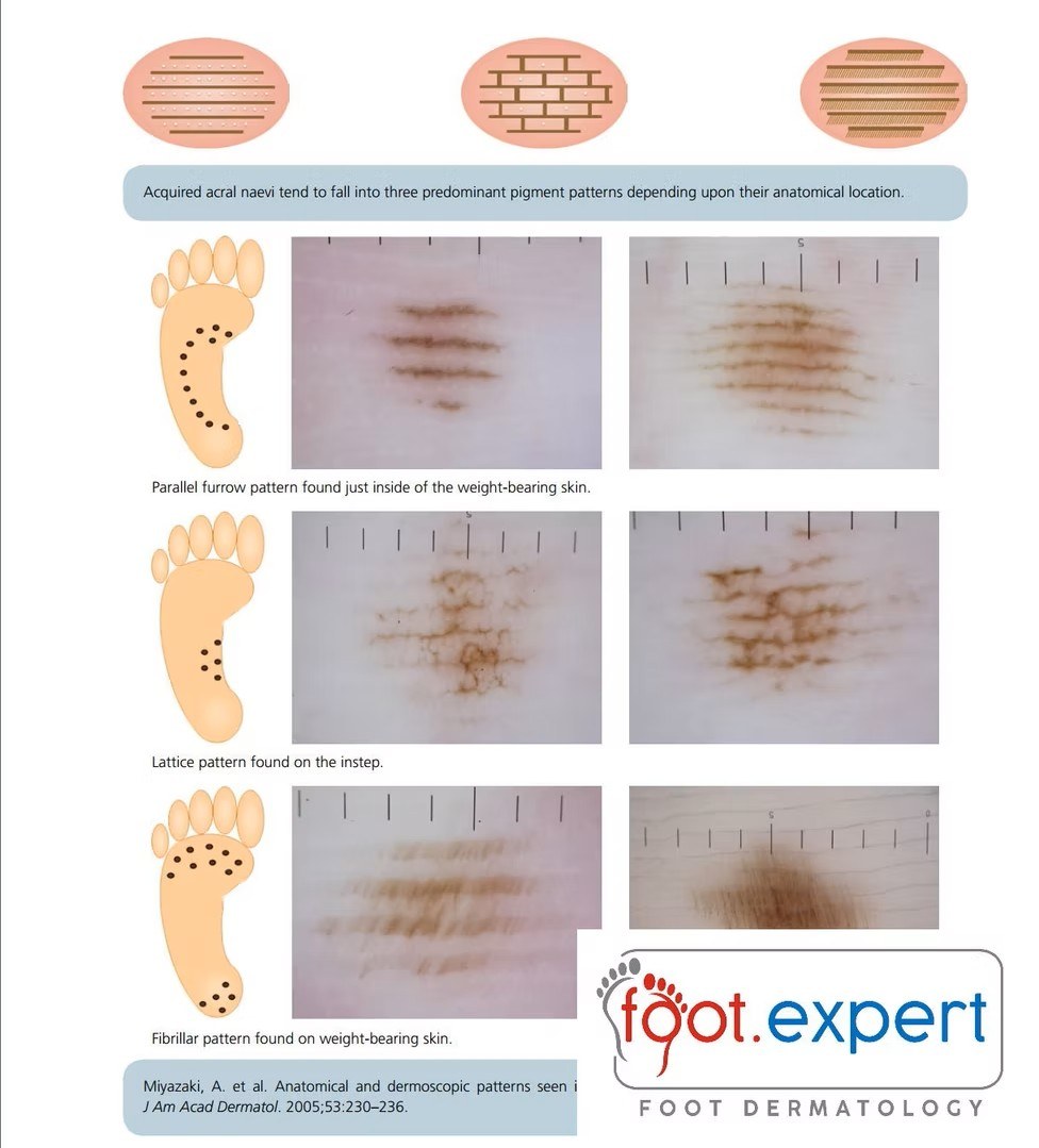
  • Regular thin lines and/or dots in the sulci (furrows) = parallel furrow pattern (PFP) and its variants. This pattern is found on the peri-weight-bearing skin of the foot, and is the most common pattern on the palms
     
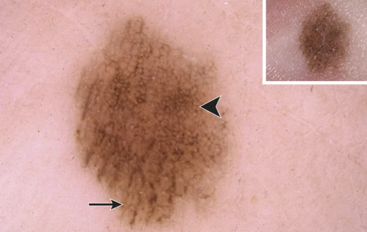
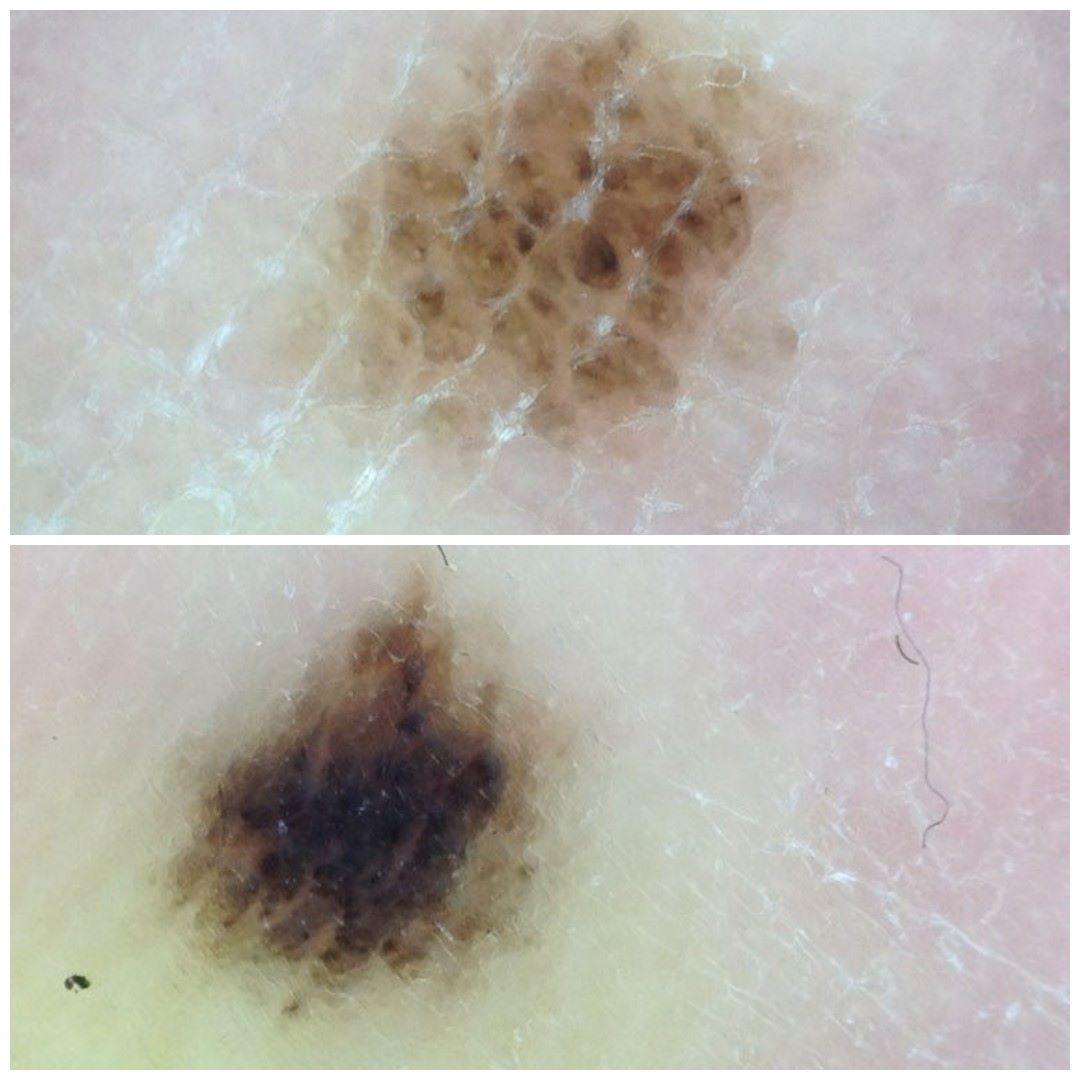
  • A ‘network-like’ pattern = the lattice-like pattern (found on the instep of the foot)
     
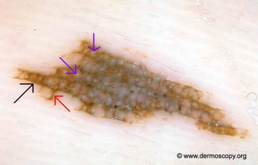
  • A regular globular pattern in combination with a parallel furrow pattern. The most common such pattern is sometimes described as 'peas in the pod' with regular globules in the wider ridges
     
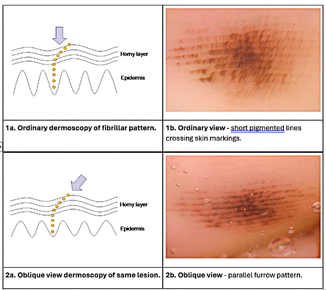
  • A typical fibrillar pattern on weight-bearing areas - oblique dermoscopy may demonstrate the true pattern is that of a parallel furrow pattern 
     
  • A homogenous pattern especially in skin of colour (a single even colour throughout the lesion)
     
  • AND, ABSENCE OF ANY RED FLAGS - some cases of ALM display benign and malignant features; any single clue for malignancy overrides benign features 

There are several variants of the parallel furrow pattern (PFP), all of which should be well-organised being comprised of thin lines and/or dots. Benign patterns include single and double dotted lines, double lines (one pattern of which has a thin line in the ridge), curved lines in raised dermal naevi, and a partial lattice-like pattern.

Of note,  congenital melanocytic naevi (CMN) at acral sites are usually larger, more asymmetric, and have a greater degree of colour variation than acquired acral melanocytic lesions. Additionally, acral CMN may evolve dramatically within the first few months, with clinical changes such as increase in size, thickness, and darkness. Such lesions often require specialist assessment.

Parallel furrow pattern vs parallel ridge pattern - the ink test

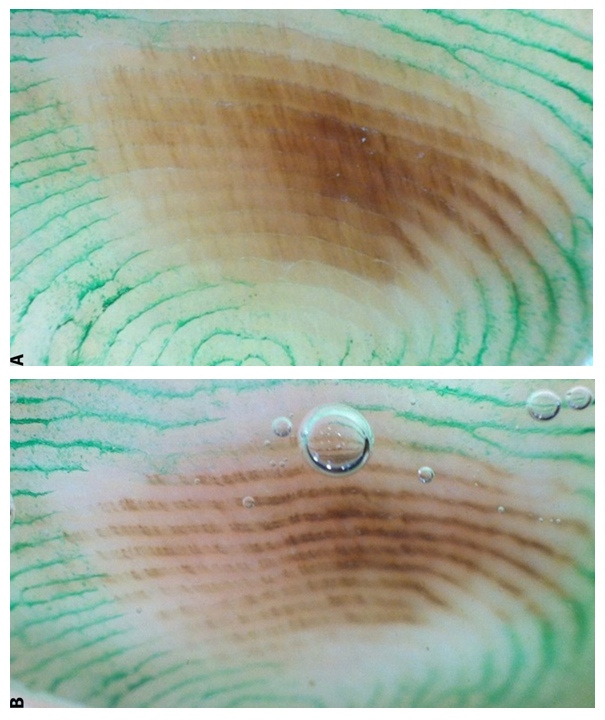
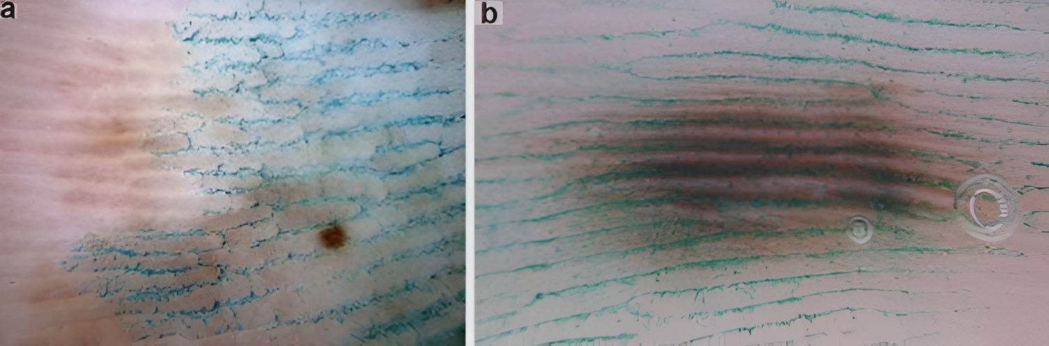
It is very important to be able to differentiate between a parallel furrow pattern (PFP), and the parallel ridge pattern (PRP), the most common pattern seen in acral melanoma. A PRP is defined as prominent band-like pigmentation on the ridges of the dermatoglyphic lines. If there is any uncertainty as to whether it is a PFP or PRP, the ink test can help differentiate:

  • Apply ink (from a bottle, not pen) onto the acral lesion; a narrow gauge syringe can help
  • Leave the ink on the skin for a few seconds
  • Gently wipe the surface using a cotton swab 
  • Examine with a dermatoscope - the ink will be sitting in the narrow furrows

Once an observer can identify a PFP from a PRP this also helps better evaluate the fibrillar pattern, which masks the true dermoscopic pattern.

Assessing the fibrillar pattern

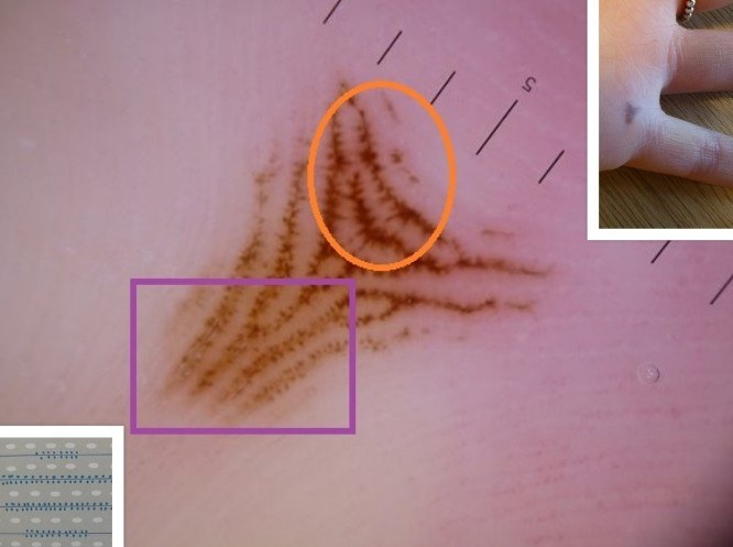
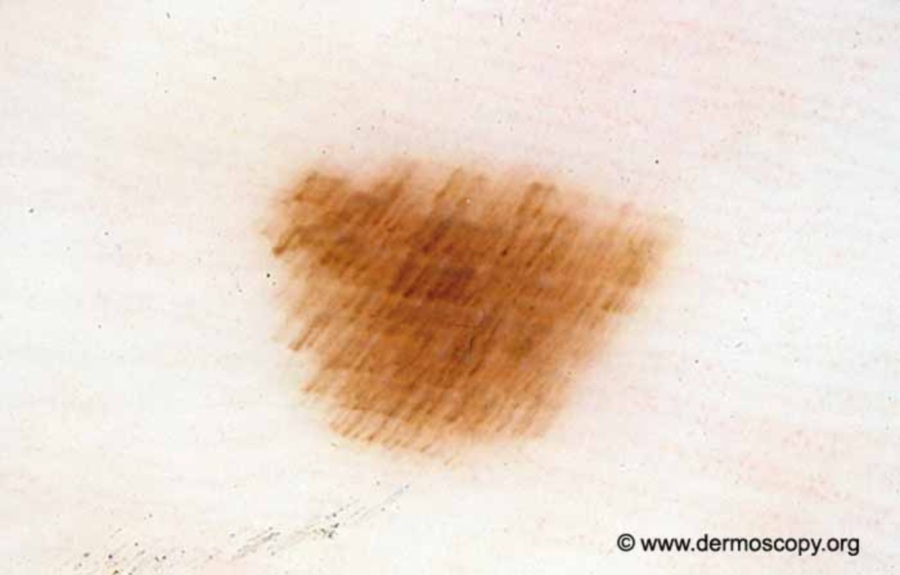
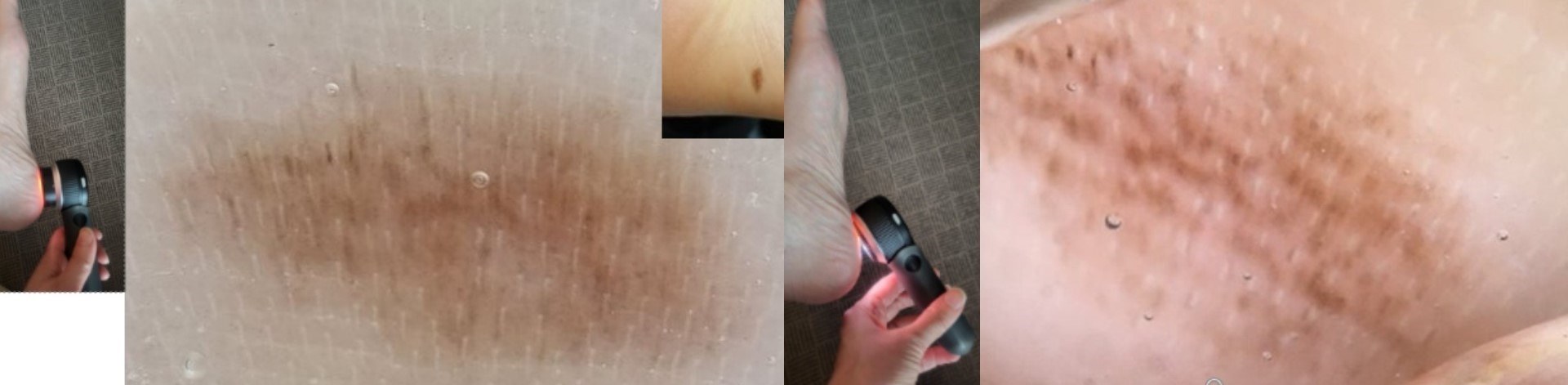
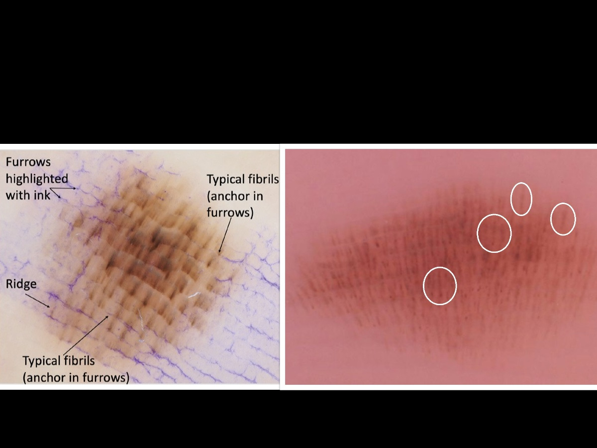
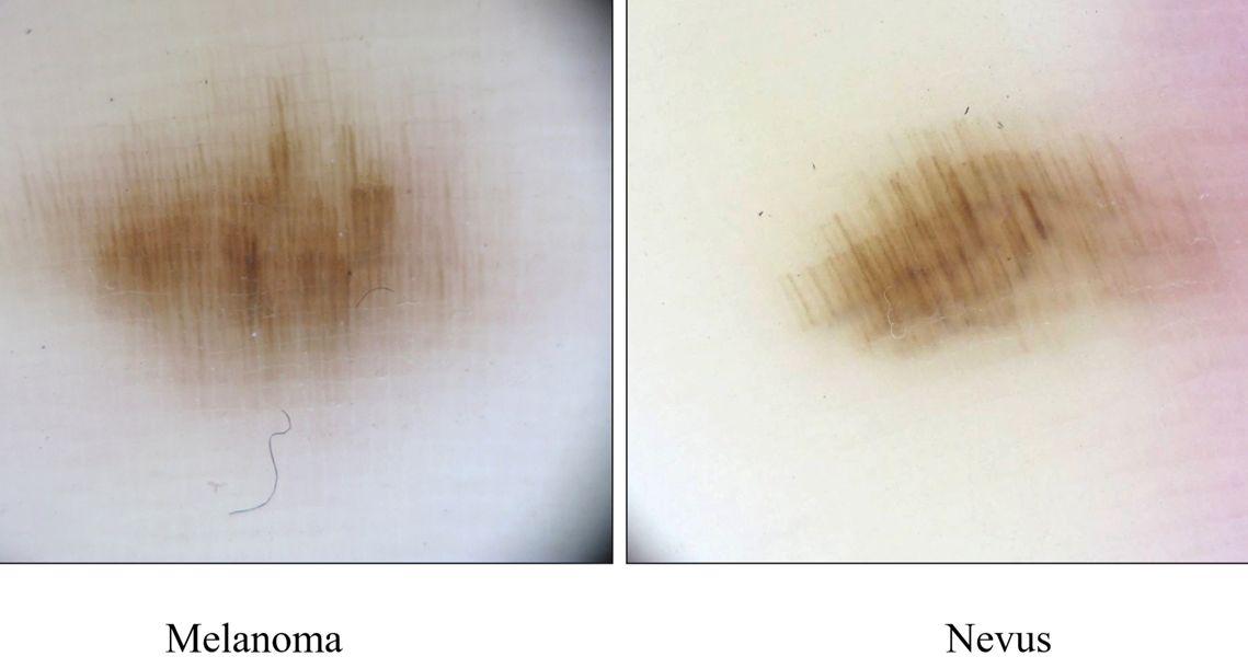
The fibrillar pattern (FP) is detected in 10–20% of plantar naevi, presenting at sites of pressure; it is uncommon on the palms. The FP is characterised by densely packed brownish fibrillar lines arranged in a parallel fashion and crossing the skin markings. Each fibril of the pattern corresponds to a melanin column in the thick cornified layer which is obliquely arranged due to the mechanical pressure from the body weight. In the majority of cases the FP is benign, with pigment originating from the furrows; occasionally FP can be malignant with pigment arising from the ridges.

  • Benign fibrillar pattern - the fibrils tend to have a similar appearance and sit obliquely across the ridges, and are anchored in the furrows
     
  • Malignant fibrillar pattern:
    • The fibrils tend to be unevenly distributed and variable in colour and thickness. In most cases, the endpoints of the fibrils are arranged randomly. Moreover, the PRP is not infrequently detected at least focally within a lesion
    • A negative fibrillar pattern is also suspicious. This pattern consists of whitish rods arranged in a parallel fashion on the dark structureless or fibrillar background. The whitish rods represent intracorneal eccrine ducts

Differentiating between benign and malignant fibrillar patterns is not always easy; the oblique dermoscopy technique can help the observer.

Oblique dermoscopy refers to resting the dermatoscope on the lesion and then tilting the scope, the lower edge of the scope must remain in contact with the edge of the lesion - looking through the dermatoscope at an oblique angle enables the observer to look underneath the stratum corneum, and may help trace the origins of the pigment, which in the case of a furrow pattern usually suggests a benign lesion. In acral melanoma the pattern does not change to a parallel furrow pattern; however, it is uncommon to identify a parallel ridge pattern. When the observer cannot determine the nature of a FP, six-monthly follow-up is recommended, although if the lesion evolves to greater than 7 mm in diameter, urgent referral is recommended.

Transition patterns

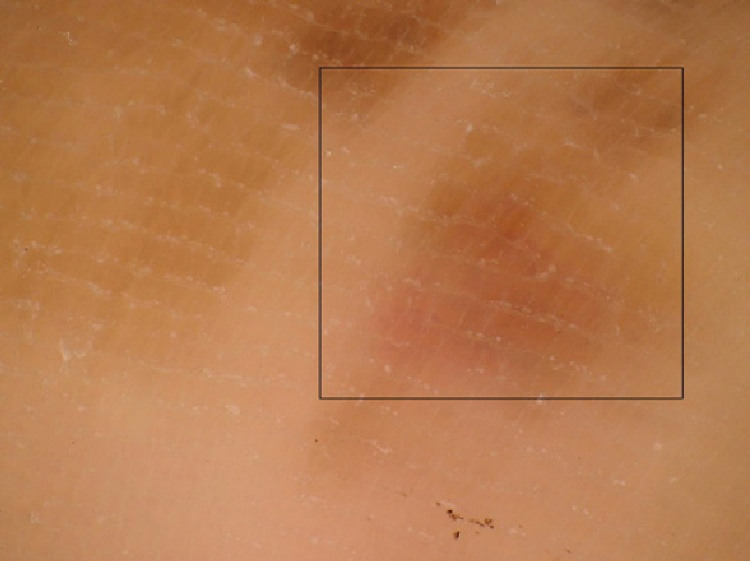
In some benign lesions more than one pattern may be noted, e.g. if a lesion involves both weight-bearing and non-weight bearing areas of skin, or if a lesion curves on to the side of the foot; in such cases the shape of rete ridges transitions from parallel to a reticular pattern. 


Acral lentiginous melanoma, excluding the nails (hover over terminologies for dermoscopic description)

For the majority of cases of ALM the observer can use the Chaos & Clues algorithm; albeit with some different interpretations, and the EFG rule used at other body sites.

  • Chaos 
     
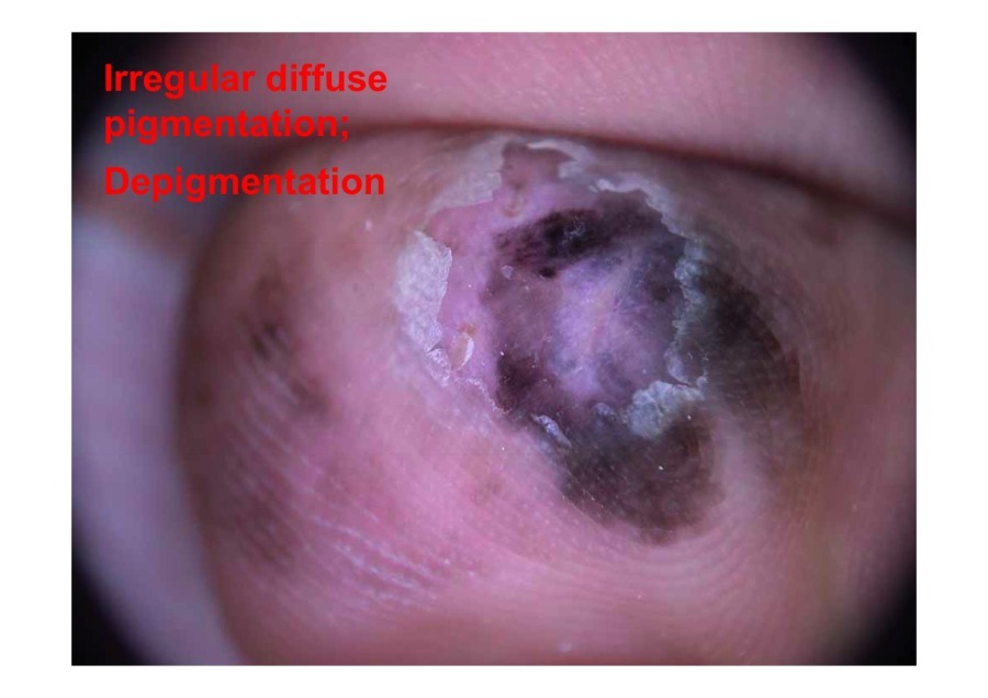
    • Colour - irregular diffuse pigmentation, or more than one colour or shade of colour (except central hyperpigmentation demonstrating a similar colour to the rest of the lesion, e.g. medium brown centre and lighter brown periphery)
       
    • Structures - a multicomponent pattern with more than one type of structure, especially if disorganised
       
  • Clues
     
    • Rather than an atypical pigment network (as on standard body sites), in acral lesions both the parallel ridge pattern (PRP) and irregular fibrillar pattern are suspect for melanoma. In the PRP the pigment bands are wider than in the PFP, and the acrosyringium are often affected
       
    • An irregular globular pattern - globules distributed in an erratic fashion, and/or variable in size or colour
       
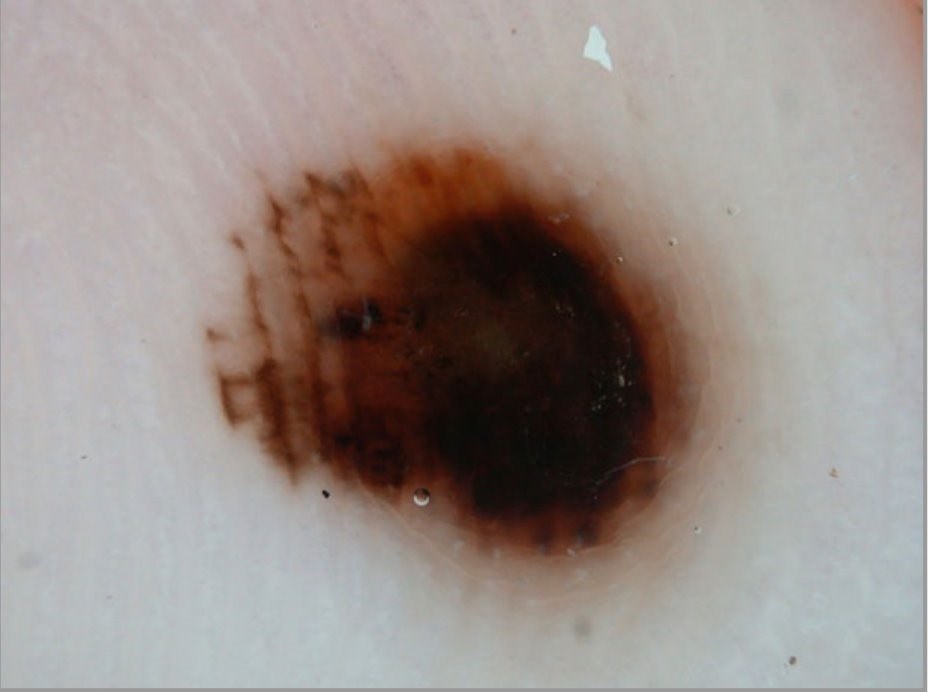
    • An eccentric structureless area (or blotch); any colour
       
    • A blue-white veil
       
    • White lines - these can either be a negative fibrillar pattern or shiny white lines (seen in polarised light)
       
    • Streaks - a serrated pattern marked by jagged or spiked pigment projections
       
    • An atypical vascular pattern - milky red areas and/or multicomponent pattern with atypical vascular structures (click here for an overview of Vessel patterns in dermoscopy)
       
  • EFG refers to ALL of elevated + firm + growing. All such lesions at acral sites should be regarded with suspicion (discounting obvious benign lesions such as myxoid cysts)

A benign lesion presenting as a parallel ridge pattern is haemorrhage (talon noir). Such lesions should be at sites of pressure / friction and have sharply defined edges, sometimes with red-black globules situated adjacent to the lesion; most importantly the pigment can be removed by pairing of the stratum corneum

The BRAFF assessment

The BRAAFF score has been shown to significantly improve the diagnostic accuracy of dermoscopy for the diagnosis of ALM. A score of 1 or higher suggests melanoma and warrants further evaluation. A score of 0 or less is less likely to be melanoma, but other factors should still be considered. The BRAAFF score should be calculated as follows:

Malignant features:
B= irregular Blotch: +1 point
R = parallel Ridge pattern: +3 points
A = Asymmetry of structures: +1 point
A = Asymmetry of colours: +1 point

Benign features:
F = parallel Furrow pattern: -1 point
F = Fibrillar pattern: -1 point 


Subungual melanoma (hover over terminologies for dermoscopic description)

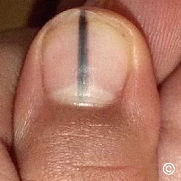
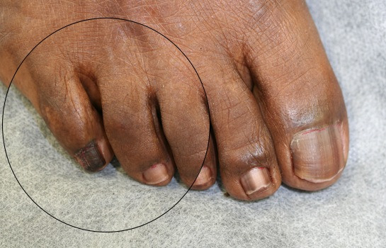
Although any digit can be affected, the big toe and thumb are the sites most commonly affected.

  • Nail matrix melanoma 
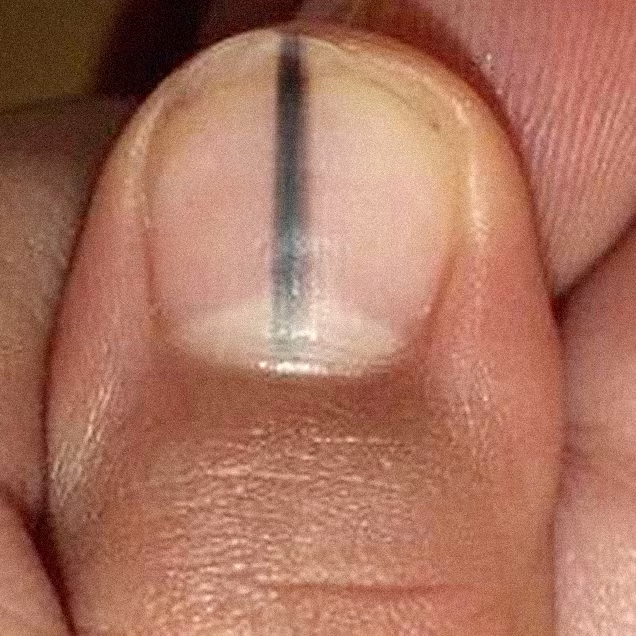
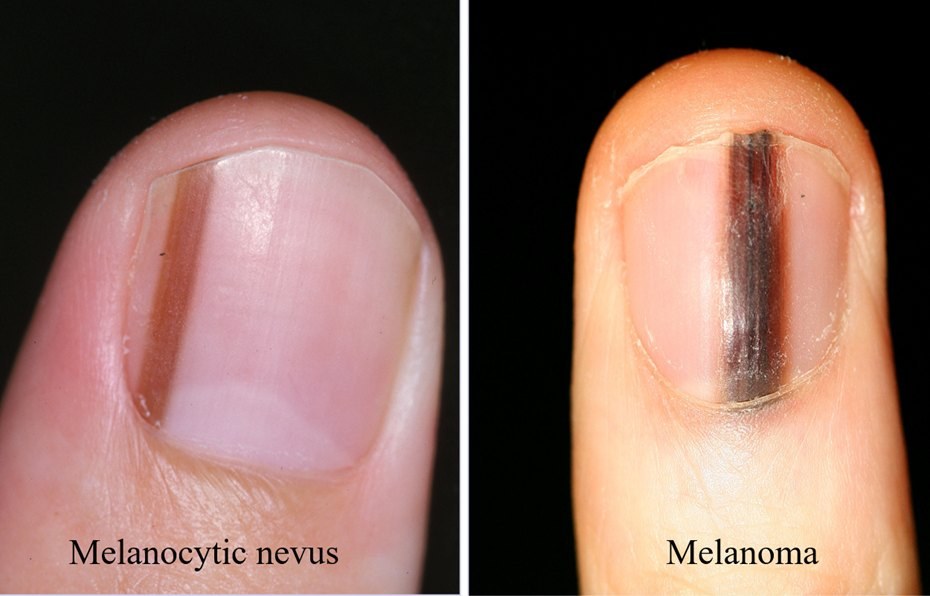
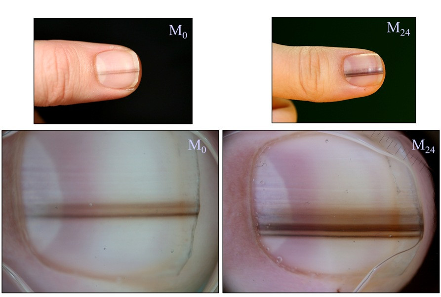
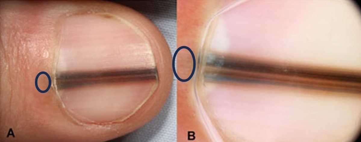
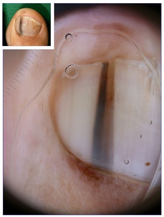
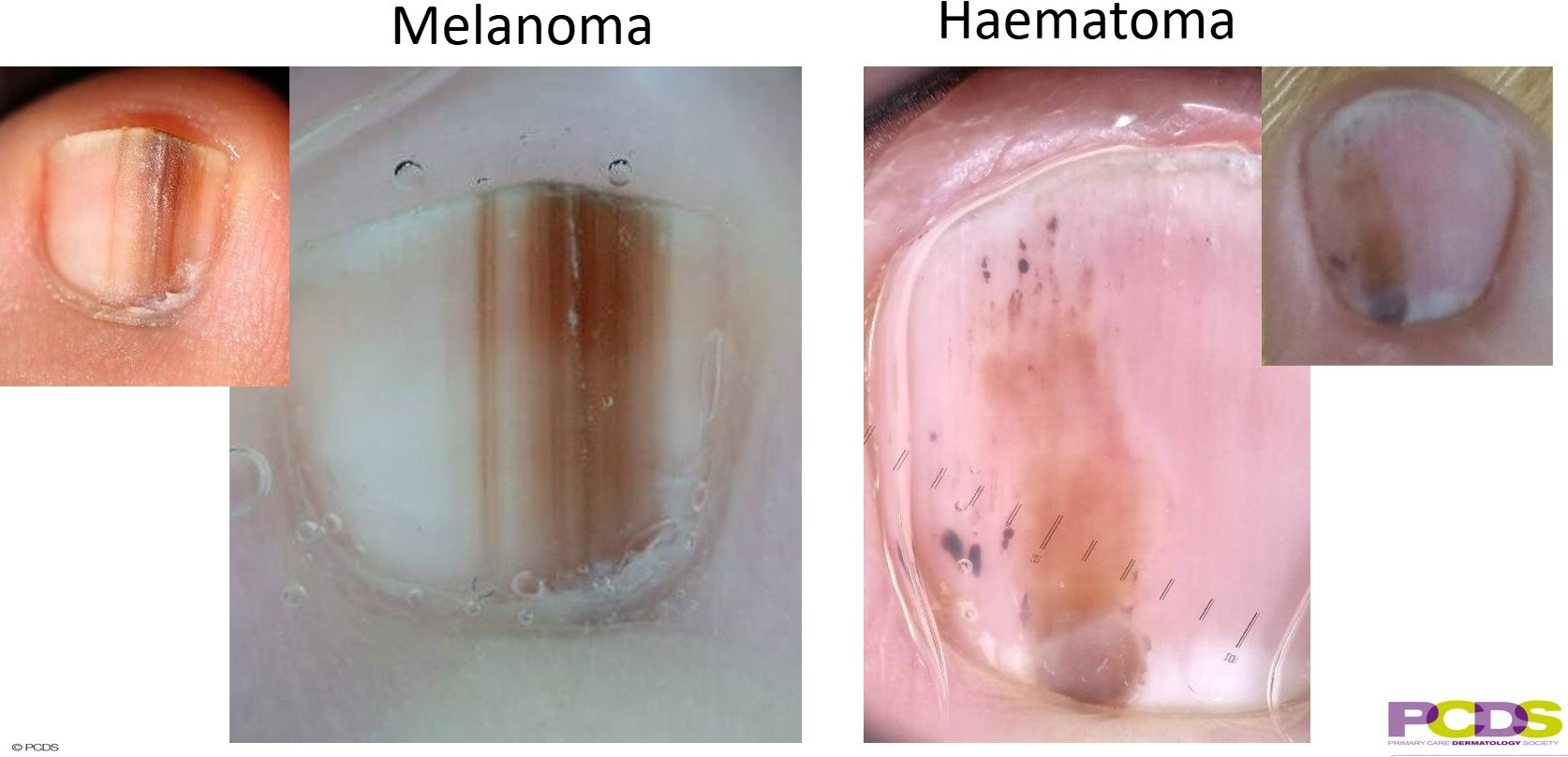
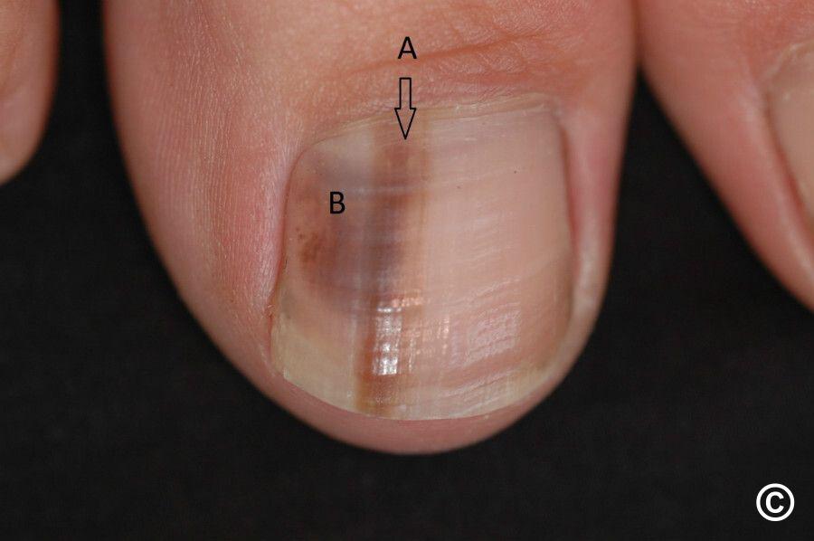
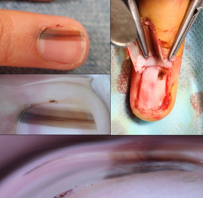
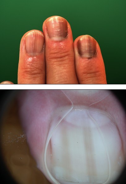
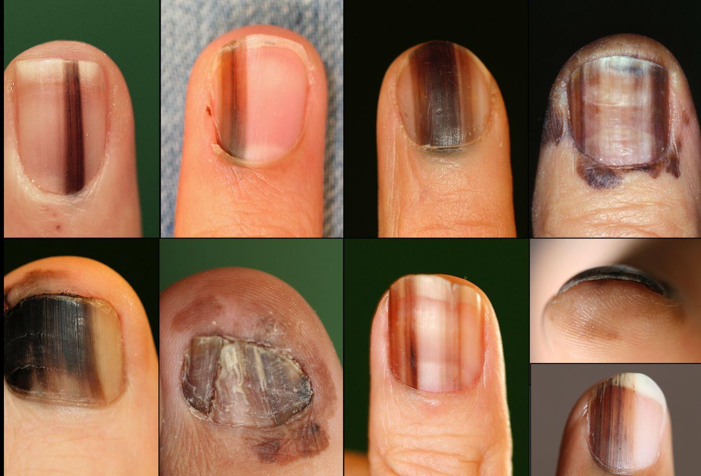
    • Usually evolve slowly, presenting with a line / band of pigment affecting one nail. Occasionally a line / band may be much lighter if the melanoma is hypomelanotic, ie containing little pigment. Given that the melanoma is in the nail matrix, the line always starts at the base of the nail and grows longitudinally, never loosing connection with the proximal nail fold. The pigmented line is melanin transferred to onychocytes which travel in the nail matrix, as opposed to melanoma in the nail plate. The early stages of nail matrix melanoma should be suspected when a single nail is affected by any of the following:
      • A black line of any diameter (unless < 3mm diameter in skin of colour)
      • A line of any colour (including hypomelanotic lesions) that is wider than 3 mm in diameter at the proximal end
      • The triangle sign - as the melanoma progresses the band widens, sometimes appearing broader at the proximal end and narrower at the distal end, which is known as the 'triangle' sign
      • A line / band containing more than one colour or one shade of colour 
         
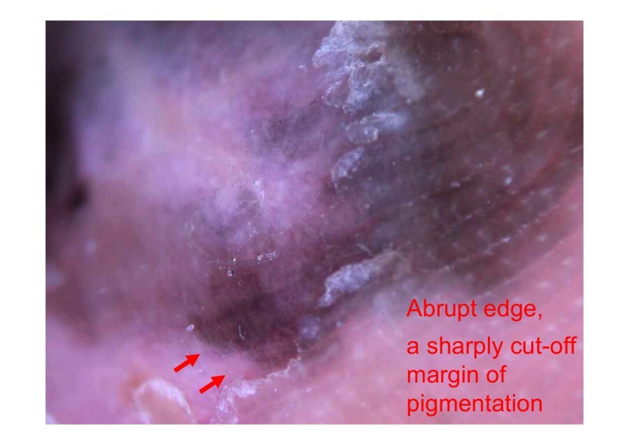
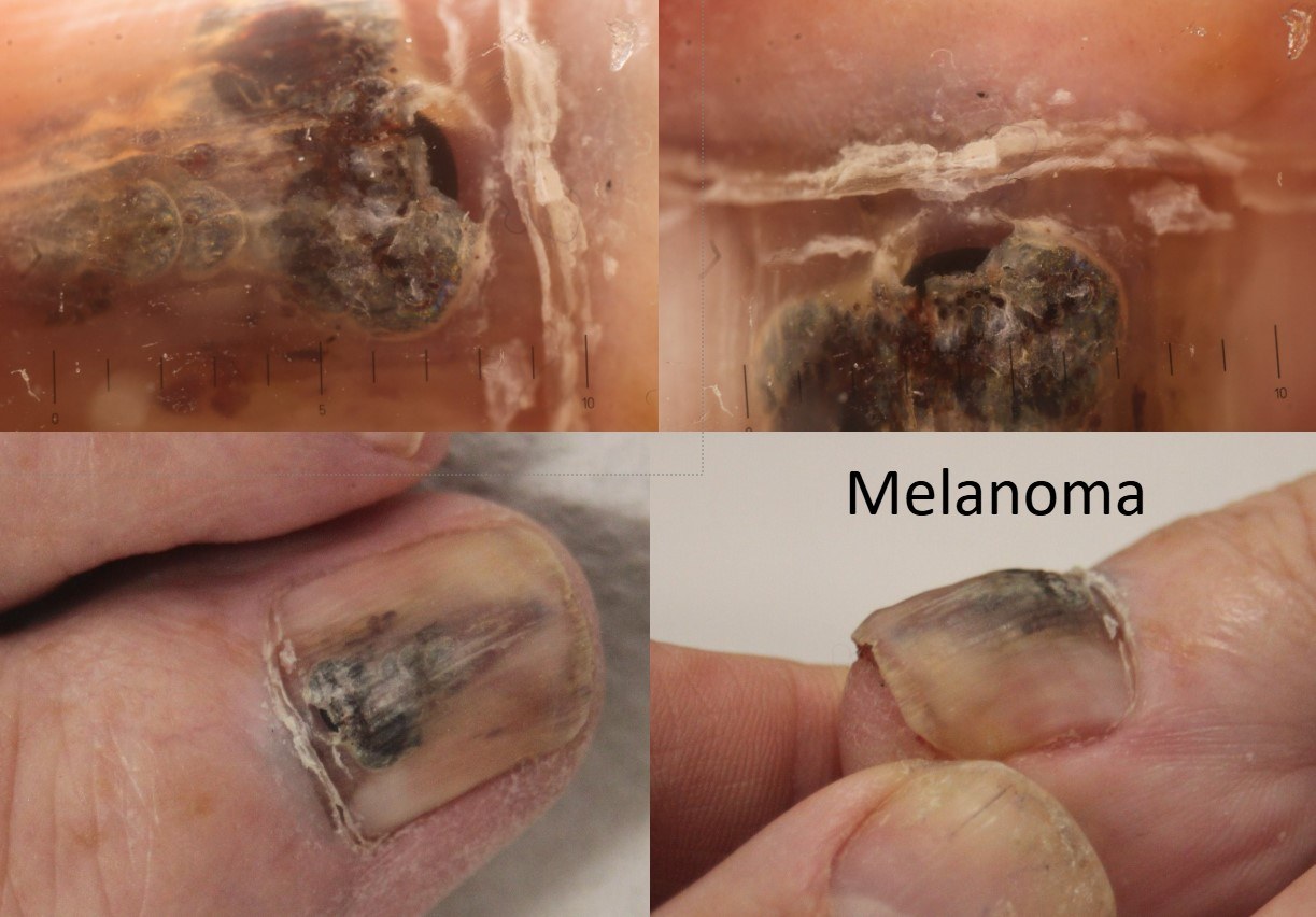
    • Extension of pigment onto the proximal or lateral nail fold (Hutchinson's sign), or the development of a nodular / ulcerative component, are usually late features. Hutchinson's sign is not always an accurate predictor of melanoma as periungual pigmentation is also present in benign disorders and Bowen's disease. Nail pigmentation may also be an illusion, especially in children with nail matrix naevi, due to transparency of the nail folds showing through the pigment
       
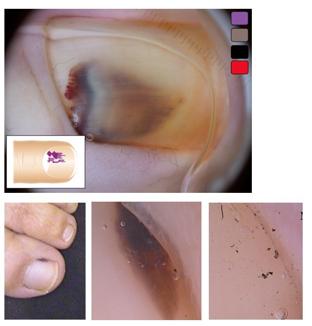
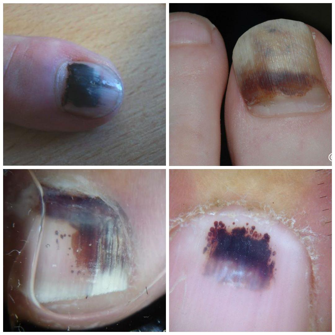
    • Subungual haematoma is very much more common than melanoma, and like melanoma is often seen in the great toe, usually with no history of trauma. A subungual haematoma is often easy to recognise:
      • Although the blood often involves the nail fold, a straight line / band is absent
      • Projections tend to have a curved appearance
      • Colours often progress as one would expect in a bruise
      • Dermoscopy often shows red clods of blood  
      • Rarely, subungual melanoma can bleed in which case both blood and the signs of melanoma described above will be present   
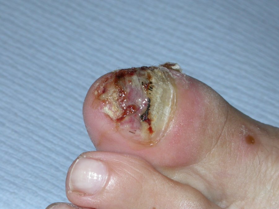
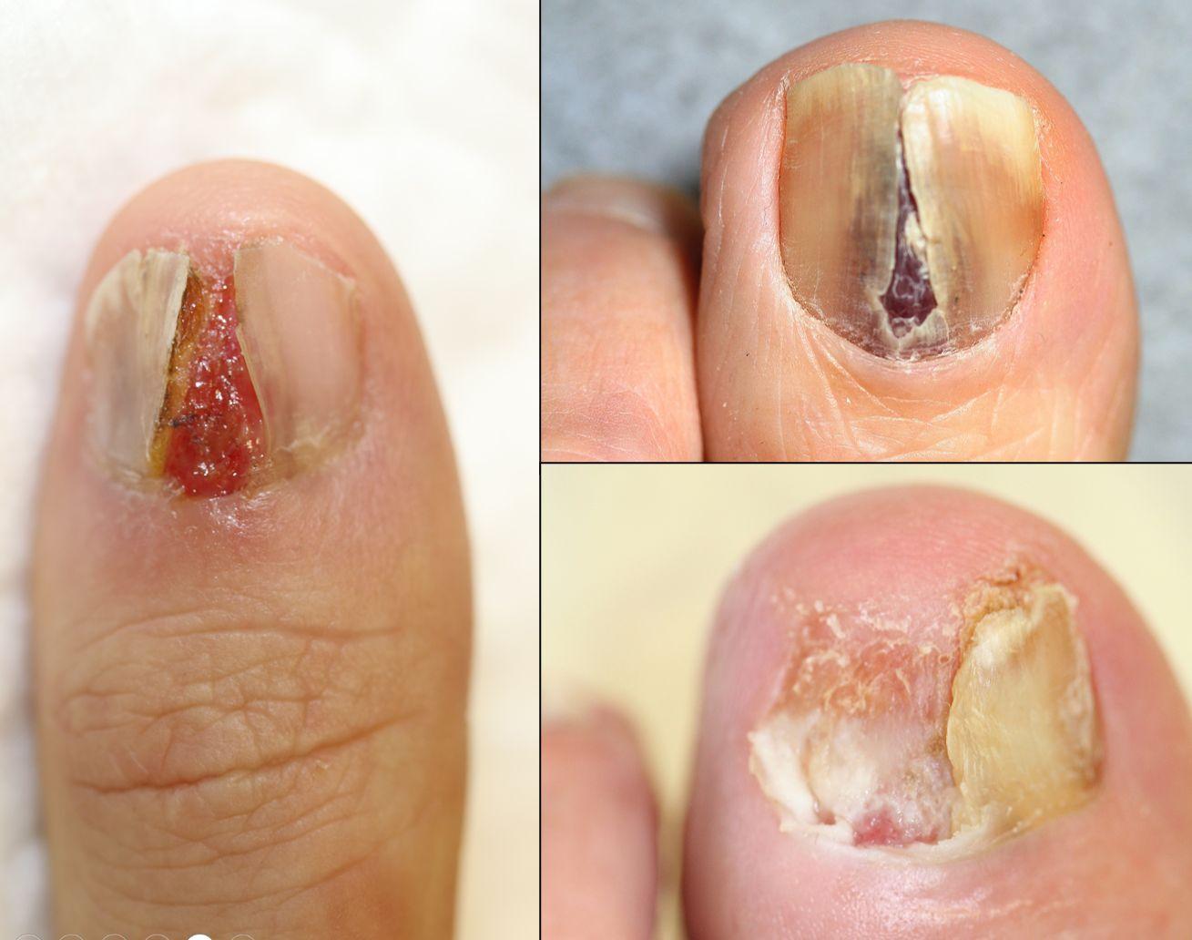
  • Nail bed melanoma 
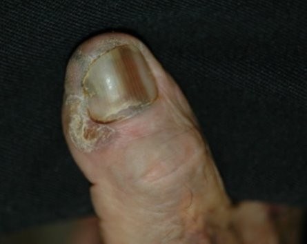
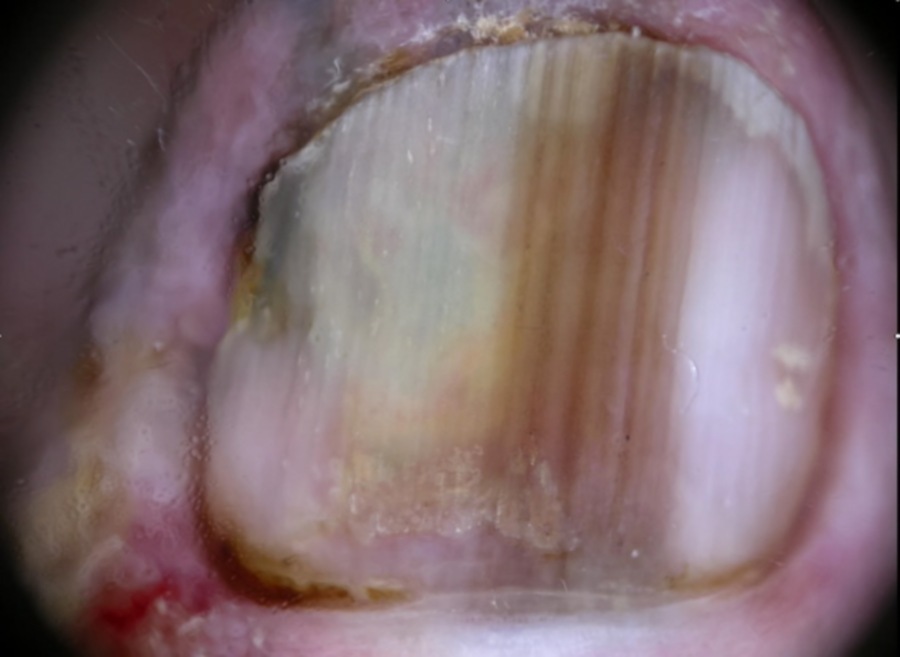
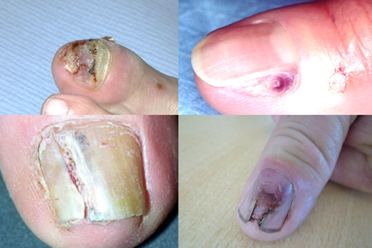
    • May have haemorrhage or pain and usually present late with nail plate destruction. The melanoma may arise as a nodule or take on the appearance of granulation tissue. While melanoma at this site can be pigmented, a disproportionate number are pink (amelanotic), making it almost impossible to differentiate with SCC, although the latter are more likely to start at the periphery. Essentially, nail destruction in combination with any of a thickening of the nail bed, a papule or nodule, or ulceration, requires an urgent referral
       
    • In contrast, fungal infection / psoriasis cause thickening, onycholysis and subungual hyperkeratosis, usually of multiple nails

There are many other causes of pigmented nails.


Clinical Images

Please refer to notes on image rights at bottom of the page with regards to individual image ownership.
Investigations

  • Suspected cases of subungual melanoma must be referred urgently to Secondary Care (2ww/USC pathway)
  • If a biopsy is needed this should always be taken from the nail matrix.  Any skin showing Hutchinson's sign must not be biopsied as such changes do not represent local in-transit melanoma and can lead to false negative results
Management

  • As discussed in the chapter Melanoma - an overview the dermoscopic examination of melanocytic lesions requires considerable experience, accordingly any lesion found to be suspicious of melanoma should be referred urgently to Secondary Care

Disclaimer - the author PCDS cannot accept responsibility for any misleading or incorrect statements, and the management of individual patients remains the direct responsibility of the individual doctor. We do however hope that visitors to this site can contact us regarding comments that are considered misleading or incorrect so that we can continue to improve the site.

Image Rights - The PCDS would like to thank Dermatoweb, DermQuest (Galderma), and others who have contributed images. All named individuals and organisations maintain copyright for the relevant images.

Quick Links

The following pharmaceutical companies have had no involvement in the content of this website or in our conference programmes


Almirall
Galderma
Glenmark
Johnson & Johnson
La Roche-Posay
LEO Pharma
Pierre Fabre
Schuco