
This website has two skin lesion diagnostic tools that have been designed to help diagnose skin cancer at an earlier stage, as well as screen out benign lesions that do not require referral for either teledermoscopy or face-to-face appointments.
The two tools are as follows:
To assist with image taking we have created two short videos on how to take good clinical images and good dermoscopic images.
To view the clinical/dermoscopic images please click on the most relevant diagnosis or group below (highlighted in bold and underlined).
1. Many of the most common benign non-melanocytic lesions can be screened out by simple clinical and dermoscopic assessment
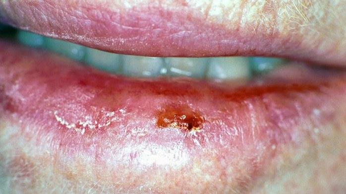
2. Lesions with variable amounts of palpable scale or erosions/crust and WITHOUT a palpable lump (papule or nodule) under the scale/crust
3. Differentiating melanoma from benign melanocytic naevi (common moles)
The clinical ABCD of melanoma equates to:
The dermoscopic assessment of melanocytic lesions requires considerable training and experience, as such dermoscopic images are not included in this section.
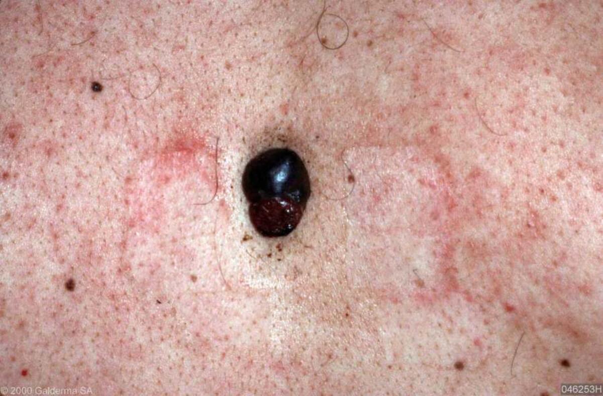
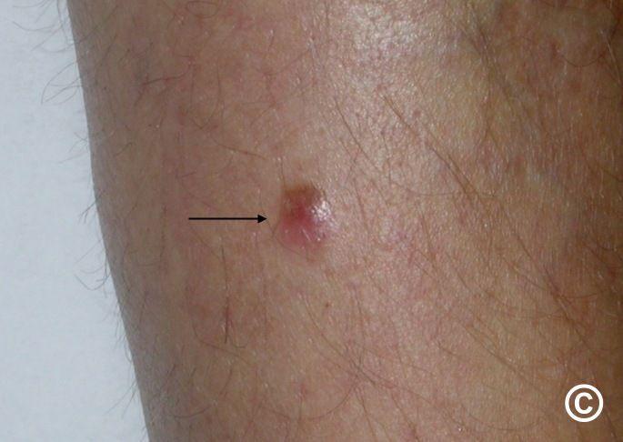
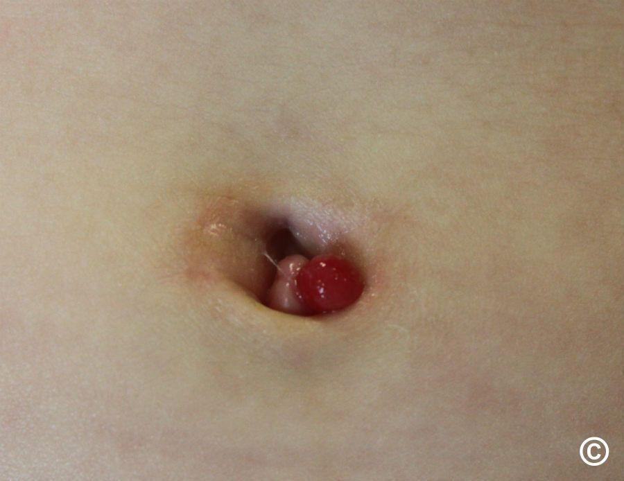
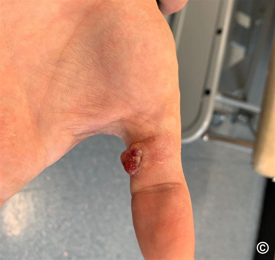
4. The EFG rule of skin cancer - EFG stands for ALL of Elevated + Firm + Growth persistent > 4 weeks
NB. Some palpable benign lesions, such as wobbly moles (naevi) and seborrhoeic keratoses, can become irritated and flare-up, arrange to review after 2-3 weeks. If not settling refer urgently as a 2WW as the differential could then include malignancy.
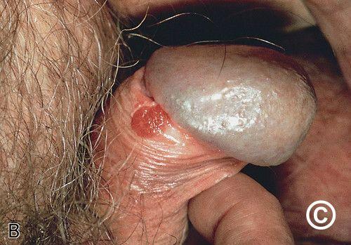
5. Other lesions of concern requiring urgent referral
6. If you can't find the lesion here refer to the Skin Lesion Diagnostic Tool and failing that send a teledermoscopy referral
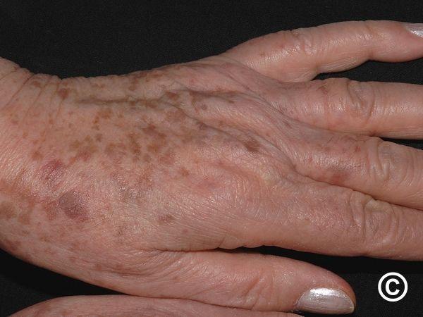
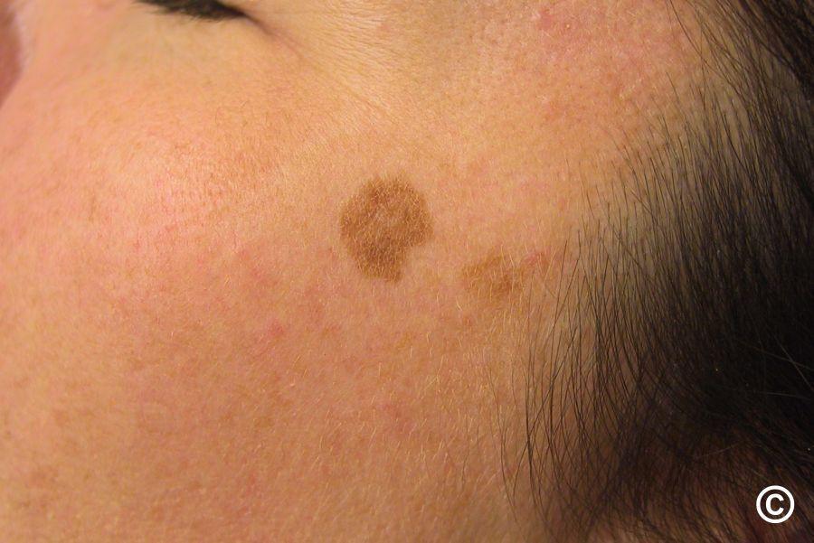
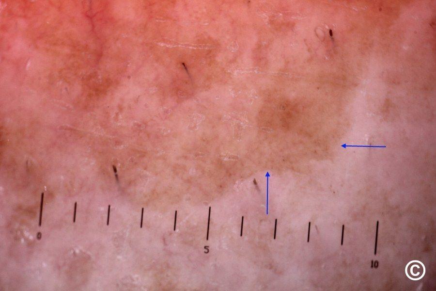
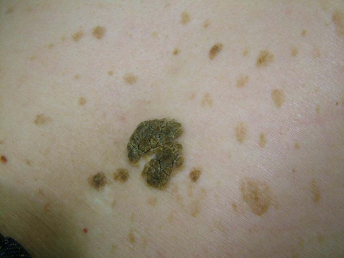
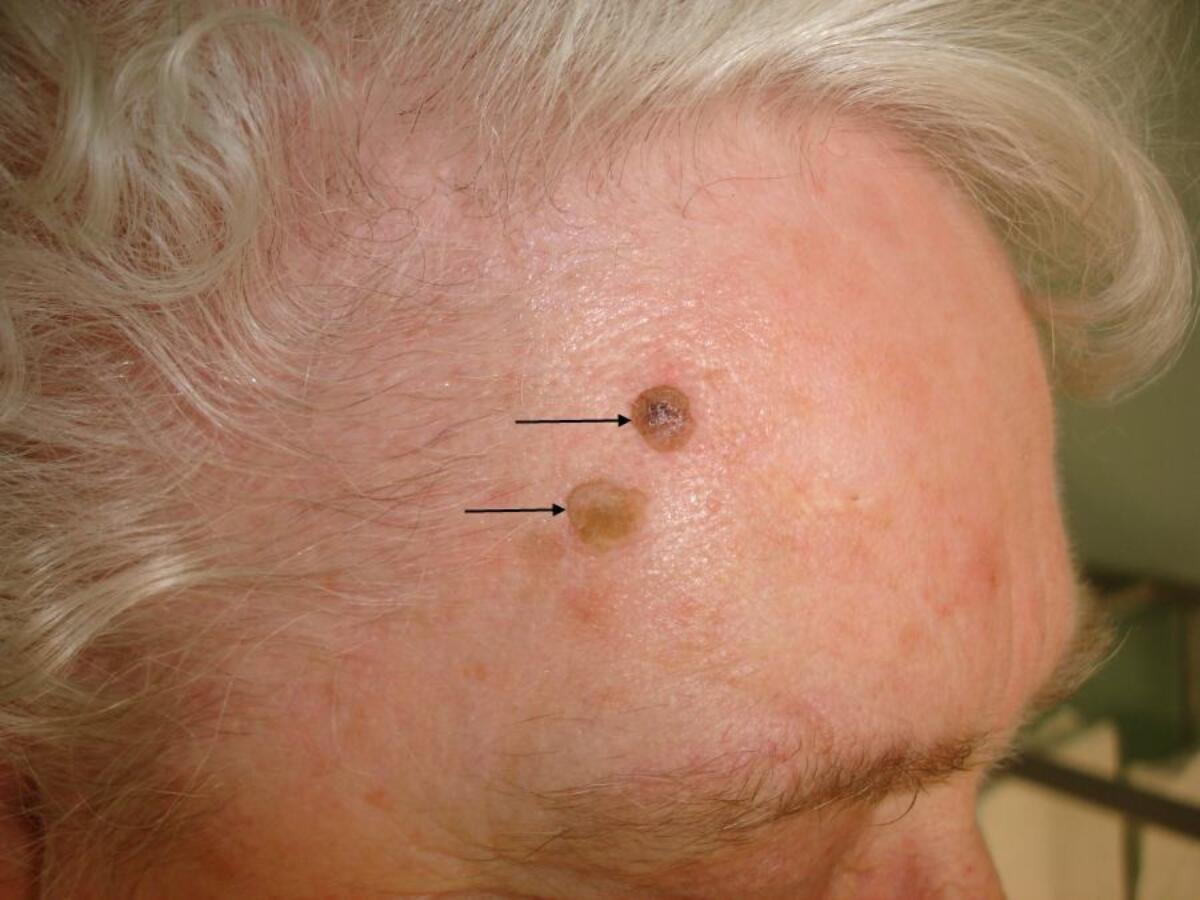
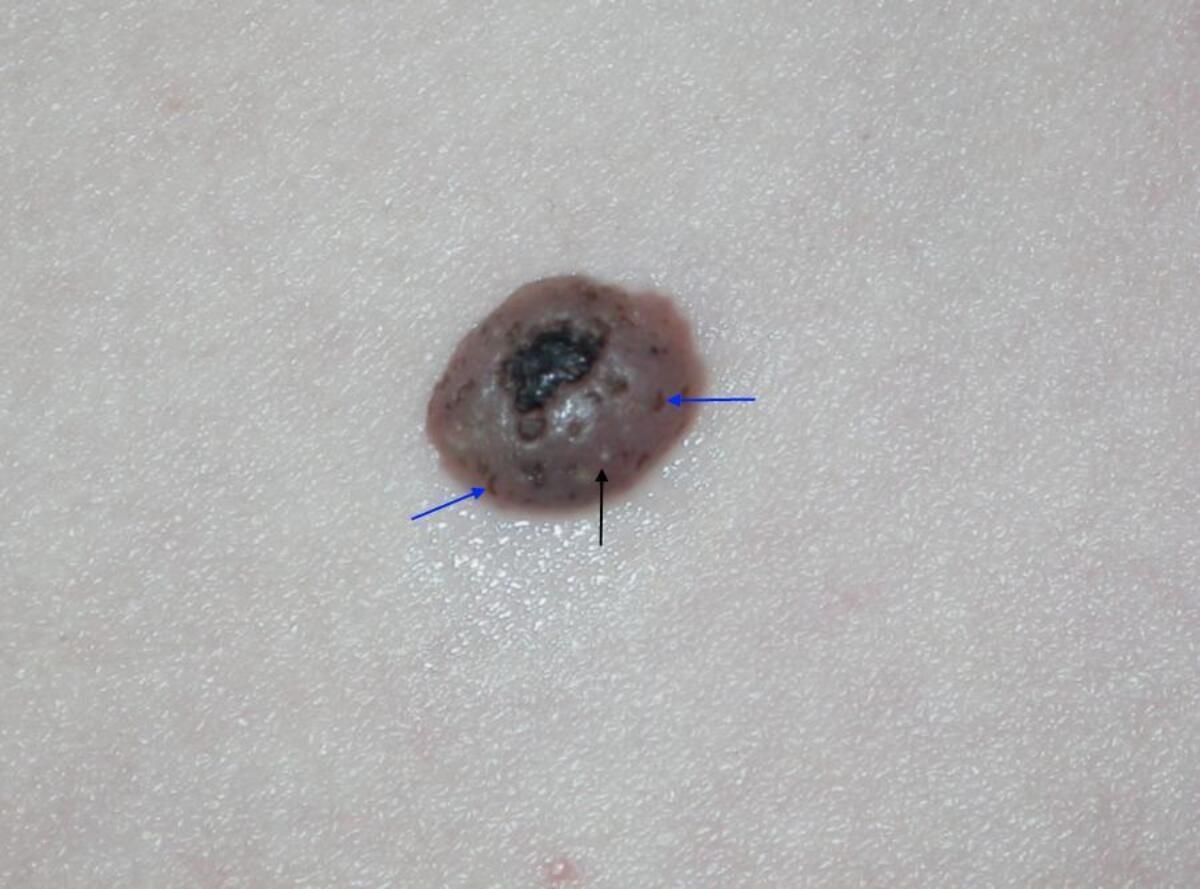
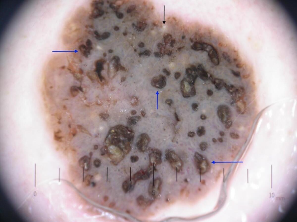
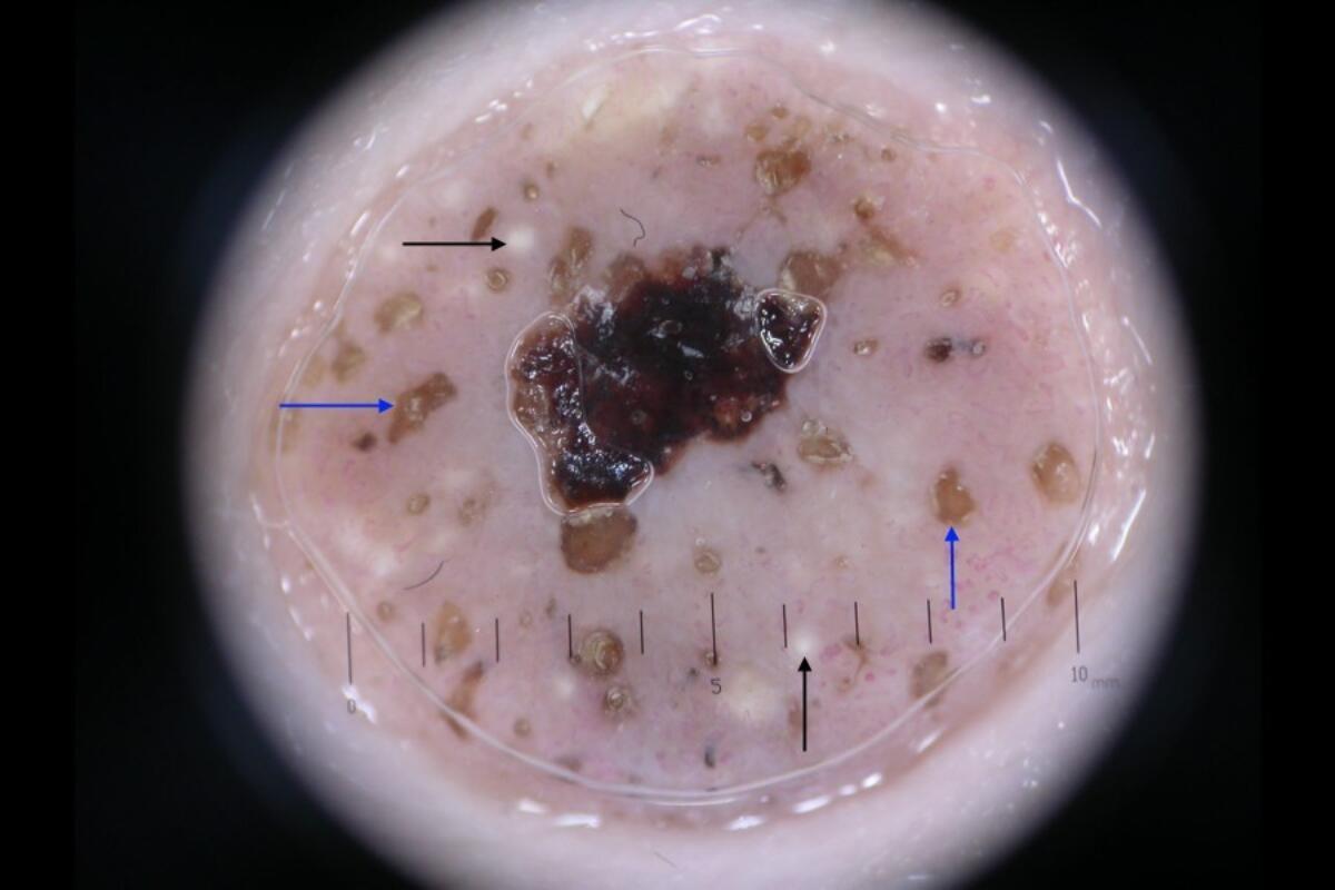
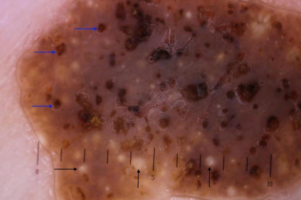

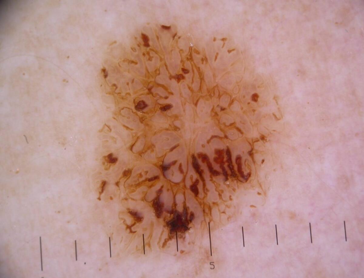
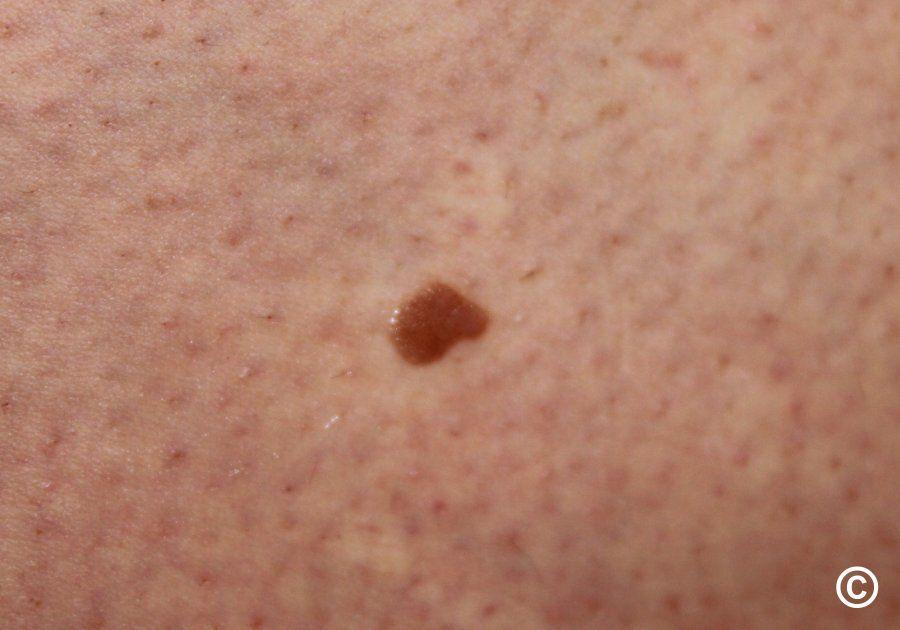
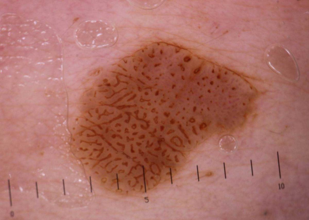
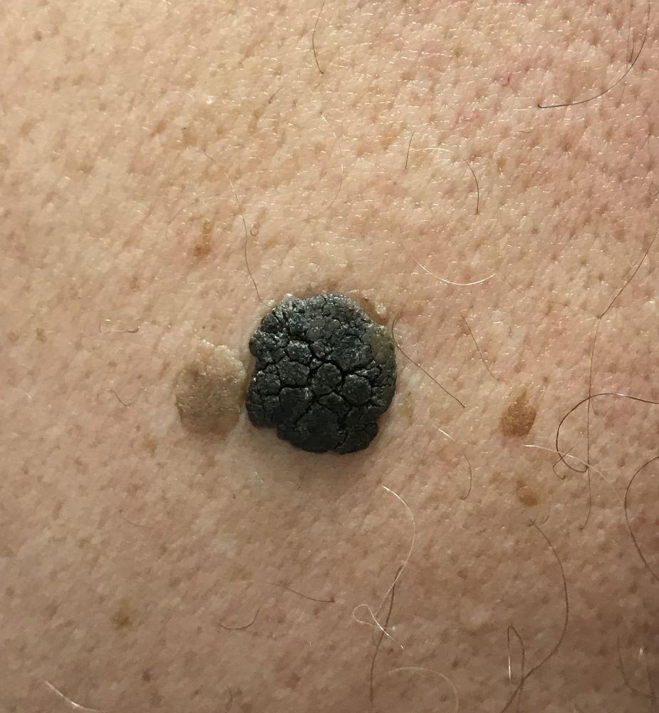
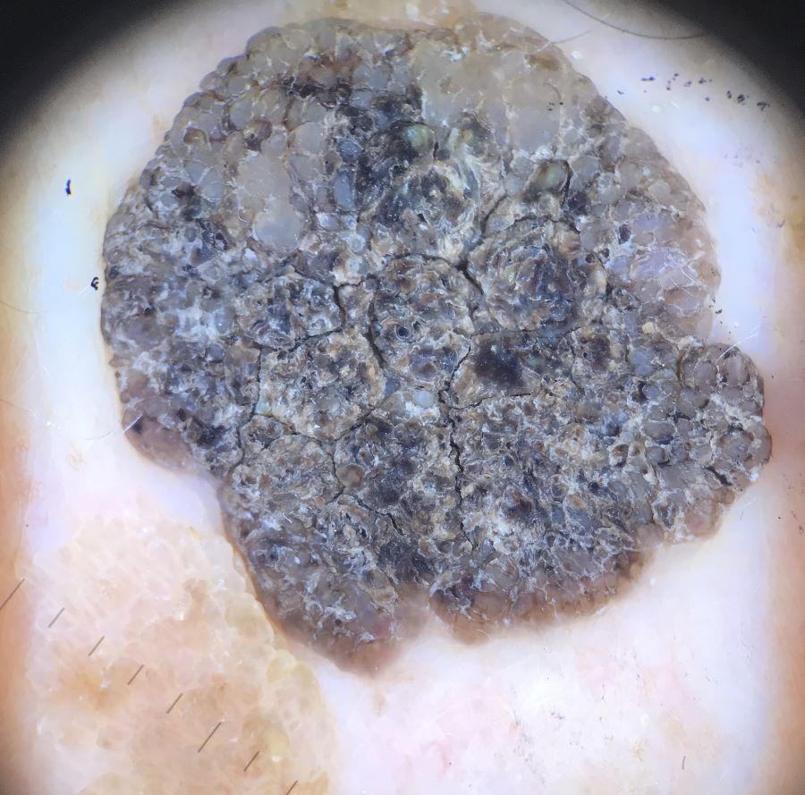
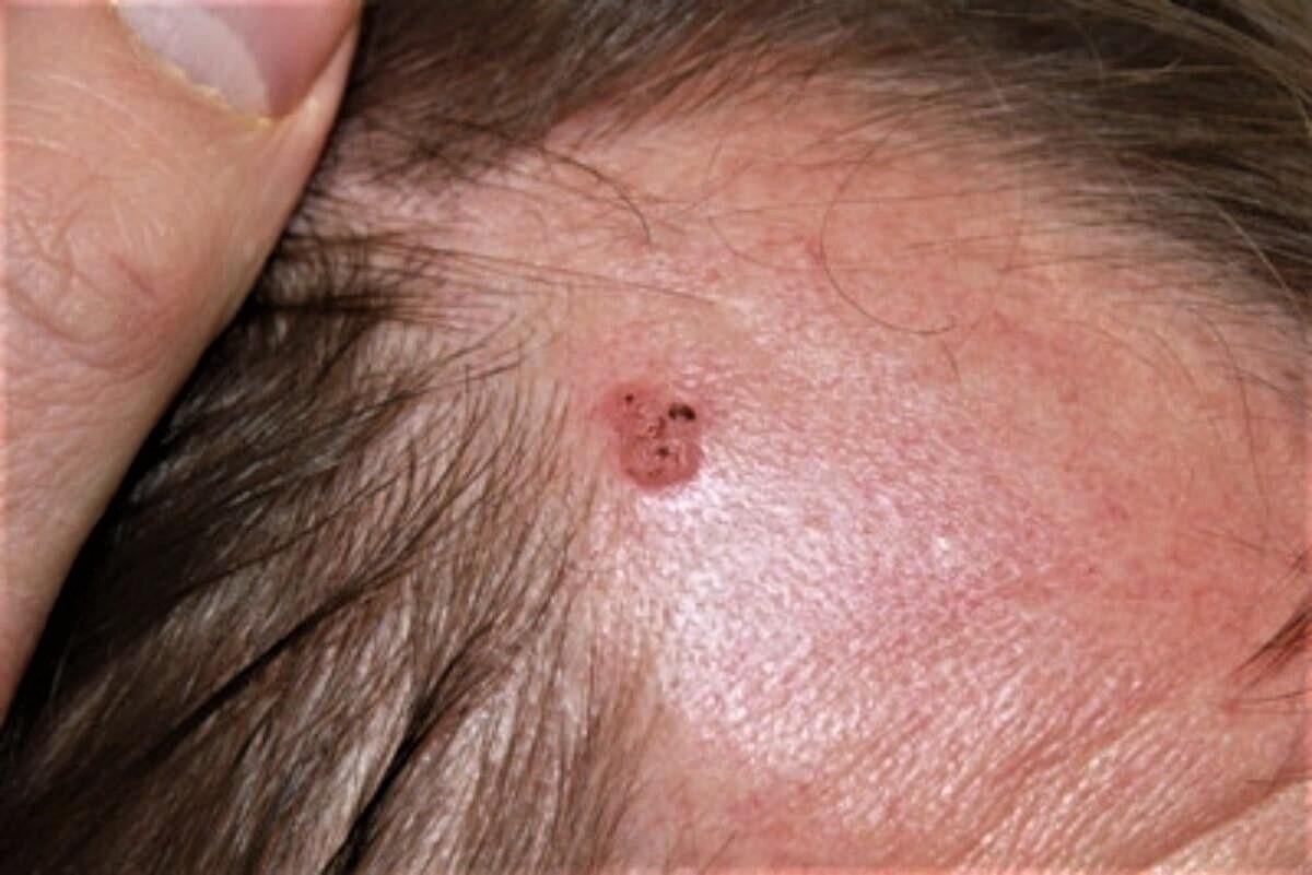
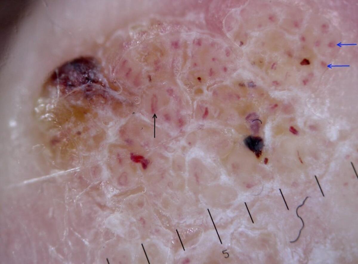
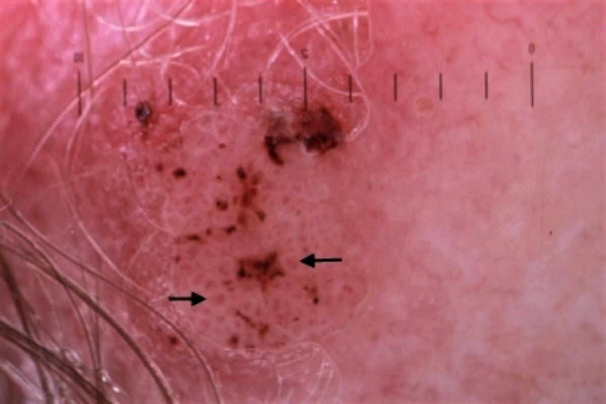
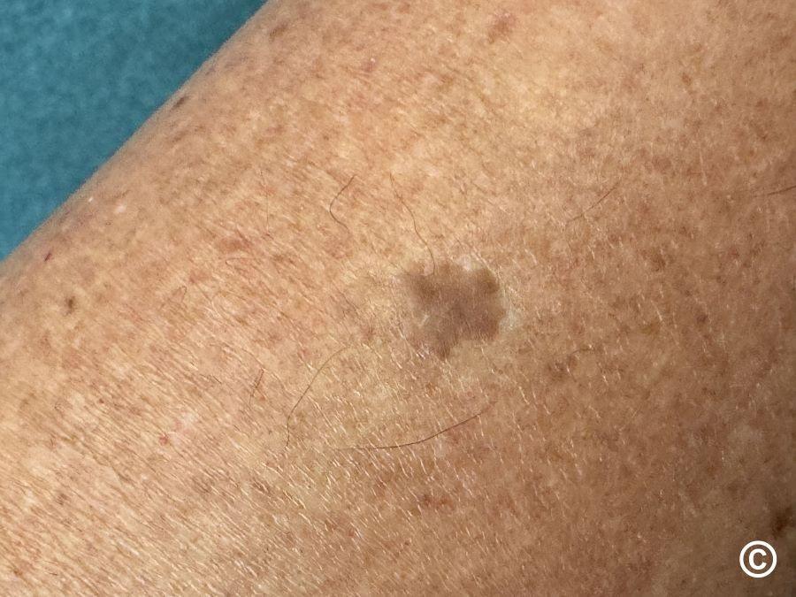
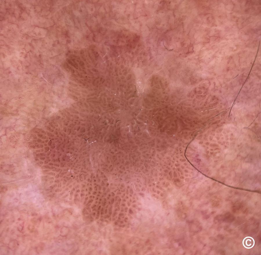
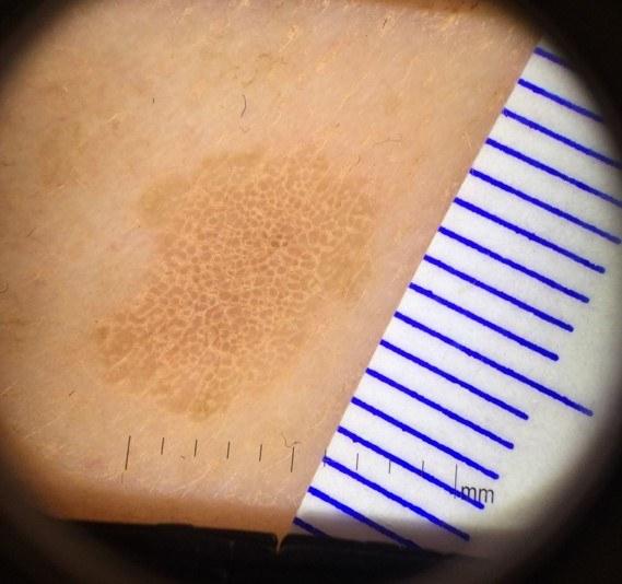
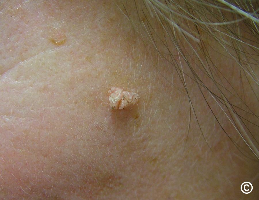
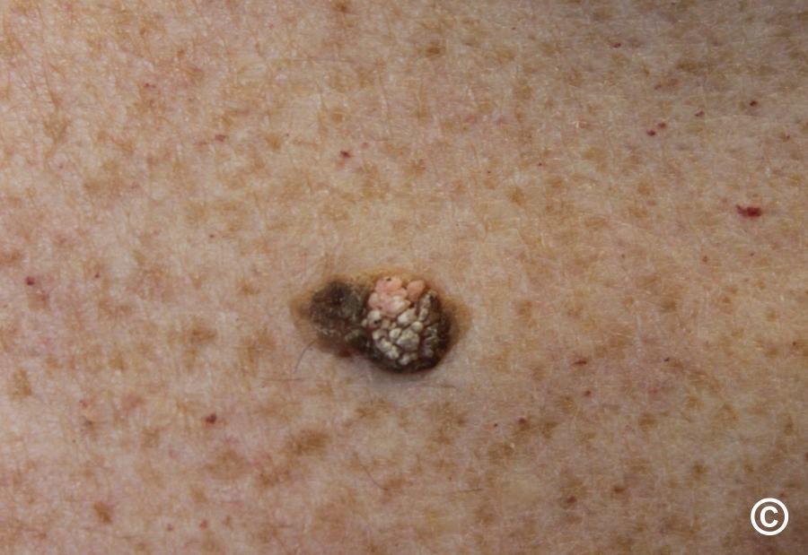
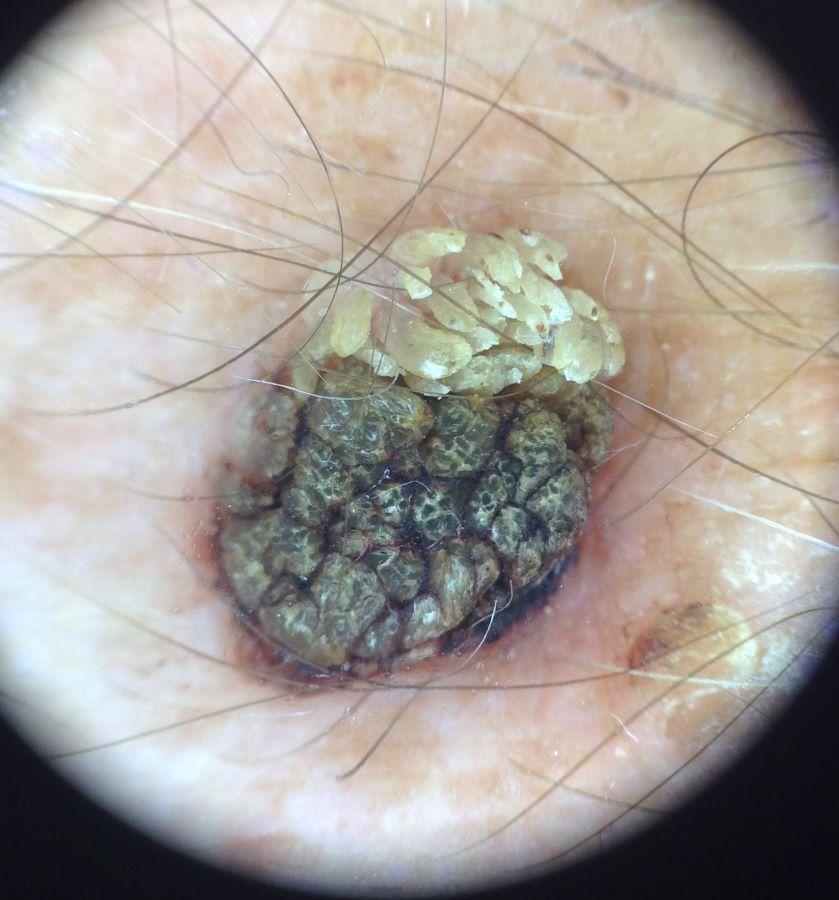
It is worth noting that a non-tender scaly or 'greasy' lesion that dermoscopically is yellow-brown is very likely to be benign.
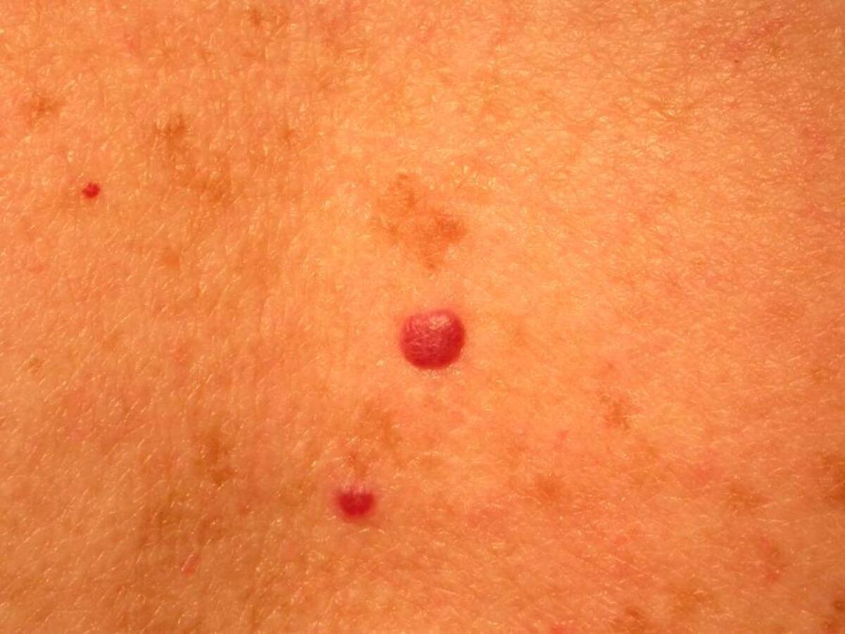
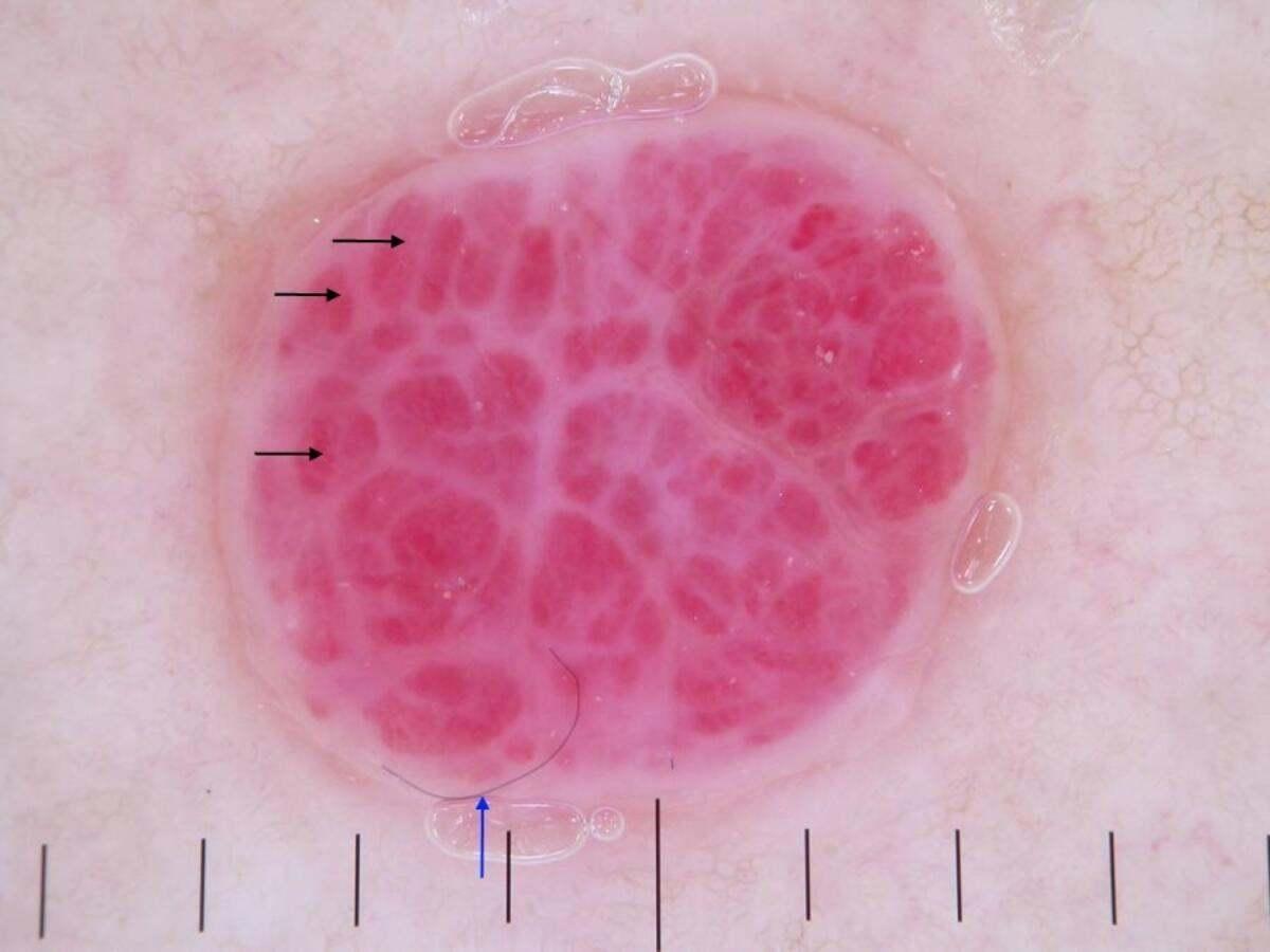
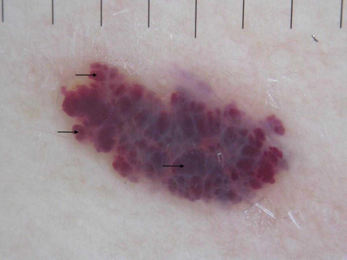


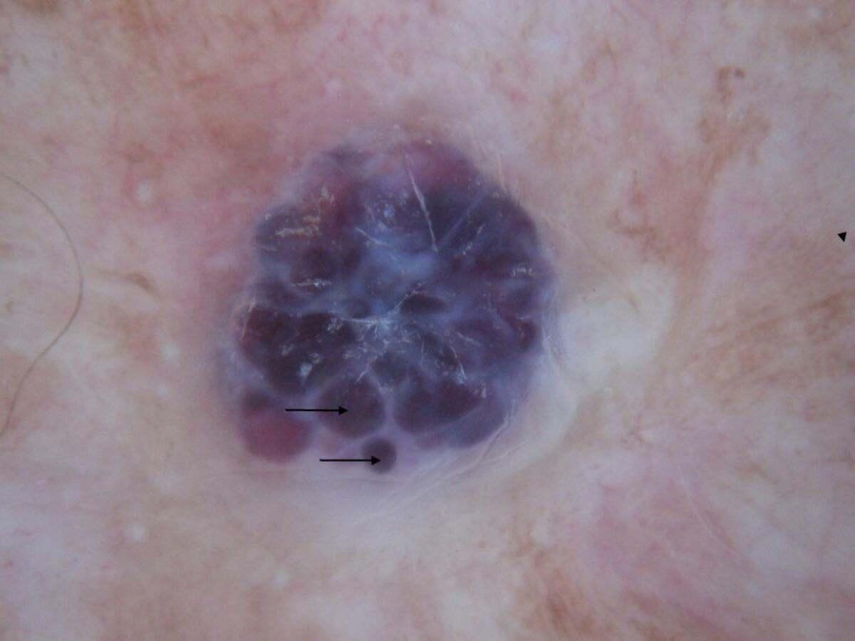
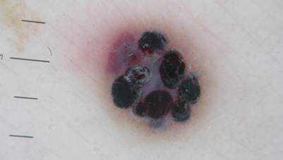
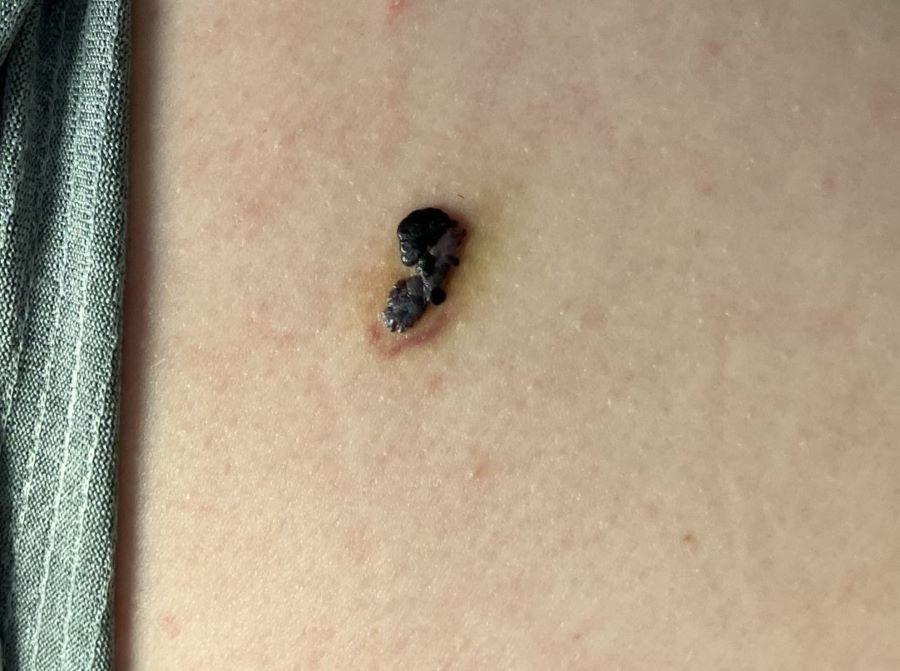
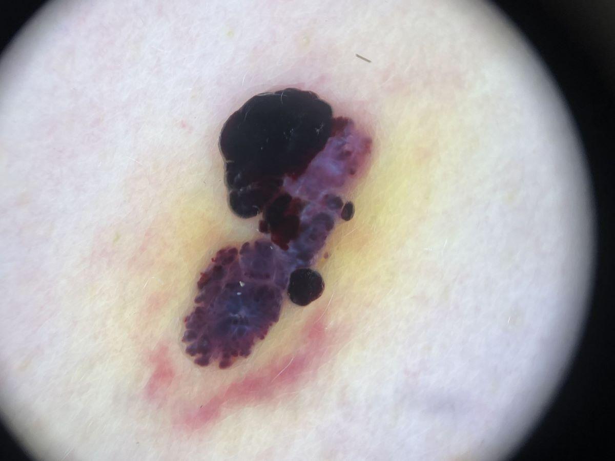
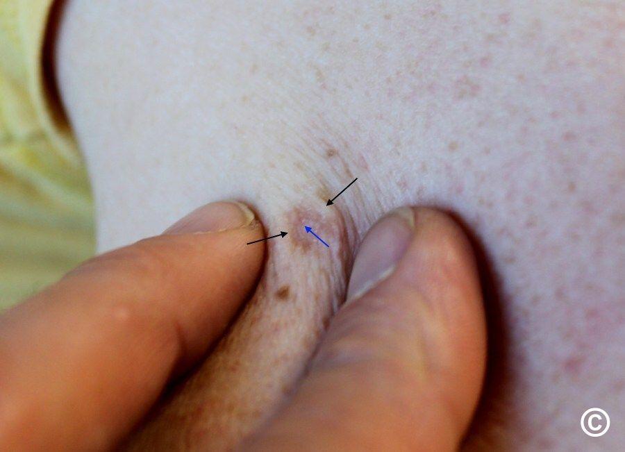
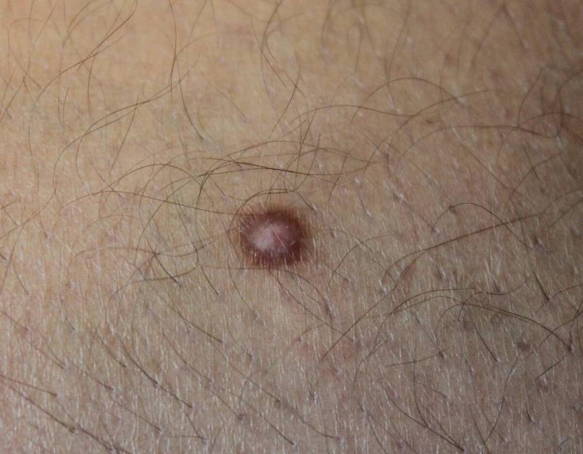

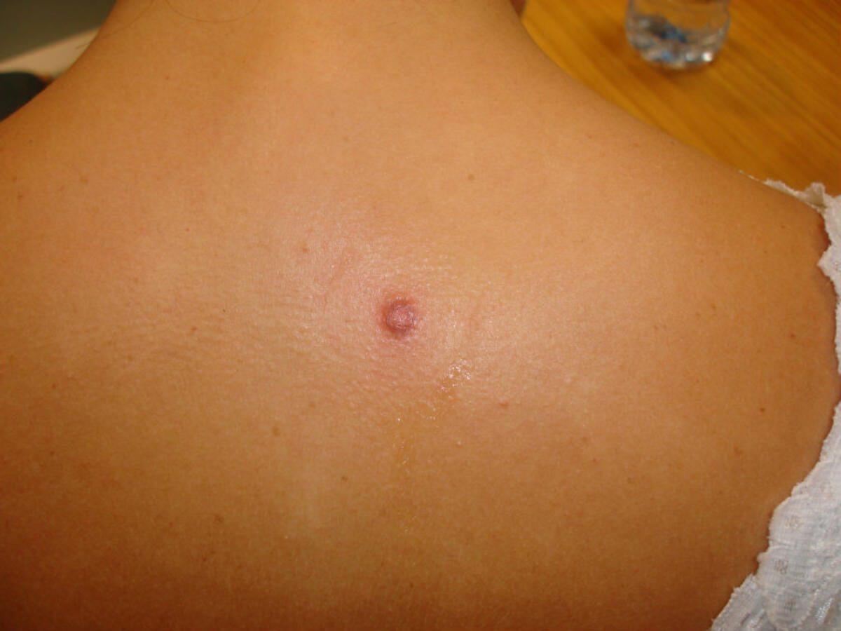
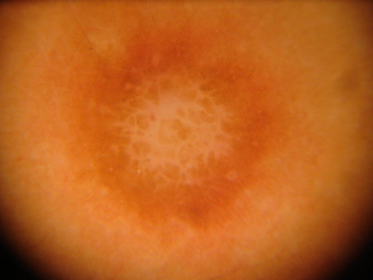
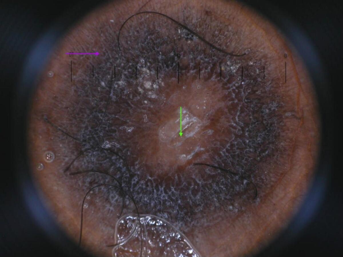
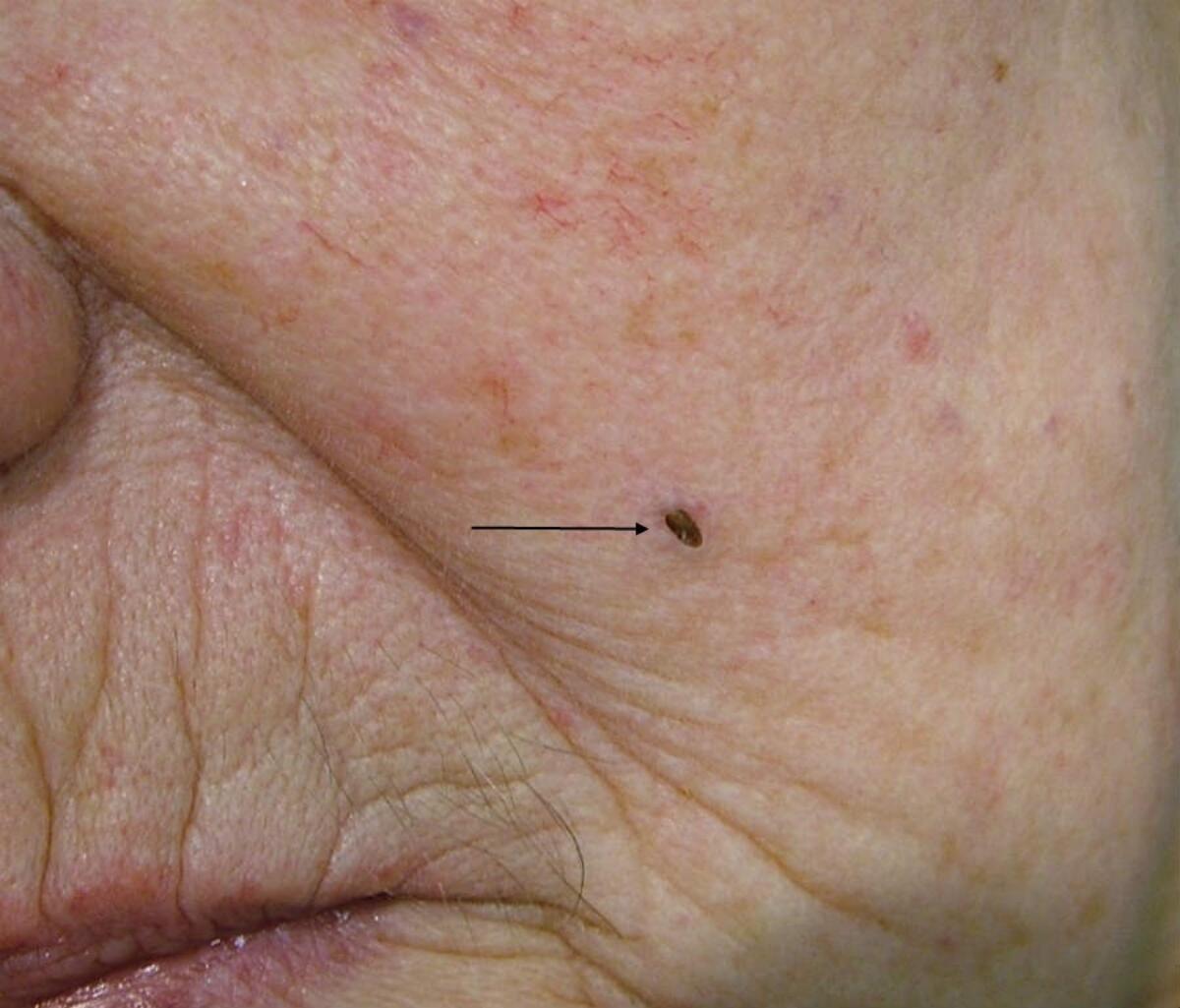
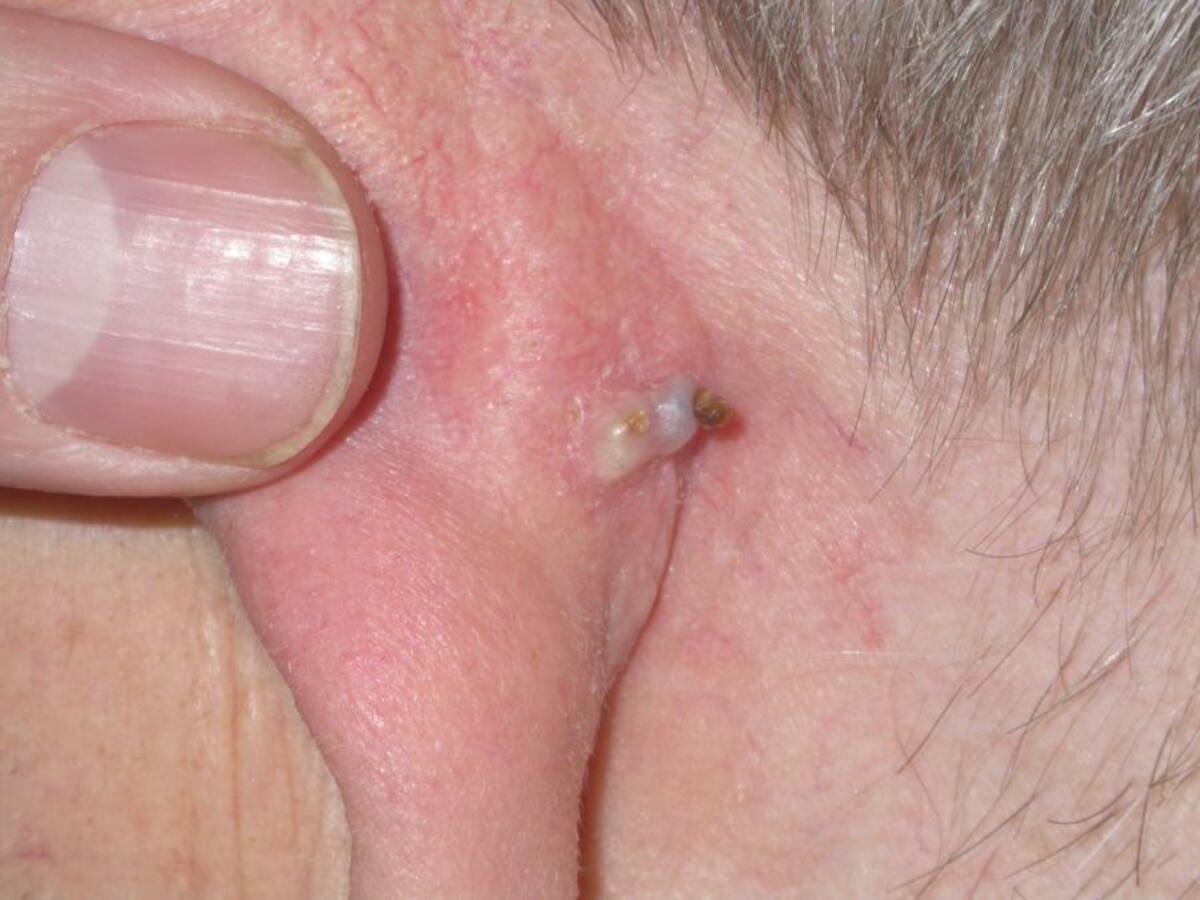
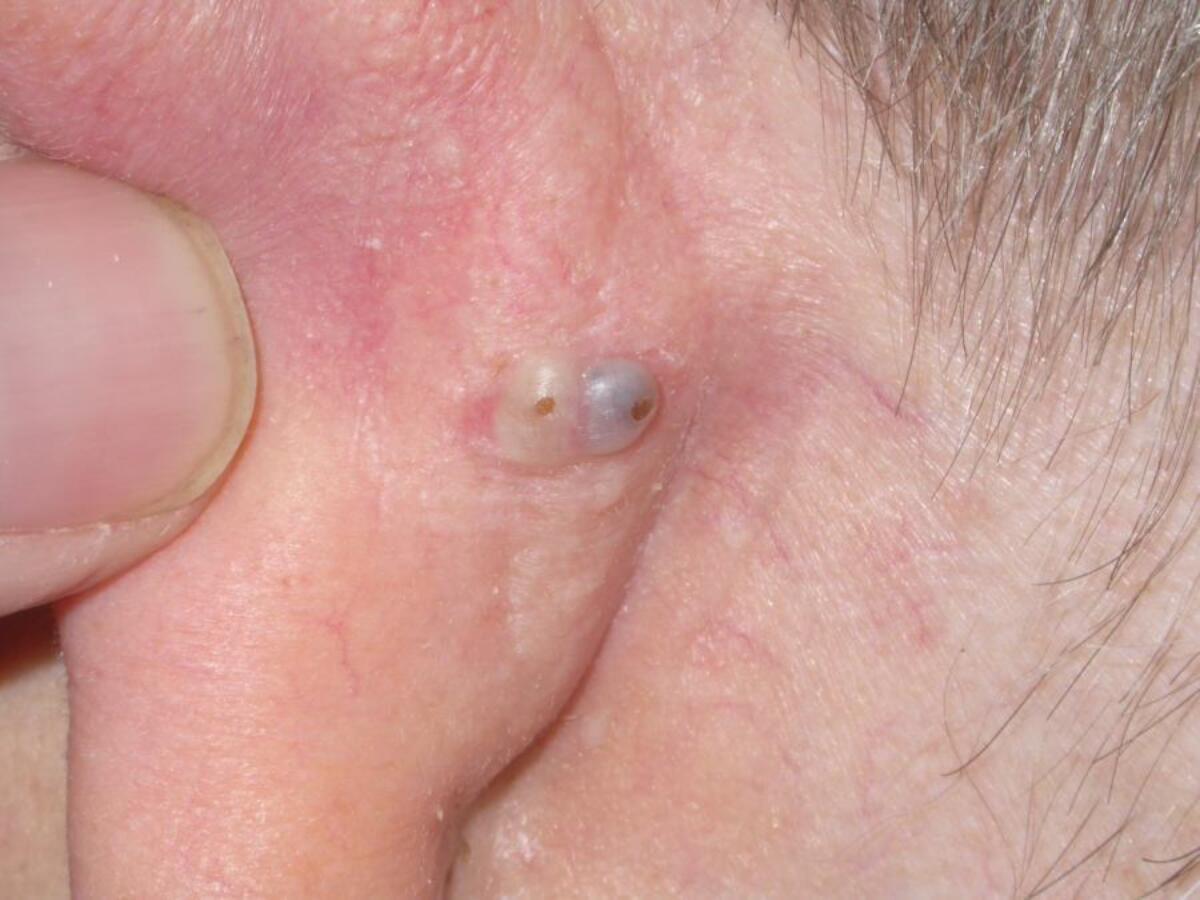
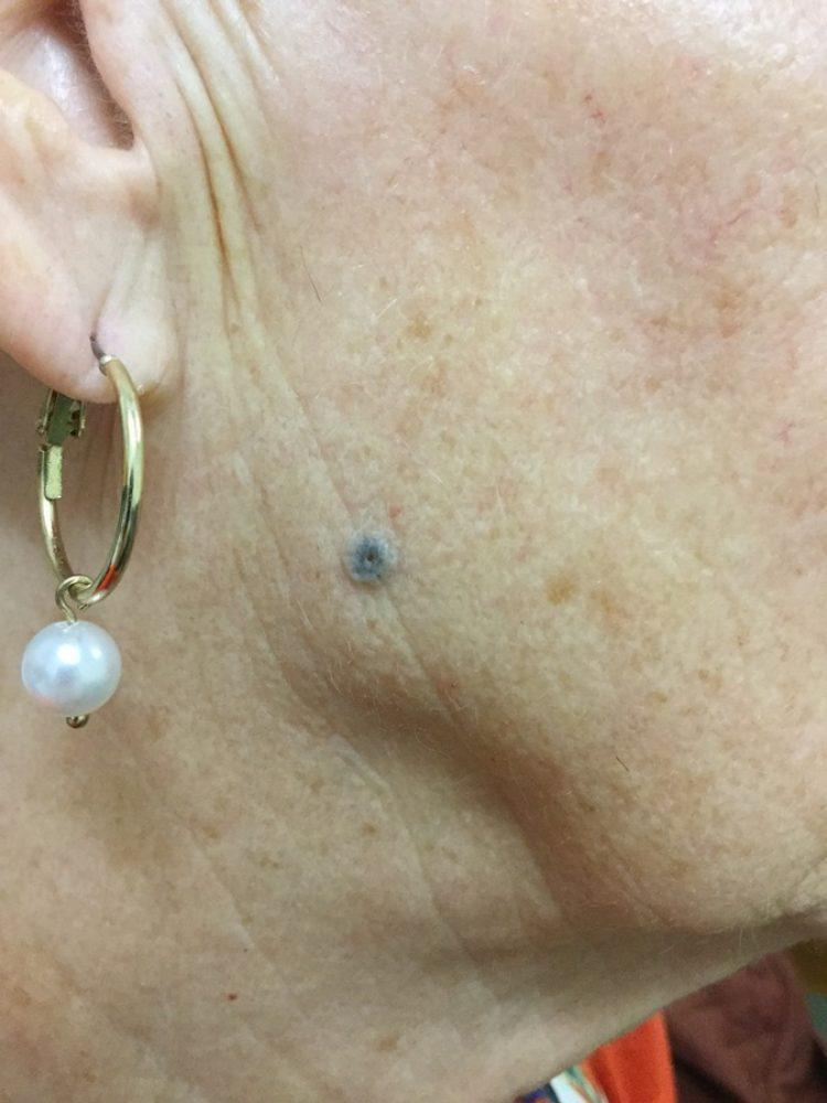
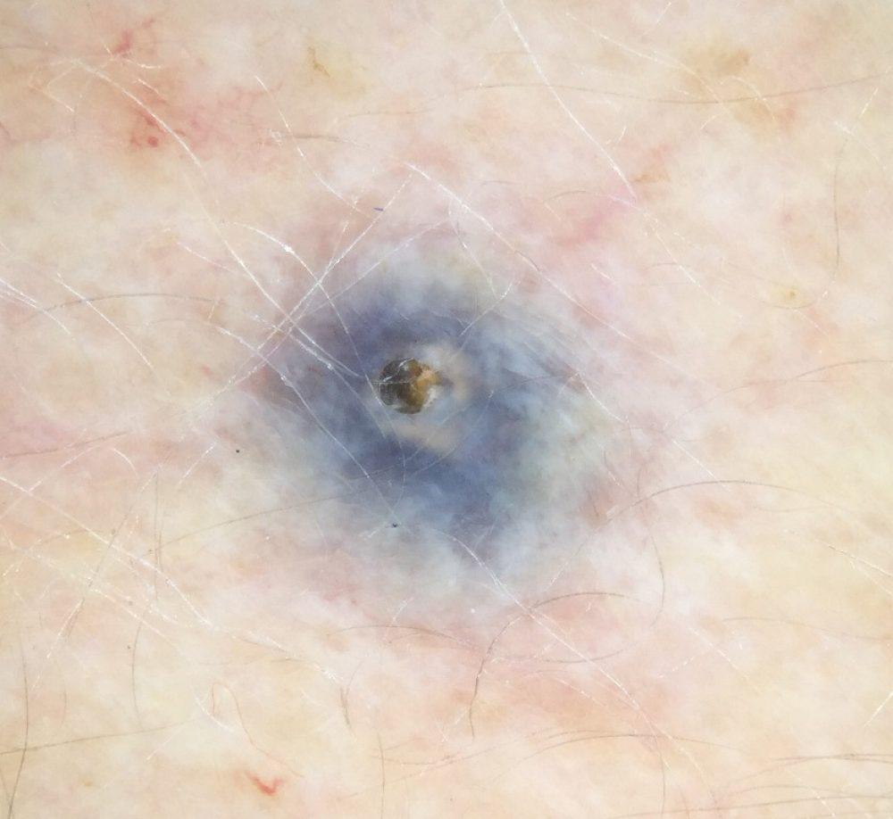
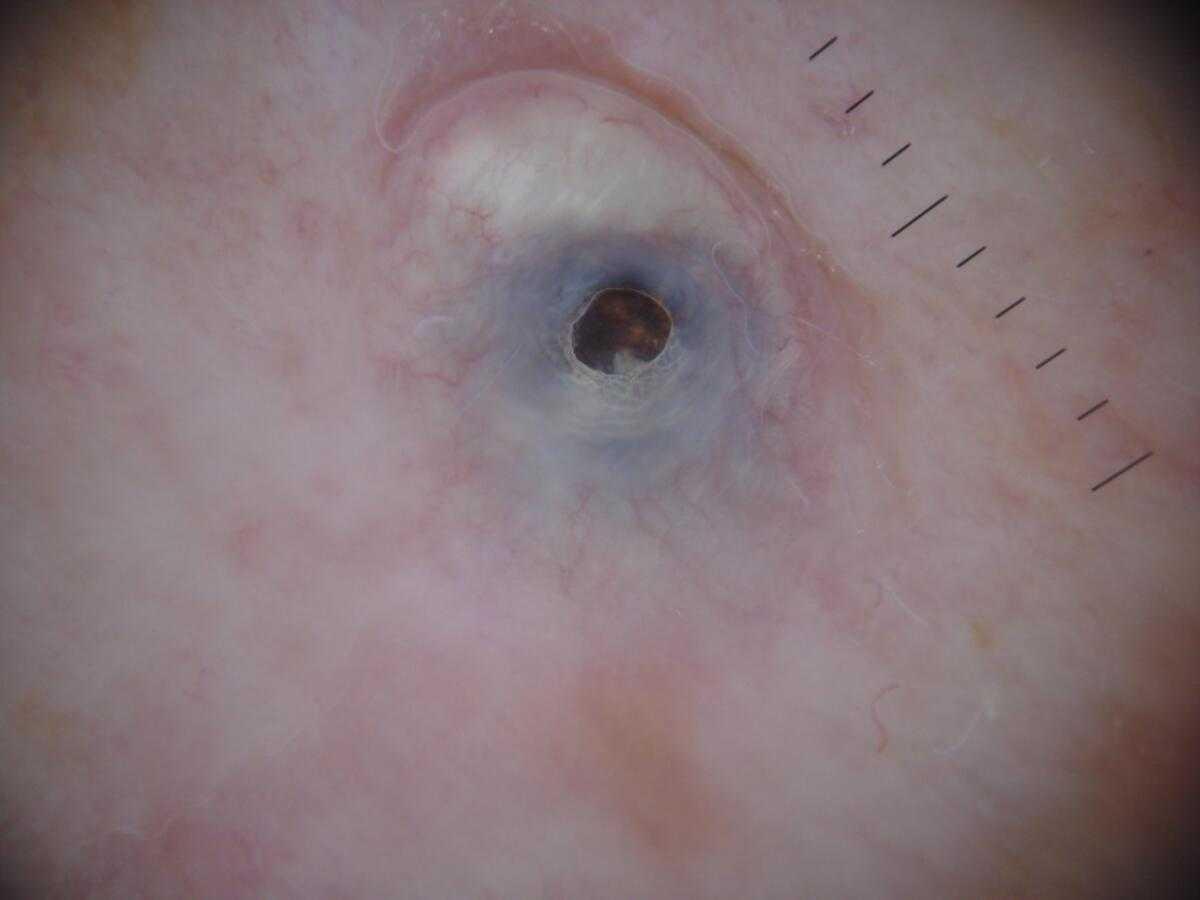
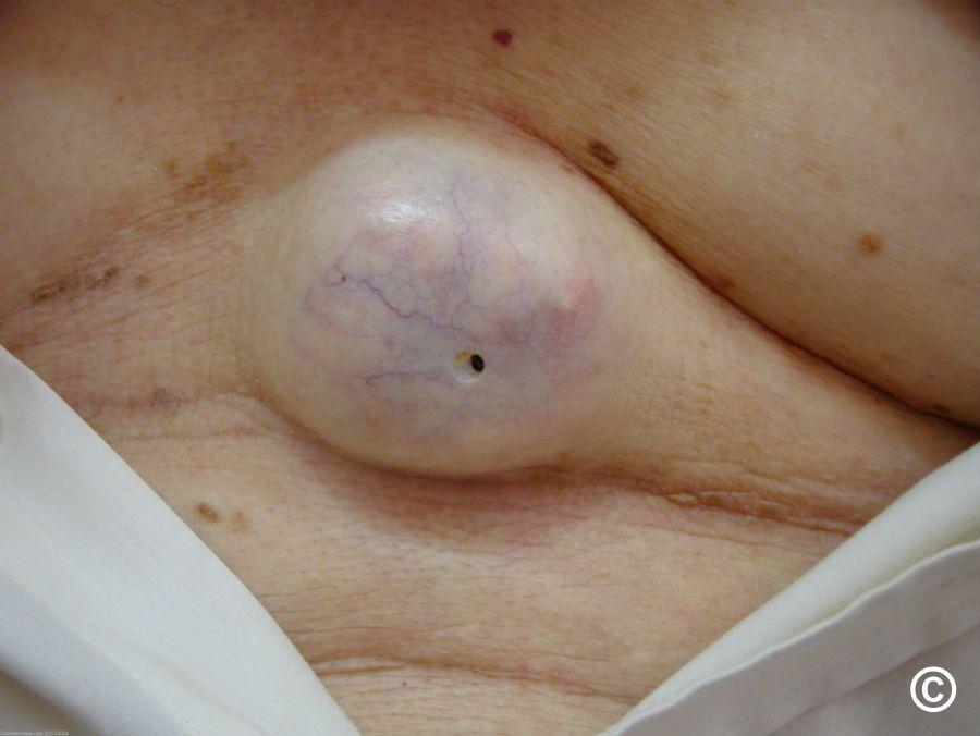
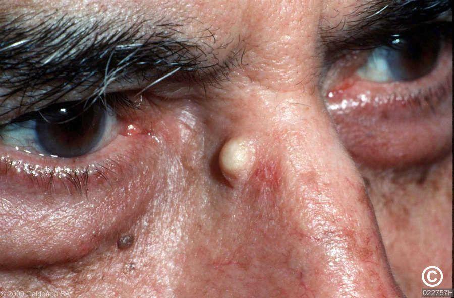
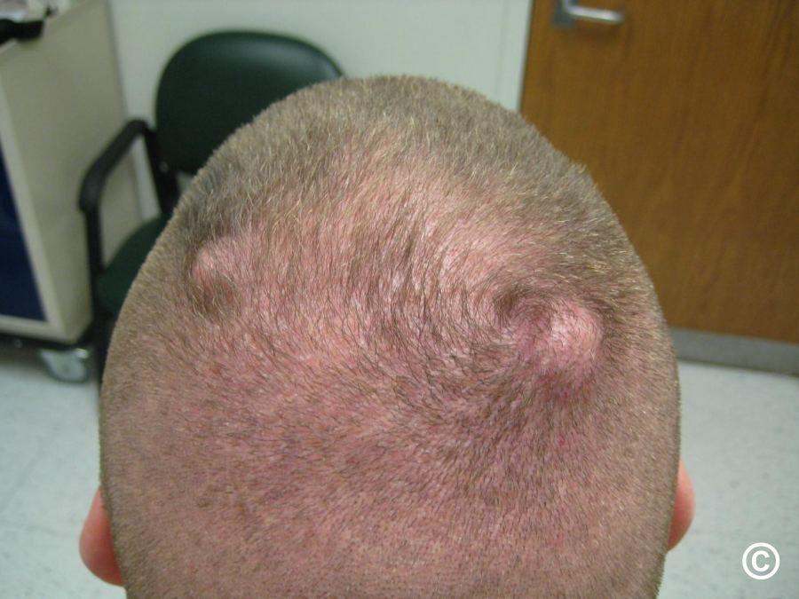
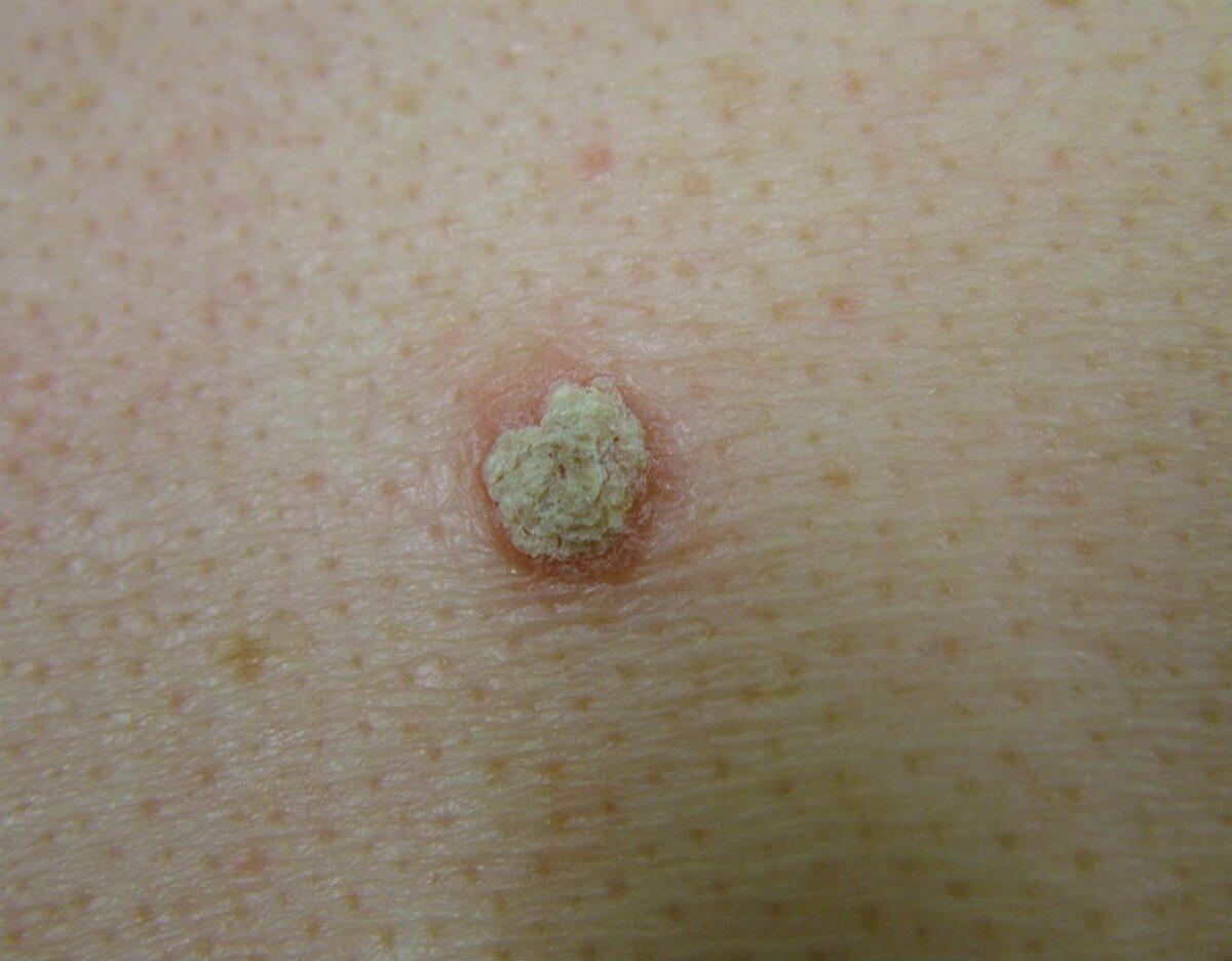
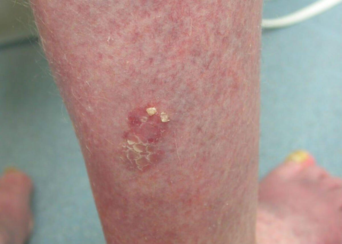
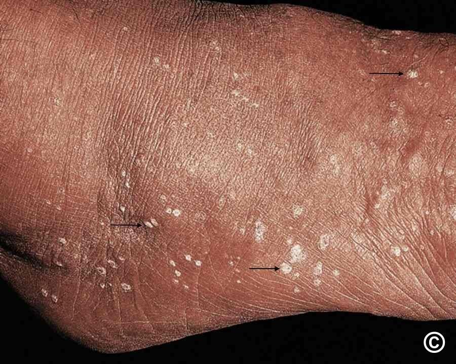

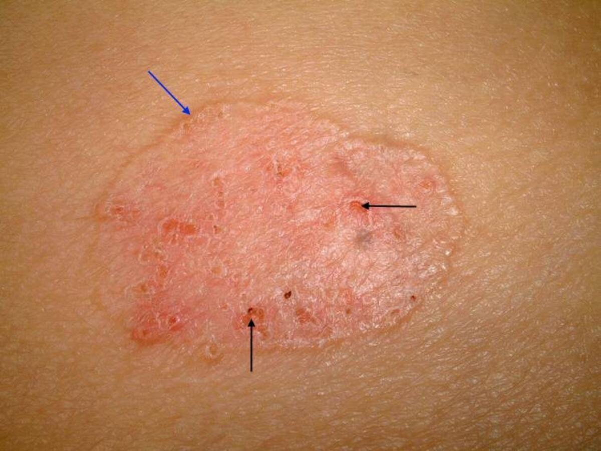

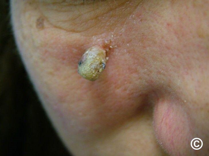
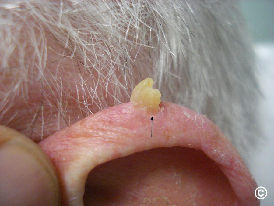
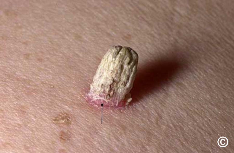
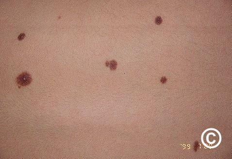
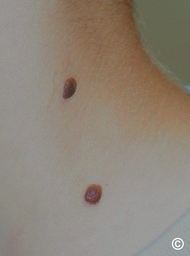
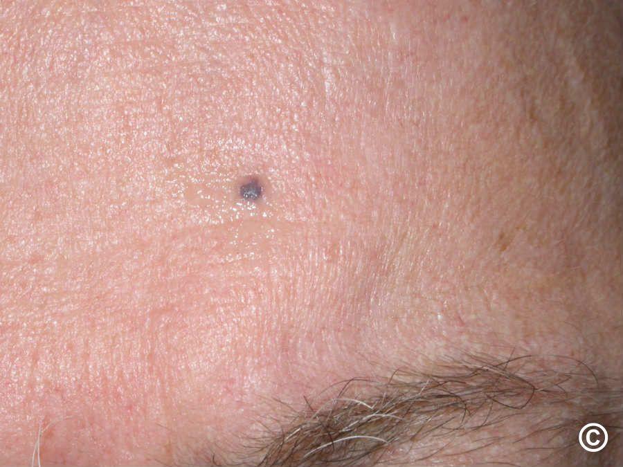
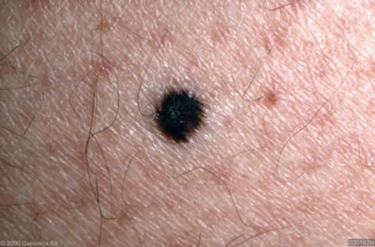
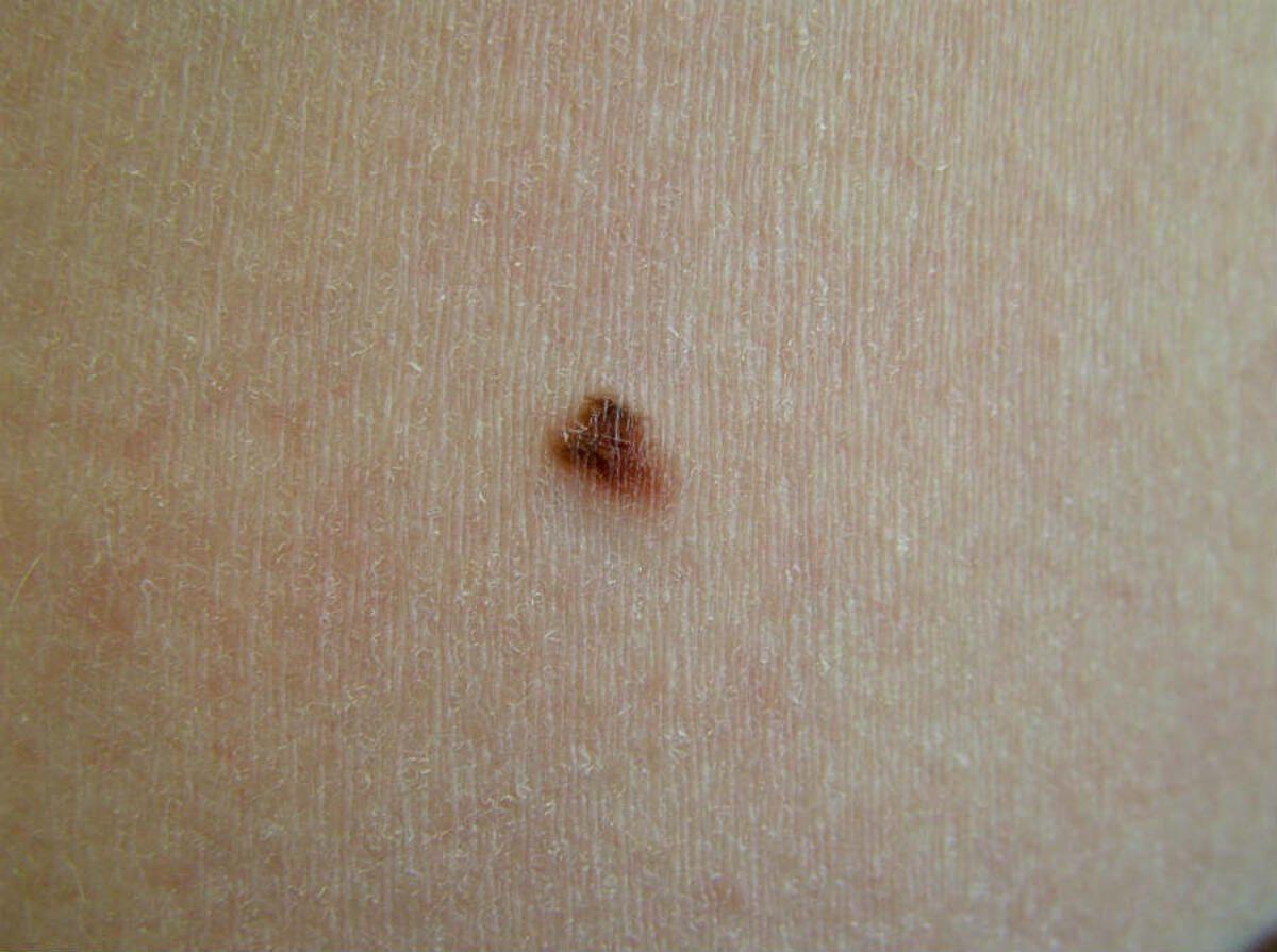
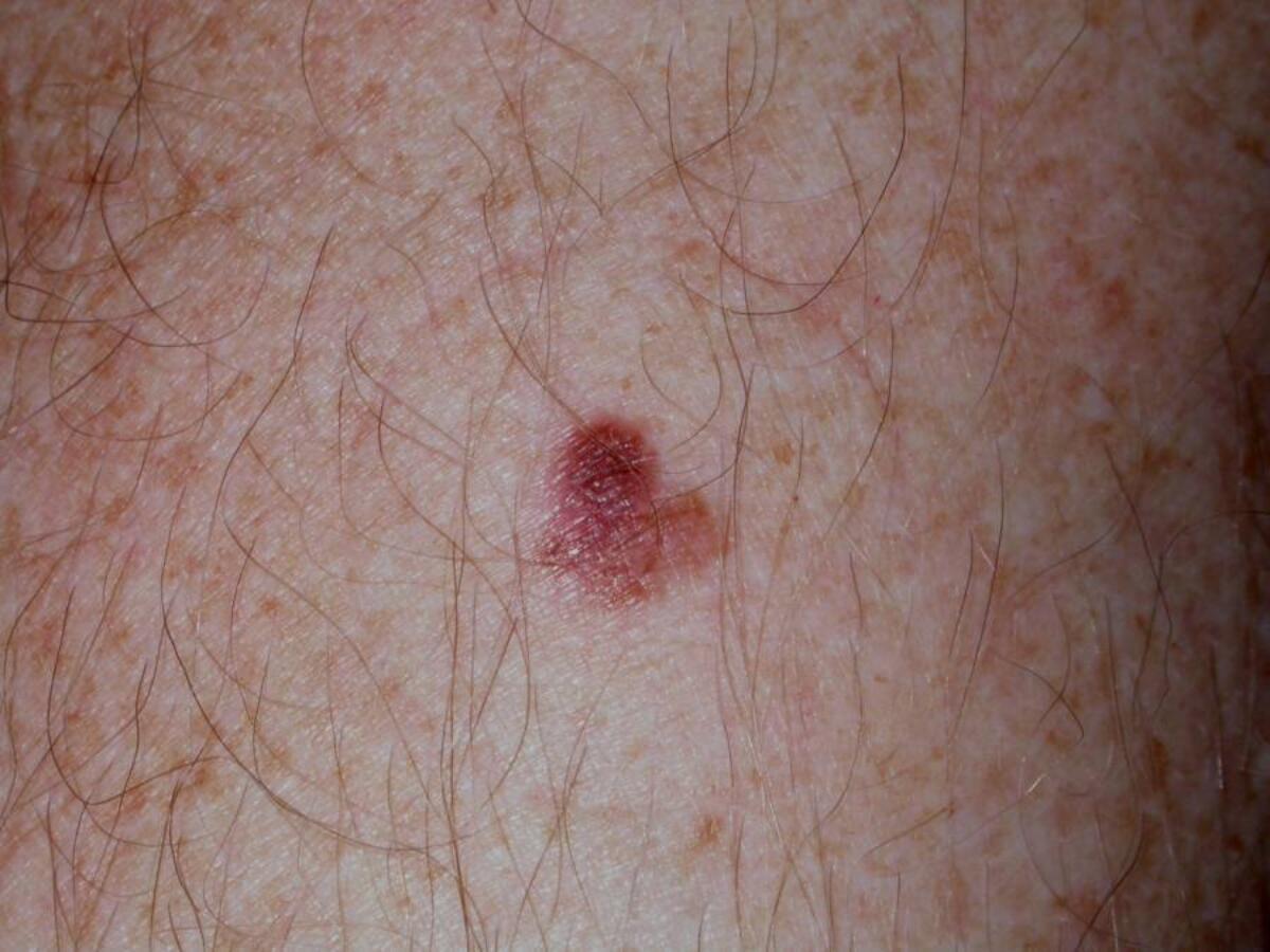
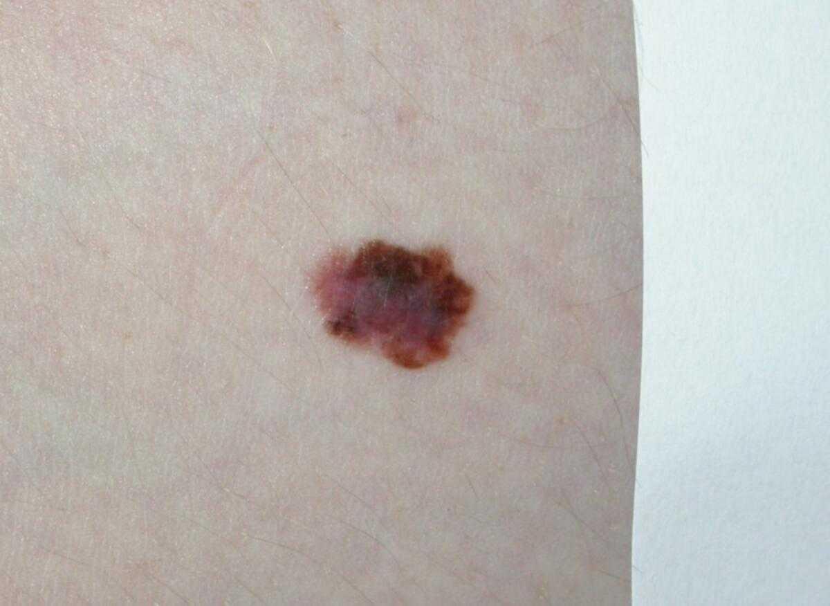
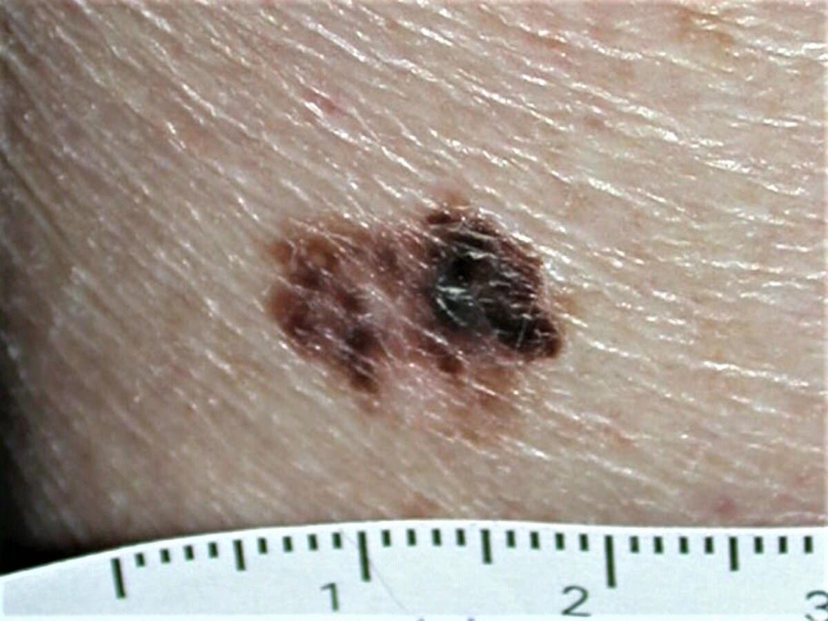
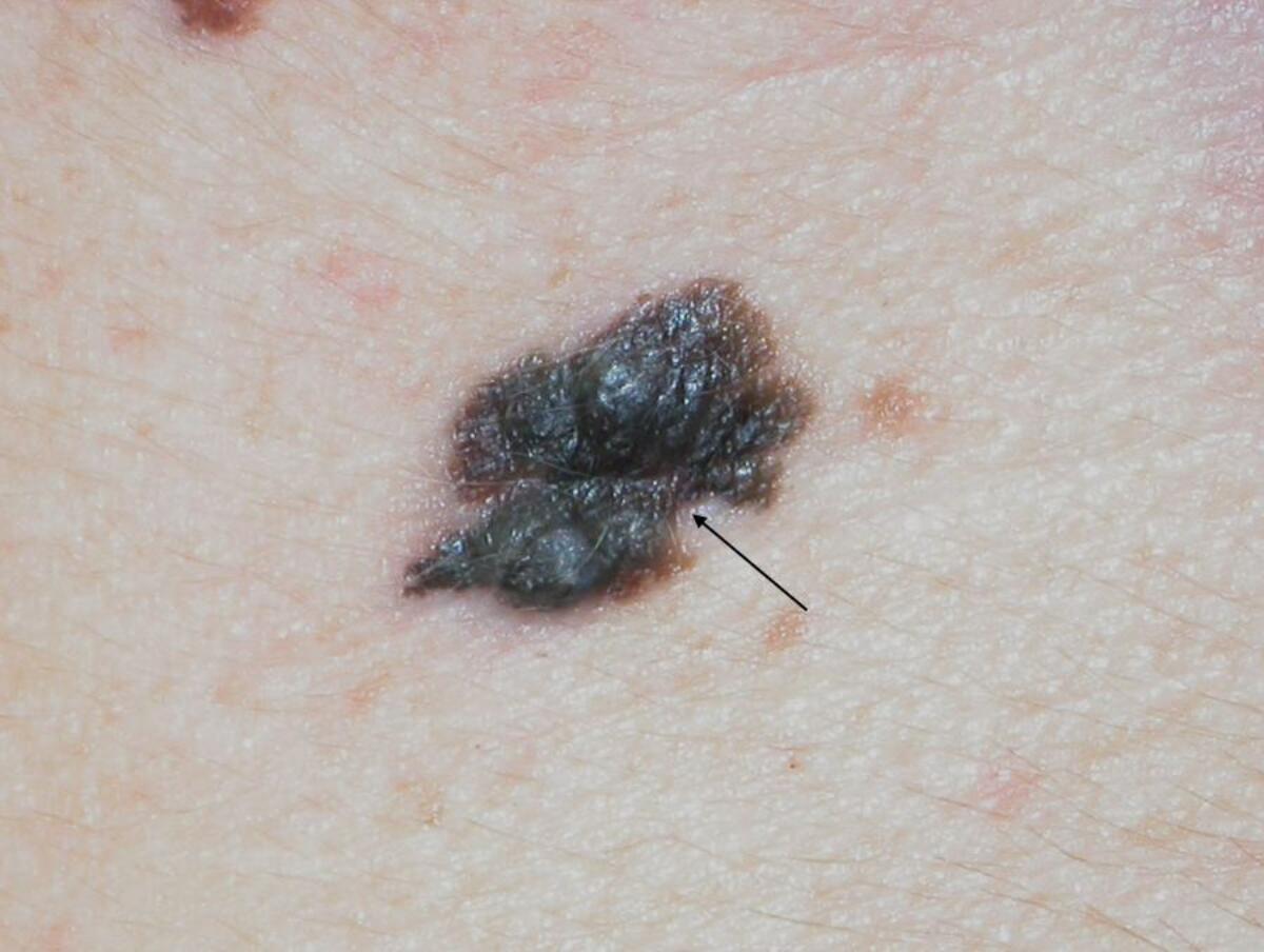
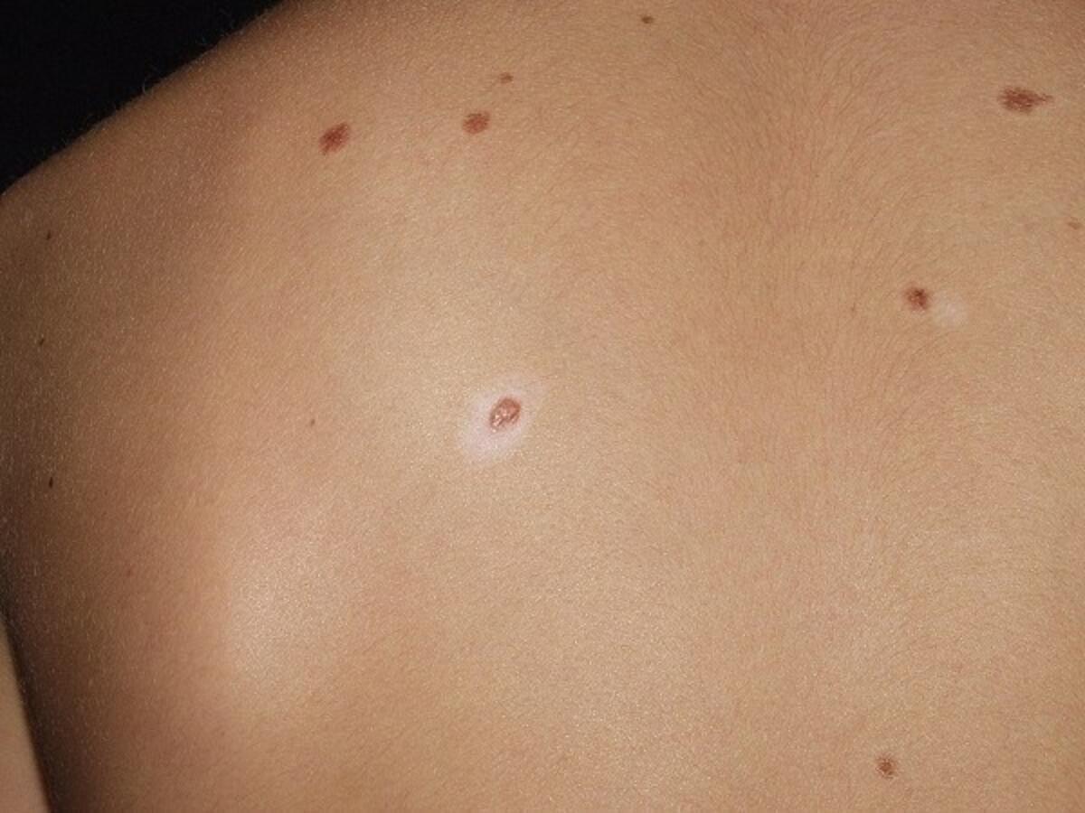
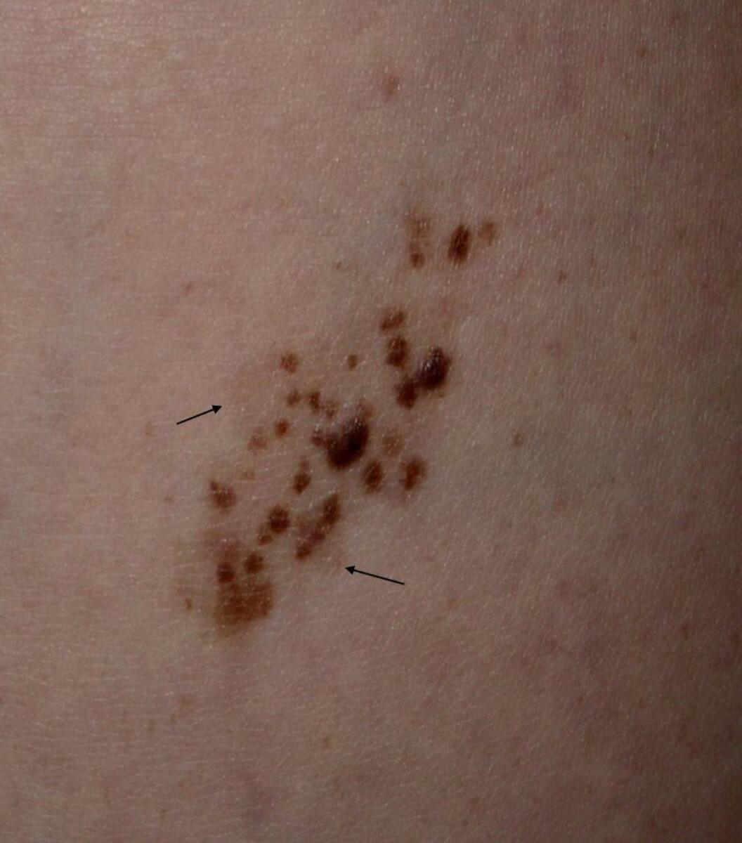
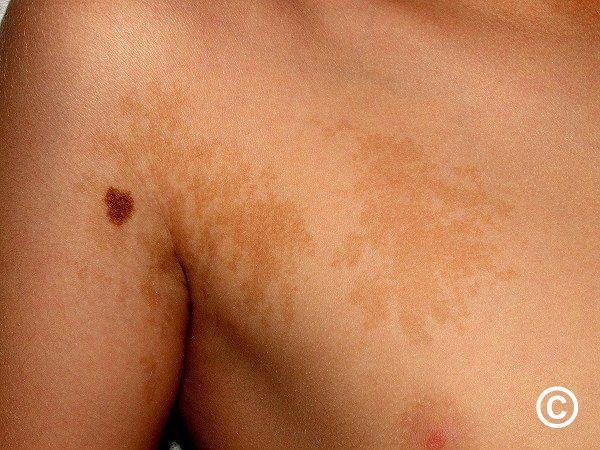
Benign melanocytic naevi (images 1-3) are usually symmetrical, have a regular border, an even colour (or have two-tones of colour that are concentric), and look like the patient's other moles. Soft and wobbly moles are usually benign. A blue naevus is a stable blue (blue/white) macule or papule, often < 5 mm in diameter. There are a wide range of benign pigmented lesions - refer to the Skin Lesion Diagnostic Tool for more detail.
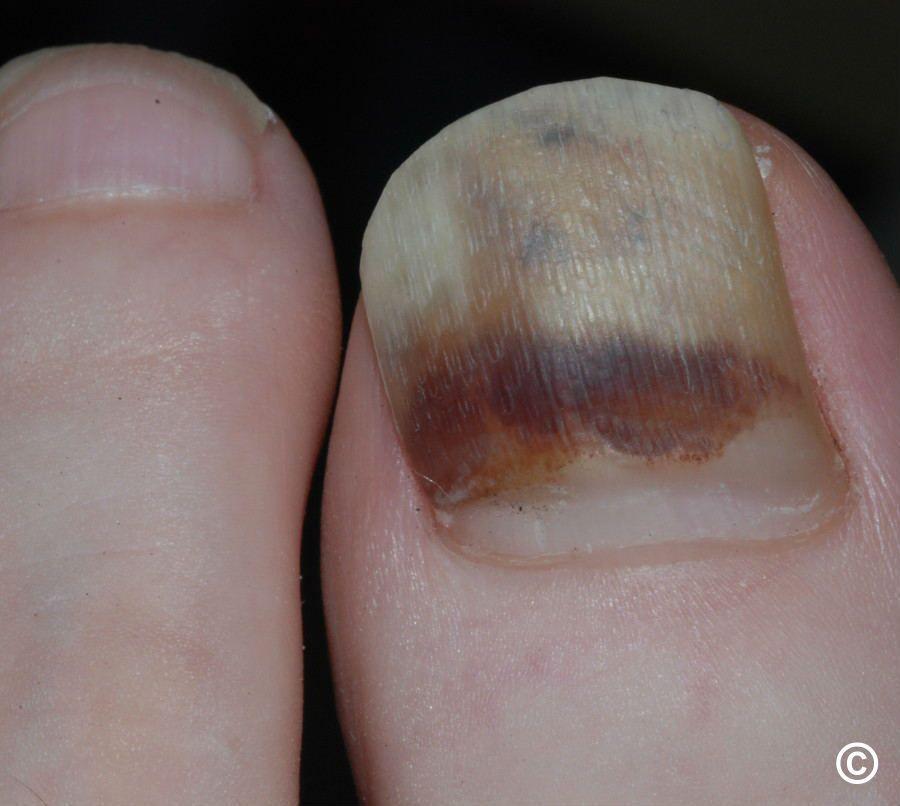
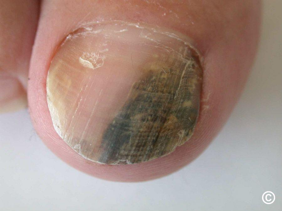
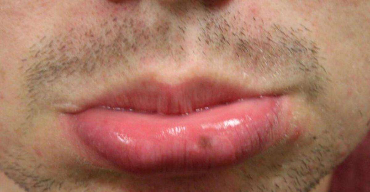
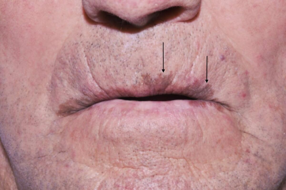
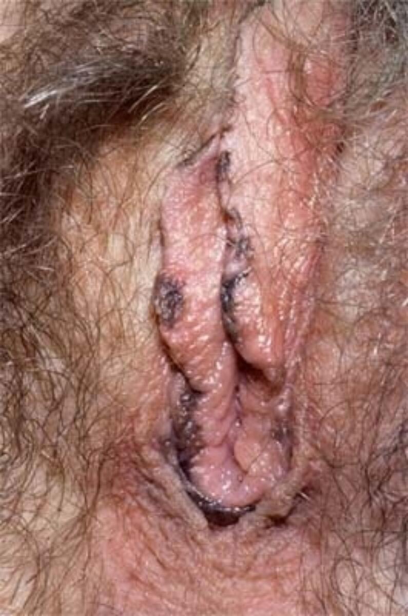
The following features are suspicious for melanoma-in-situ (including lentigo maligna) and superficial spreading melanoma (images 4-9):


The following pharmaceutical companies have had no involvement in the content of this website or in our conference programmes







