Peeling skin conditions
LAST UPDATED: Jul 18, 2025
Introduction
While there are many conditions that can cause skin peeling, this chapter, which is set out as below, focuses on the less common, chronic peelings conditions such as the Peeling Skin syndromes.
Clinical findings
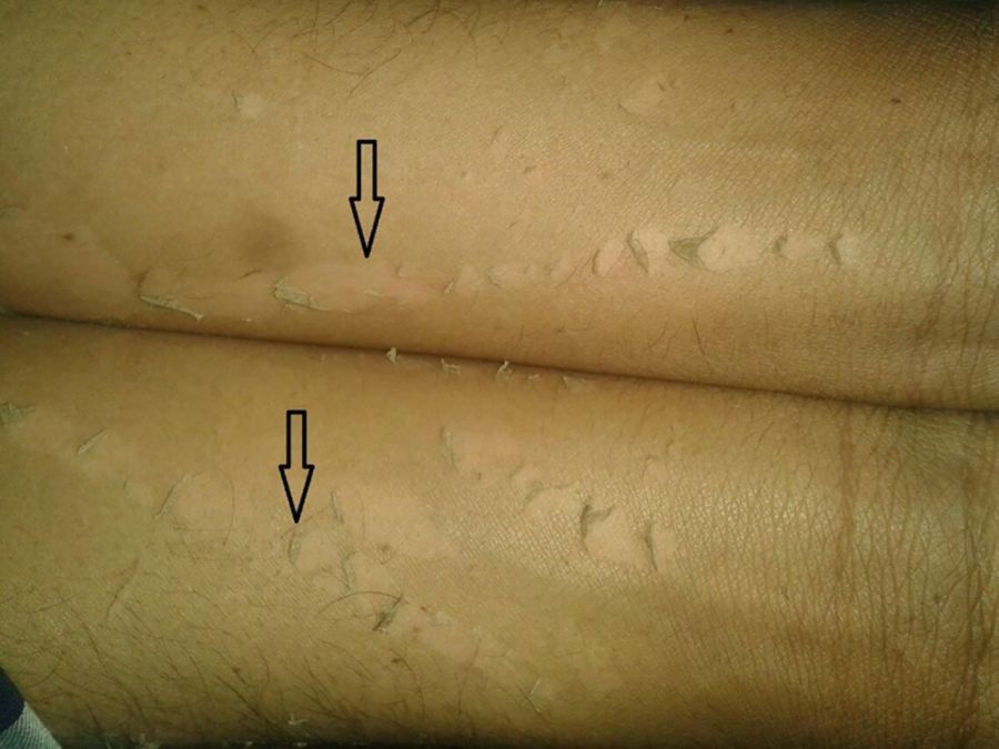
Conditions associated with acral peeling
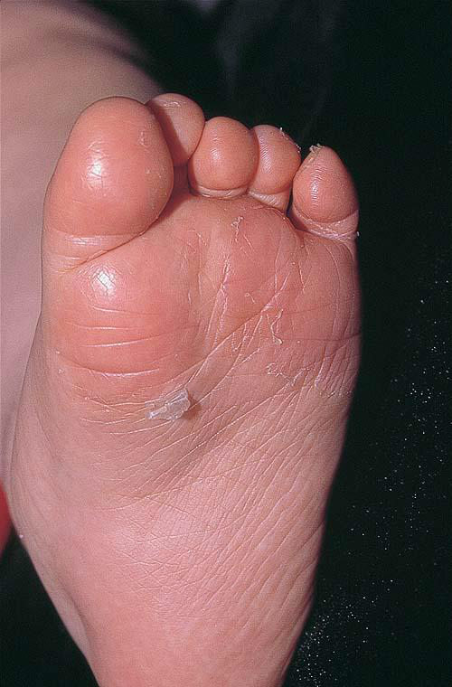
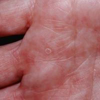
Keratolysis exfoliativa
- A common condition, mainly seen in young adults over summer months, and probably more often in those who sweat more
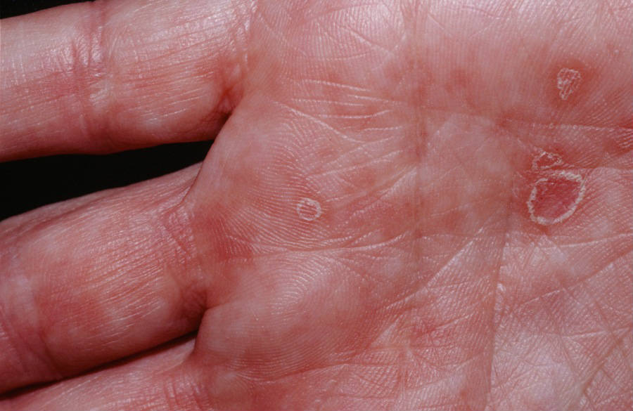
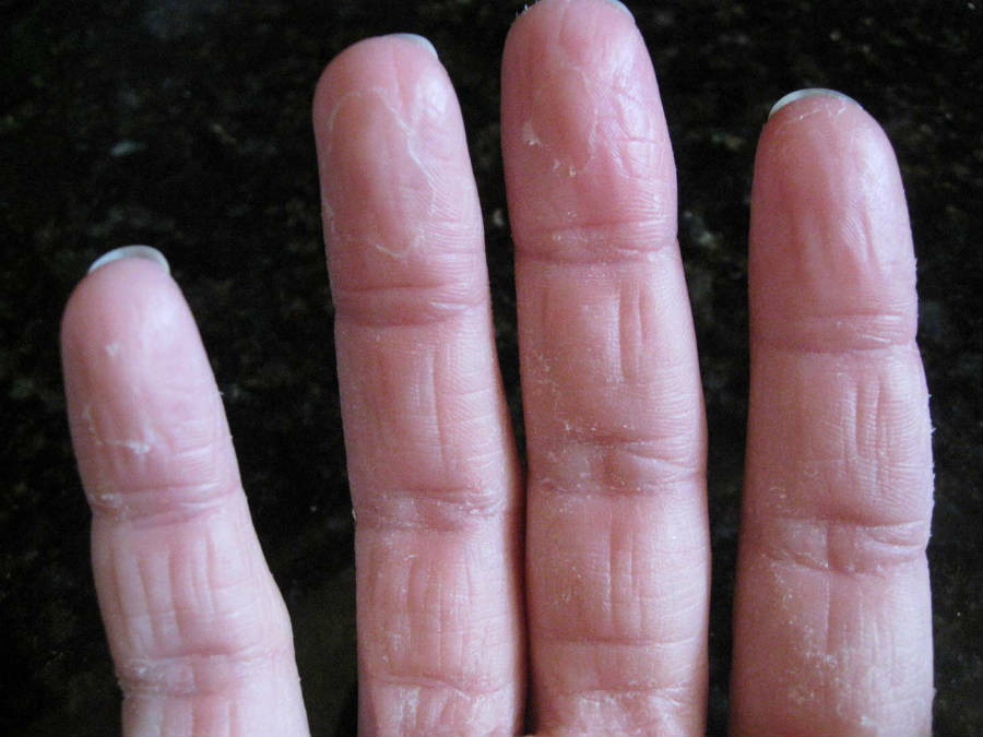
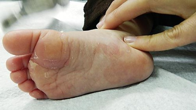
- Lesions present as small white rings or very superficial blisters on the fingers or palms, which soon peel off. The soles are less frequently affected
- The condition is self-limiting, and can be improved with the use of emollients
Other common causes
Acutely unwell children
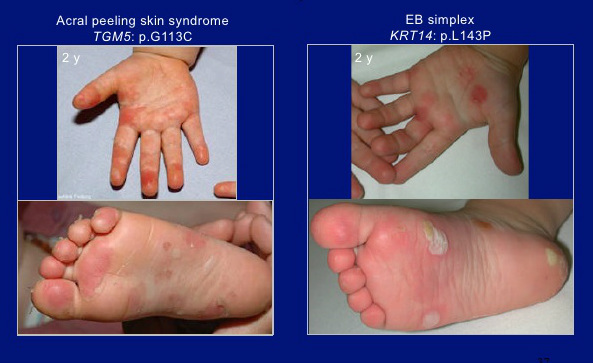
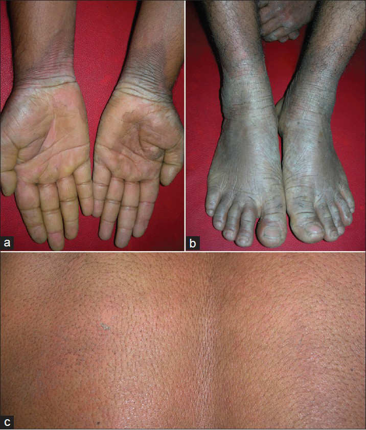
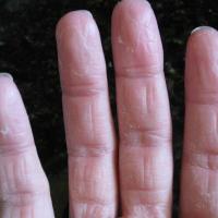
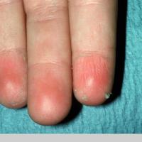
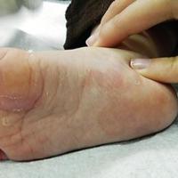
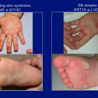
The Acral Peeling Skin syndrome (APSS)
- Congenital or familial acral peeling is extremely rare
- It is an autosomal recessive condition. This means that a person with APSS has inherited a defective copy of the gene from both parents
- The signs and symptoms of peeling skin usually appear soon after birth, but they may also develop later in life
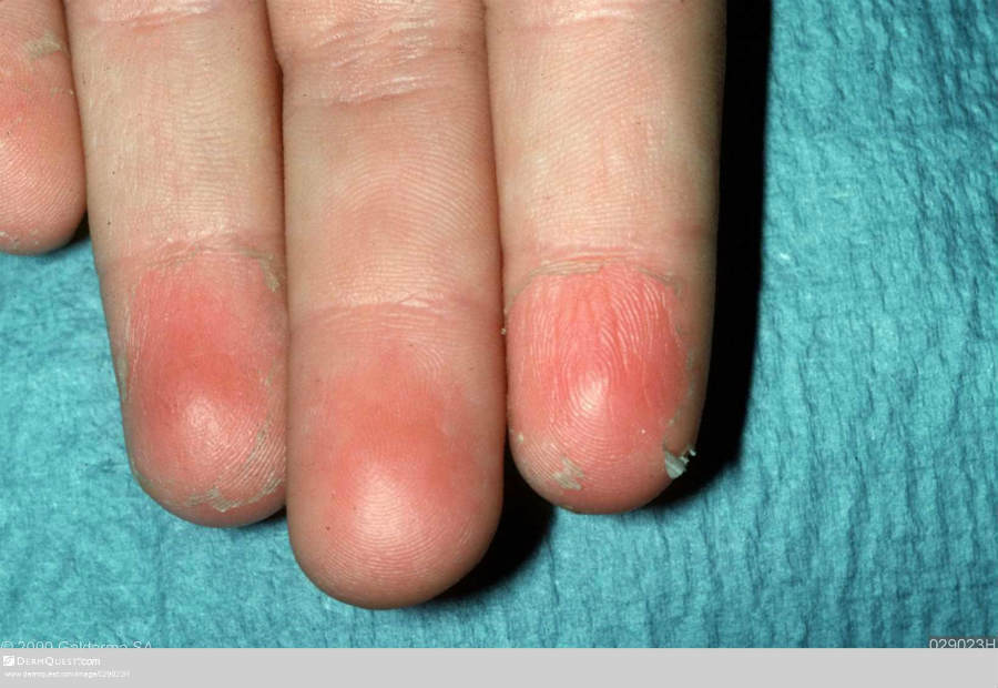
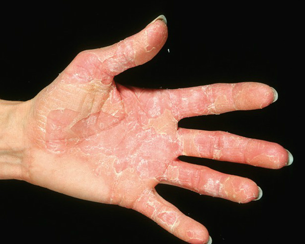
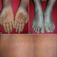
- The main symptom is painless peeling of the skin on the hands and feet. Patients may also experience itching and erythema. Symptoms can be made worse with exposure to water, perspiration, heat, or friction
- One of the main differential diagnoses is localised epidermolysis bullosa simplex
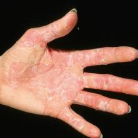
Oudtshoorn disease (syn. keratolytic winter erythema)
- A rare autosomal dominant condition that was first described in the Oudtshoorn district of Cape Provence, South Africa
- Symptoms tend to present anywhere from infancy through to early adult life
- Unlike with the Peeling Skin syndromes, the main exacerbations are during cold, dry climatic periods
- The condition is characterised clinically by intermittent and recurrent centrifugal peeling with erythema, particularly of the palms and soles
- In more severe cases, similar patches can be found extending up the limbs to the buttocks and trunk. Lesions can also be annular or polycyclic
Conditions associated with generalised peeling
In addition to the Staphylococcal Scalded Skin syndrome, Stevens Johnson syndrome / toxic epidermal necrolysis, and pustular psoriasis, generalised skin peeling can follow on from a number of widespread inflammatory and bullous eruptions. The list that follows are all rare conditions.
Peeling Skin syndromes (PSS)
- The Peeling Skin Syndrome (PSS) refers to a group of rare autosomal recessive conditions characterised by episodic or persistent, superficial, asymptomatic, spontaneous peeling of the skin and histologically by a separation of the stratum corneum from the stratum granulosum at the subcorneal level
- Symptoms usually present at birth or in early childhood, and then becomes persistent or episodic. Symptoms tend to be exacerbated in the summer
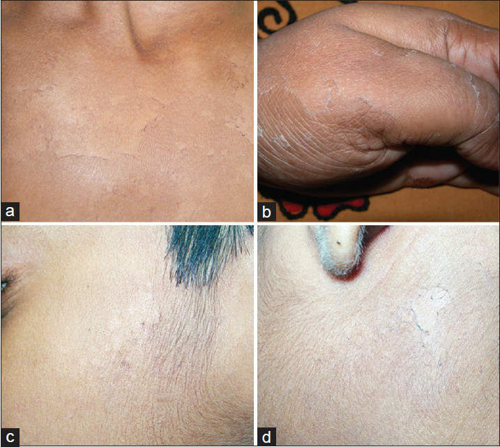
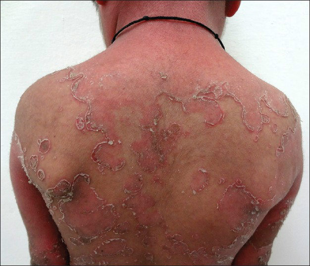
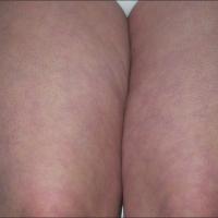
- PSS presents with either an acral (as described earlier) or generalised distribution (or both). In the generalised form, the peeling is widespread, with the palms and soles, usually, but not always, spared. Some cases remain difficult to classify
- PSS can also be classified as non-inflammatory (type A) or inflammatory (type B):
- Type A (non-inflammatory) PSS - generalised asymptomatic peeling of the trunk, limbs and occasionally the face. Histological examination shows an orthokeratotic epidermis with a separation that occurs either within the lower part of the stratum corneum or just above the granular layer
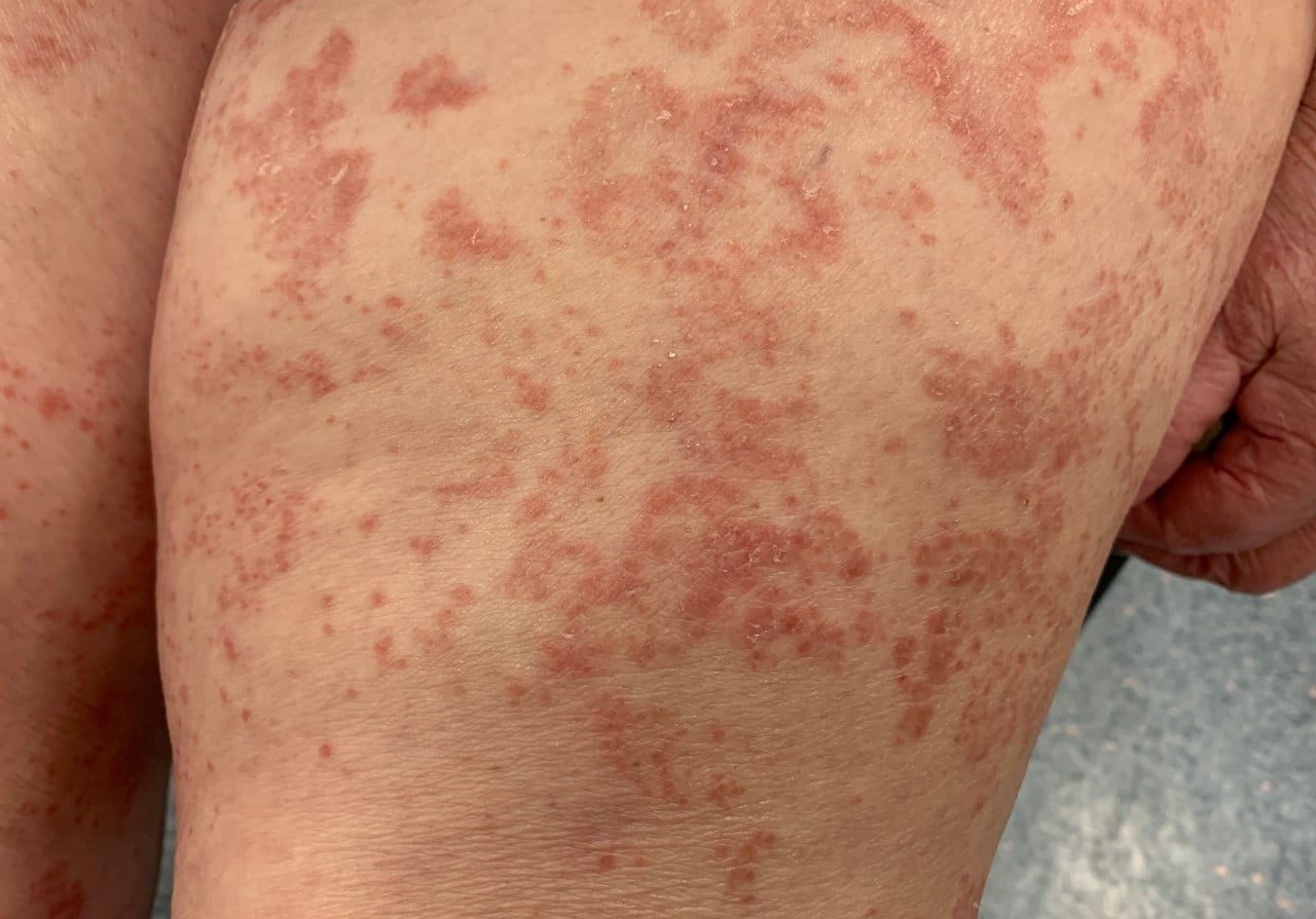
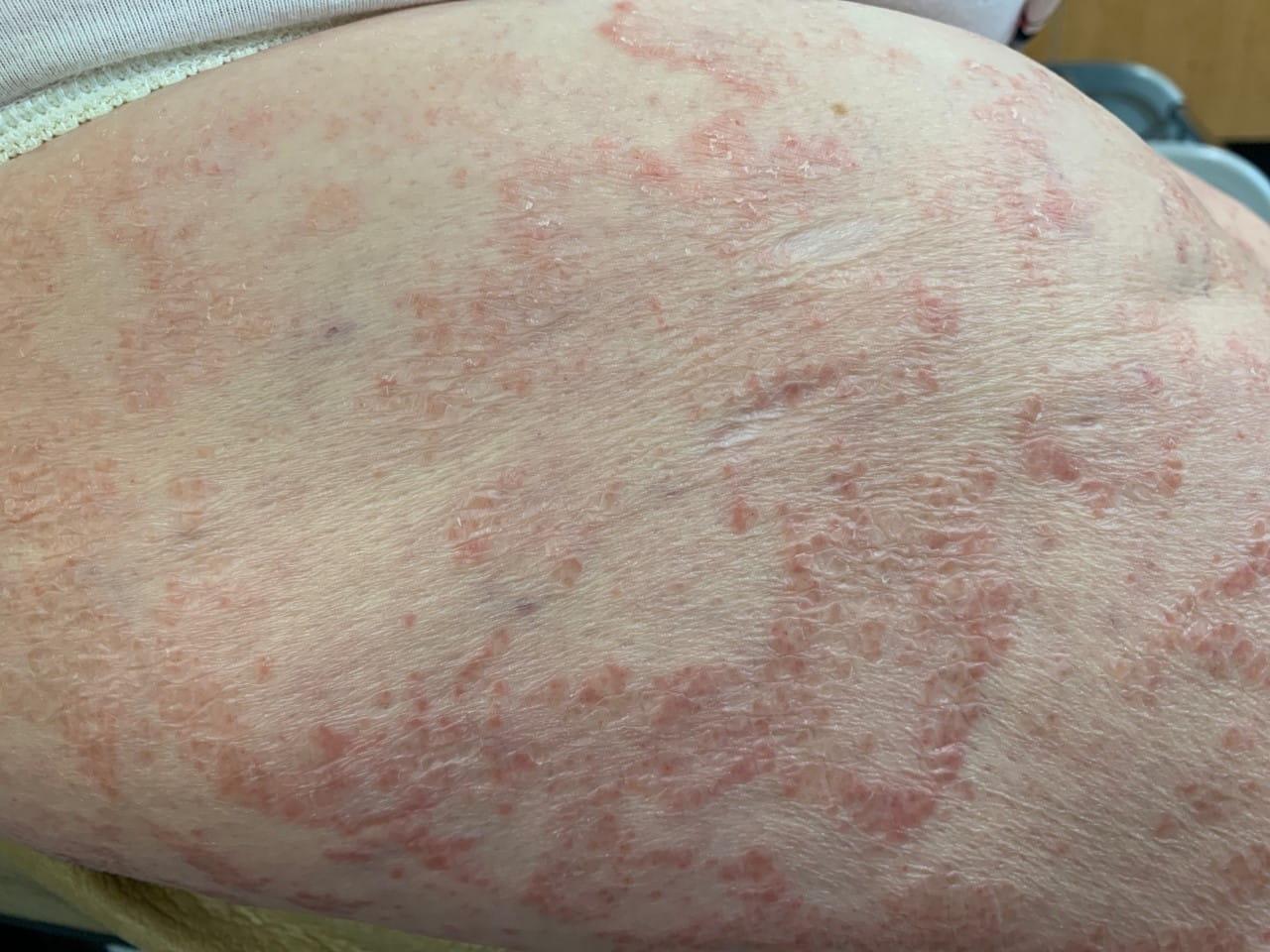
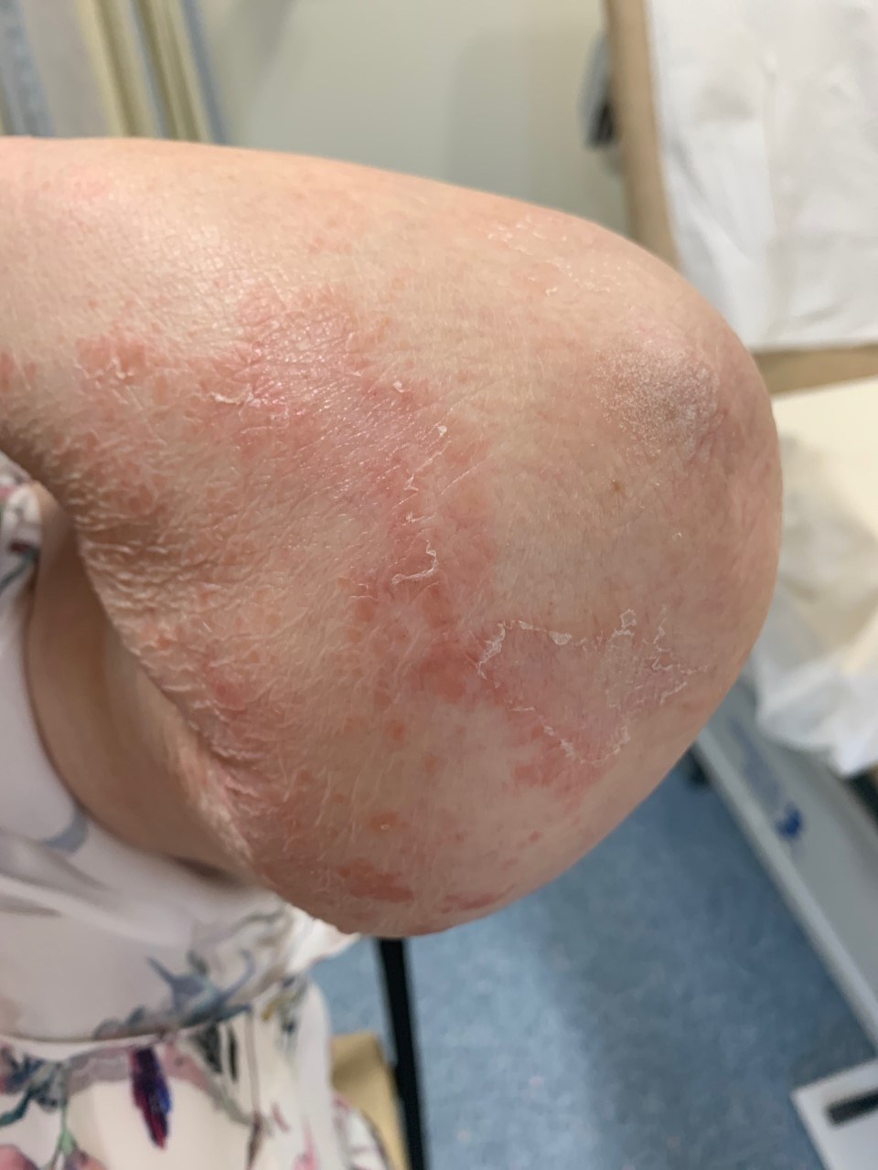
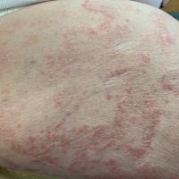
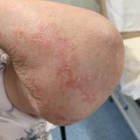
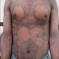
- Type B (inflammatory) PSS - characterised by erythematous migratory patches with a peeling border, pruritus, and a tendency towards atopy. Histology can show an absence of the stratum corneum or a few layers of parakeratosis, which tend to be separated from the stratum granulosum. Psoriasiform acanthosis and perivascular infiltration with mononuclear leucocytes can also be seen
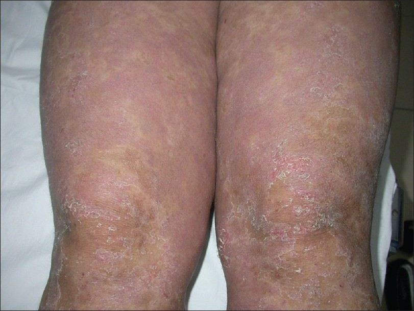
Erythrokeratoderma
- Is the association between localised hyperkeratotic plaques, distinct areas of erythema, and sometimes peeling
- The clinically and genetically heterogeneous group of erythrokeratodermas encompasses several rare genetic skin disorders, including autosomal dominant erythrokeratoderma variabilis and progressive symmetric erythrokeratoderma
- The majority of patients present in infancy
- The skin lesions of erythrokeratoderma variabilis and progressive symmetric erythrokeratoderma show many similarities:
- Erythrokeratoderma variabilis (EKV)
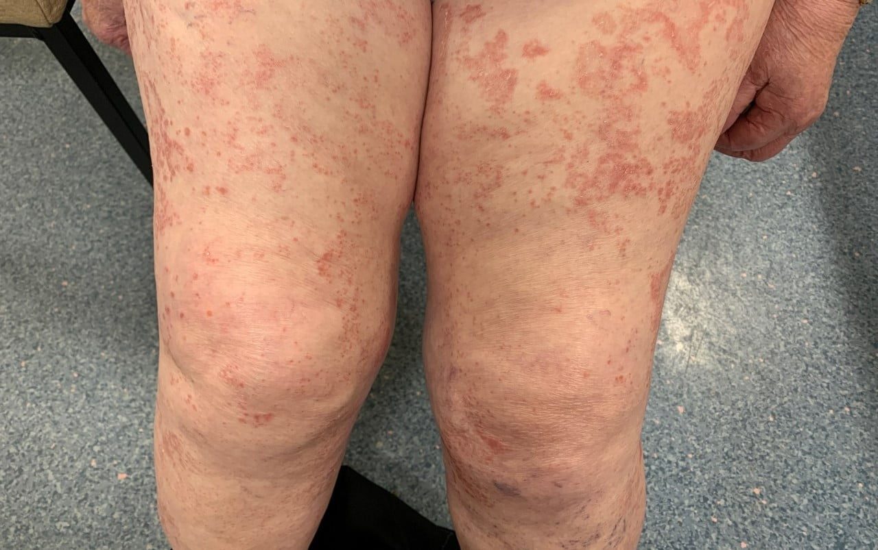
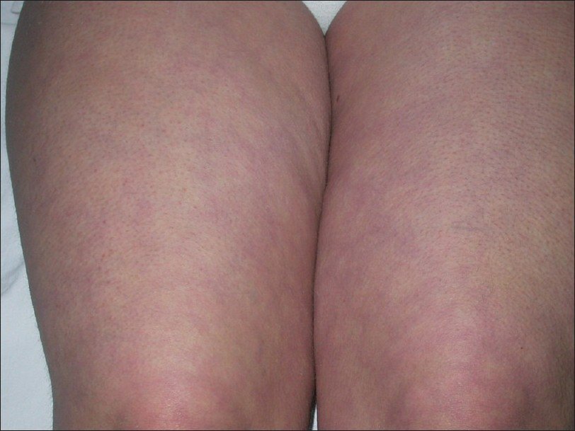
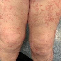
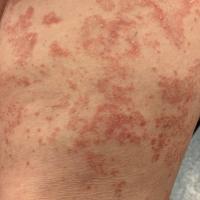
- Most cases present initially with well-circumscribed, annular or polycyclic erythematous patches. Episodes usually persist for minutes to hours, although may last for days. Fine scaling or peeling may be present. There is a very marked variation in the number, size, shape, distribution and location of the lesions
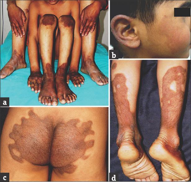
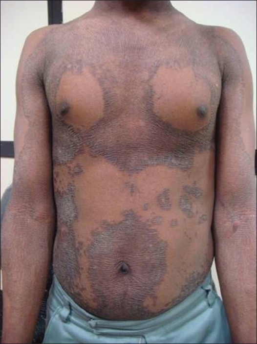
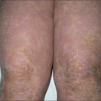
- Over time the hyperkeratosis develops, which may be generalised or localised with well-defined, yellow-brown, thickened, rough, hyperkeratotic plaques, which have accentuated skin markings. The most commonly affected pattern is a symmetrical involvement of the extensor surfaces, lateral trunk and buttocks. The flexures, face, and scalp are generally spared
- In about 50% of cases the hyperkeratosis involves the palms and soles, and this is often associated with peeling
- The plaques are relatively stable and last for months to years, but they can also clear completely
- Lesions are most prevalent during childhood. Improvement and periodic clearing of the skin are not unusual as the patient ages
- In the early stages of EKV, before the hyperkeratosis develops, it can look similar to a more extensive form of Oudtshoorn disease (described above), although the latter will flare over the winter with predominant involvement of the palms, soles, and limbs
- Progressive symmetric erythrokeratoderma (PSEK)
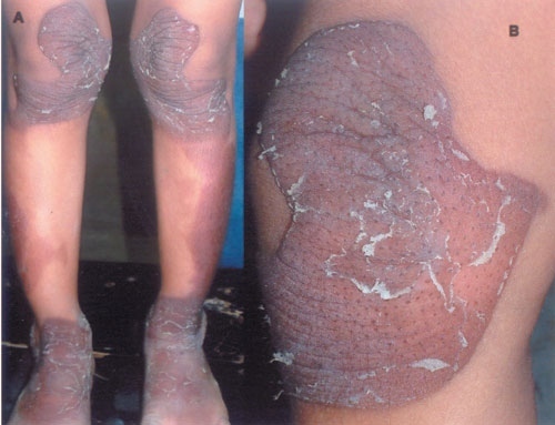
- The condition causes fixed, orange-red, hyperkeratotic plaques, which gradually extend through childhood. The most commonly affected sites are the cheeks, upper trunk, buttocks and extensor surfaces
- In contrast to EKV, there is no migratory erythema
- The palms and soles are often affected
Ichthyoses
- These conditions are generally associated with scaling as opposed to peeling, although Netherton's syndrome is characterised by variable degrees of erythema, with episodic skin peeling, and later a characteristic serpiginous migratory annular / polycyclic rash with double-edged scale
- Refer to the chapter Ichthyosis
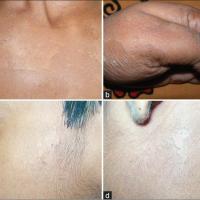
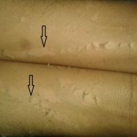
Clinical Images
Please refer to notes on image rights at bottom of the page with regards to individual image
ownership.
1
2
3
4
5

6
7
8
9
10
11
12
13

14
15

16
17
18

19
20
21
Management
-
There is no known treatment that can cure these conditions although emollients can provide symptomatic relief
-
Treatments sometimes provided in Secondary Care include systemic retinoids (mainly acitretin), and occasionally phototherapy
Disclaimer - the author PCDS cannot accept responsibility for any misleading or incorrect statements, and the management of individual patients remains the direct responsibility of the individual doctor. We do however hope that visitors to this site can contact us regarding comments that are considered misleading or incorrect so that we can continue to improve the site.
Image Rights - The PCDS would like to thank Dermatoweb, DermQuest (Galderma), and others who have contributed images. All named individuals and organisations maintain copyright for the relevant images.