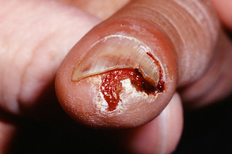
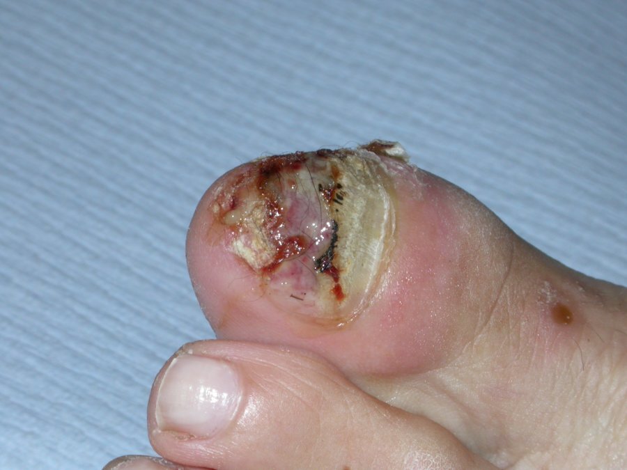
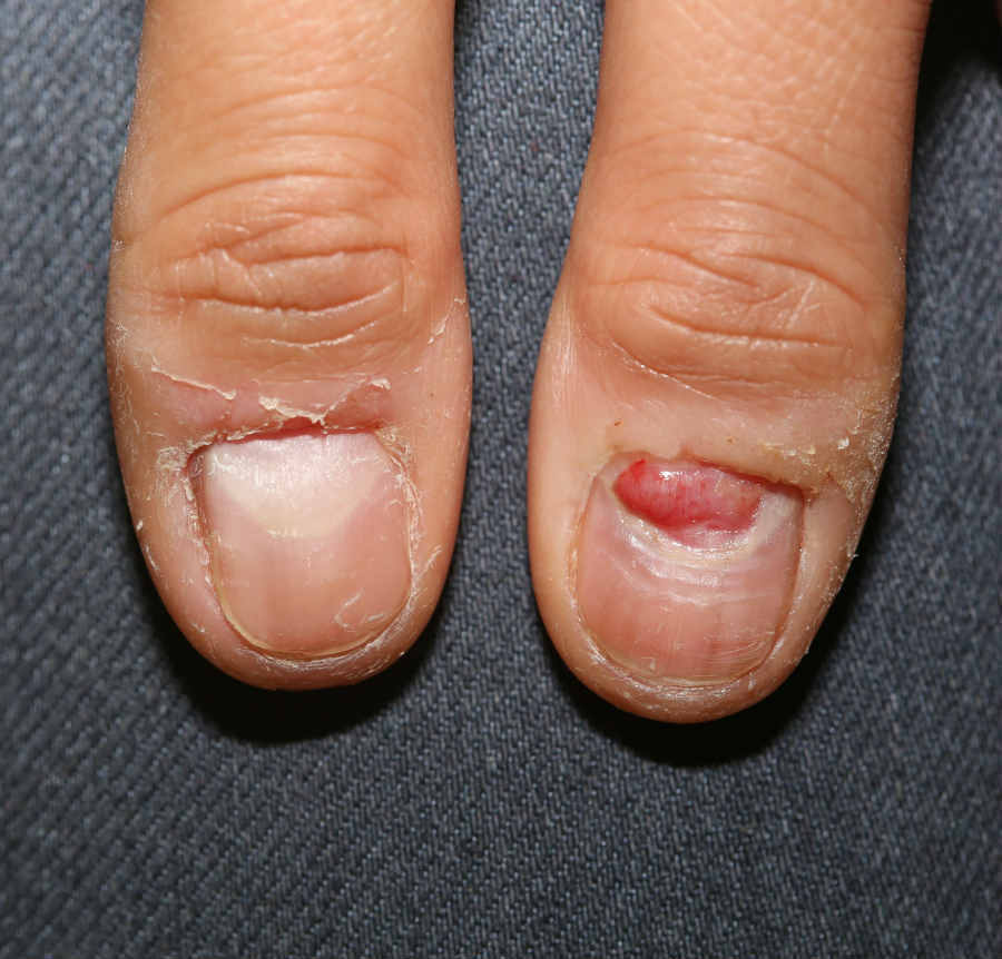
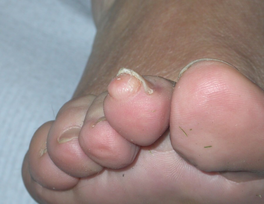
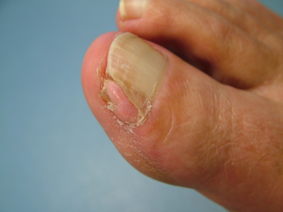
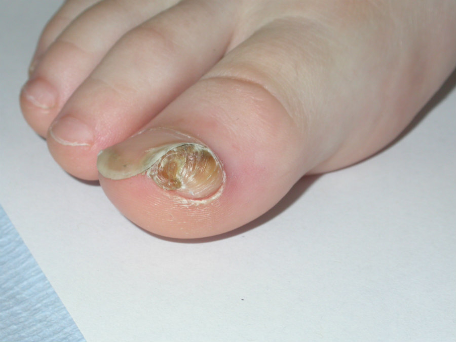
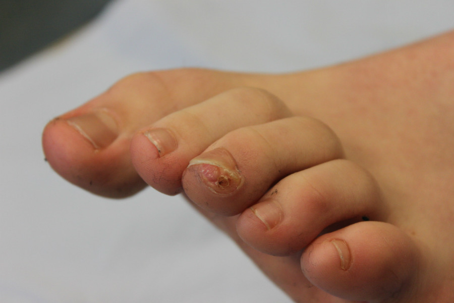
Nail disorders
LAST UPDATED: Jul 16, 2026
Introduction
This chapter, which is set out as below, provides an overview of nail disorders. For detailed information on paronychia please refer to the related chapter.
History
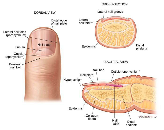
Anatomy of the nail (figure 1)
The nail (also called the nail plate) is firmly attached to the nail bed; it is less adherent proximally, apart from the posterolateral corners. Approximately one‐quarter of the nail is covered by the proximal nail fold, and a narrow margin of the sides of the nail plate is often occluded by the lateral nail folds. Underlying the proximal part of the nail is the white lunula; this area represents the most distal region of the matrix. The nail matrix is a localised region beneath the proximal nail, which produces the major part of the normal nail plate.
Terminology used in nail disorders
-
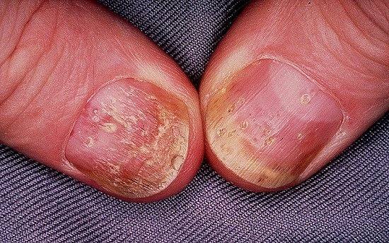
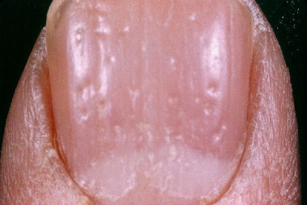
Pits - punctate surface depressions in the nail plate, more commonly affecting fingers than toes
-
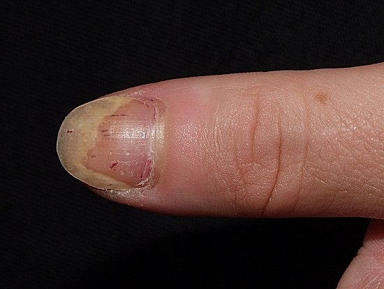
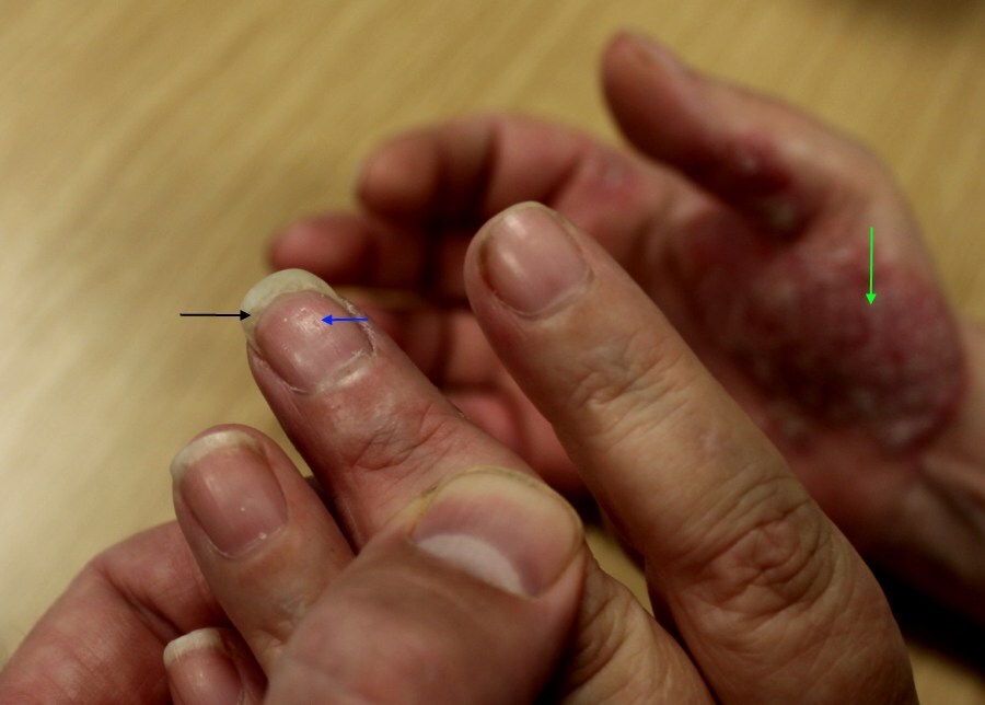
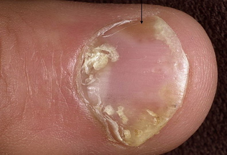
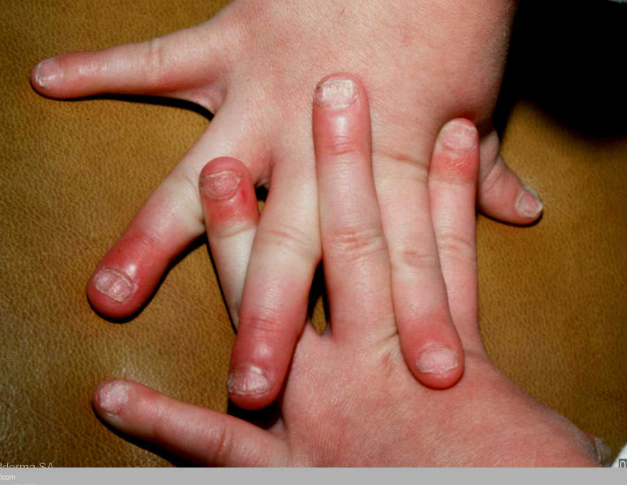
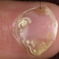
Onycholysis - the distal and/or lateral separation of the nail plate from the nail bed. Psoriatic onycholysis can be considered the reference point for other forms of onycholysis and is typically distal (ie affecting the free margin of the nail), with variable lateral involvement. Isolated islands of onycholysis present as ‘oil spots’ or ‘salmon patches’ in the nail bed. All the other common causes of onycholysis are associated with diminished adherence of the nail plate to the nail bed as a primary (idiopathic) or secondary event: the latter include trauma, fungal infection, eczema, and drug reactions
-
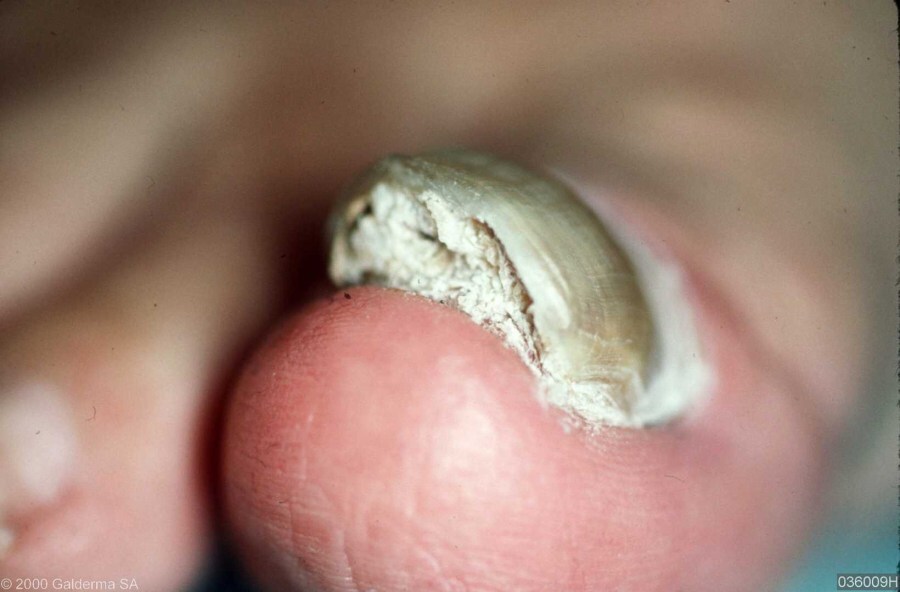
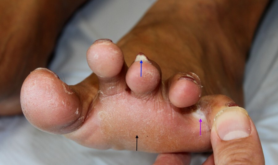
Subungual hyperkeratosis - excessive scaling under the nail. This is often due to a fungal infection or psoriasis. Warts, and rarely a squamous cell carcinoma, can produce a scaly mass under the nail
-
Longitudinal ridges - the presence of lines or ridges that run along the length of the nail
-
Transverse ridges - the presence of lines or ridges that run across the nail
-
Melanonychia - black or brown pigmentation of the nail plate
-
Leukonychia - white discoloration of the nail plate
-
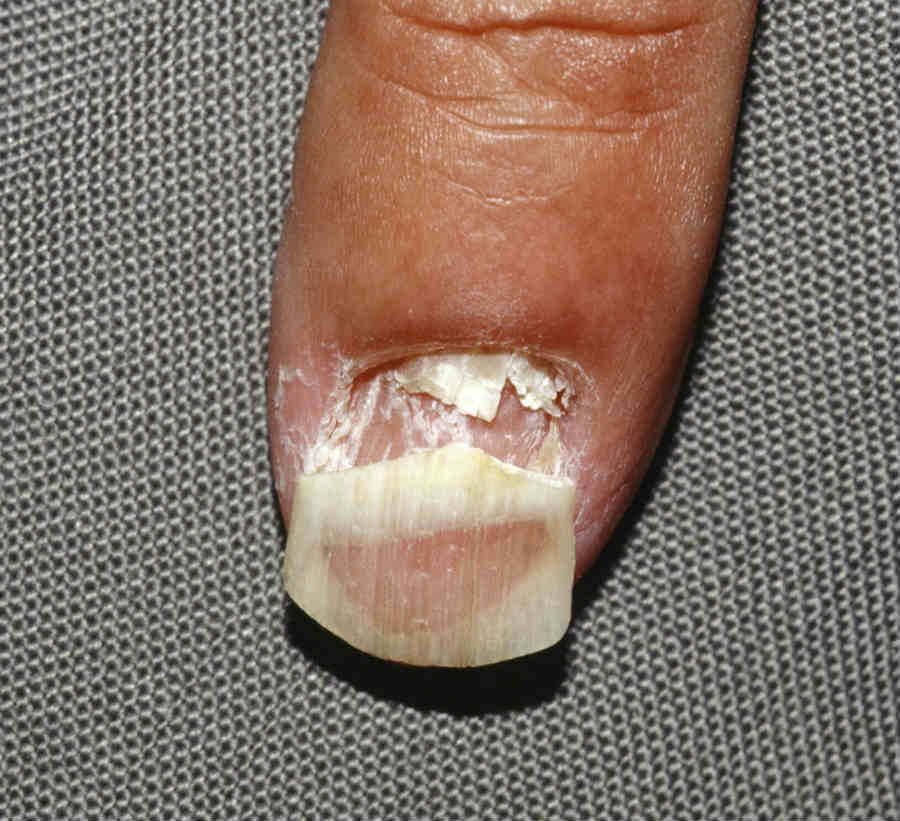
Pterygium - the winged appearance achieved when a central fibrotic band divides a nail proximally in two. The most common cause is lichen planus, it can be idiopathic
-
Paronychia - inflammation of the nail fold
Clinical findings
This section is set out as follows:
- Congenital nail disorders (a brief introduction)
- Red-brown-black nail discolouration
- Yellow or white-yellow nails with or without onycholysis, thickening of the nail, or, subungual hyperkeratosis
- White nail discolouration (leukonychia)
- Other colour changes
- Nail pitting
- Longitudinal ridges
- Transverse ridges
- Various nail disorders
- Skin lesions
- Drug-induced nail disorders
Congenital nail disorders
- The clinical findings range from the complete loss of nails (anonychia), eg in some cases of congenital ectodermal dysplasia, to thickened and discoloured nails seen in pachyonychia congenita. Congenital nail disorders are very rare and are not considered further on this website
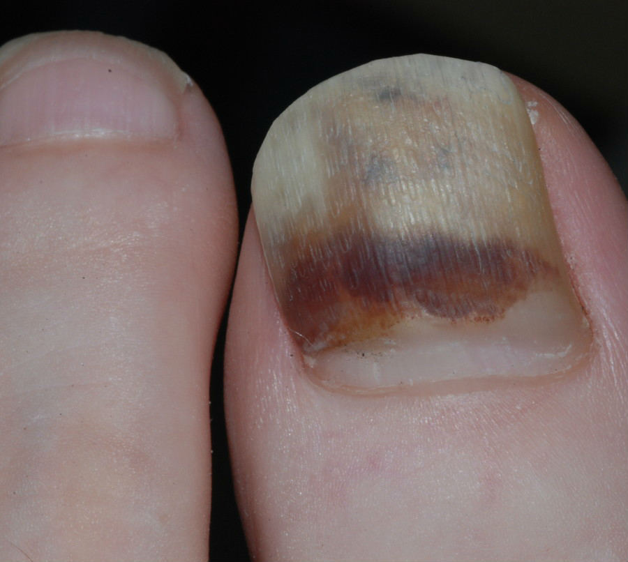
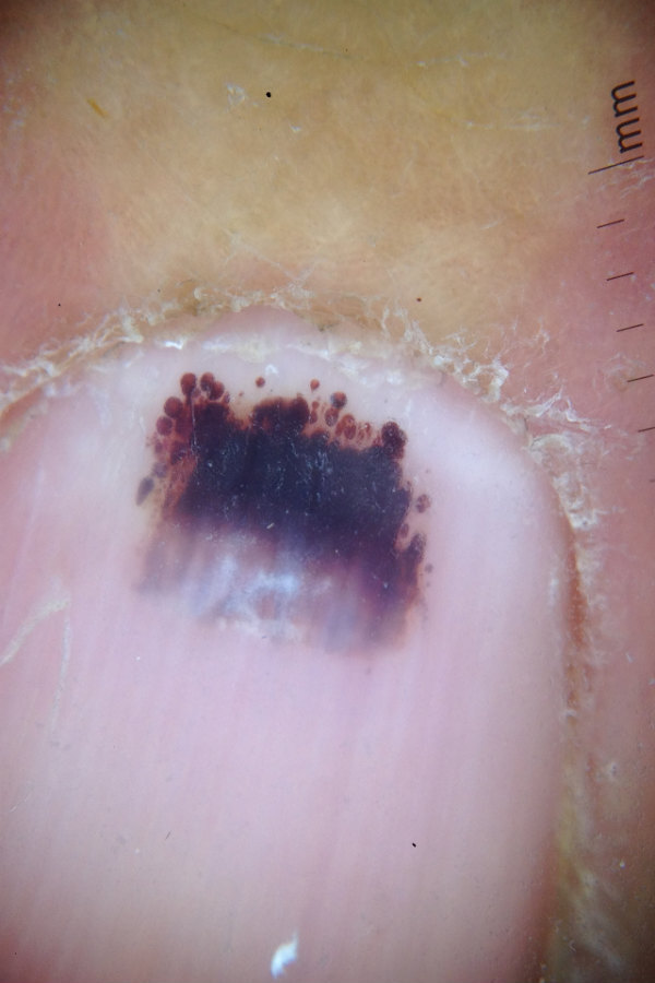
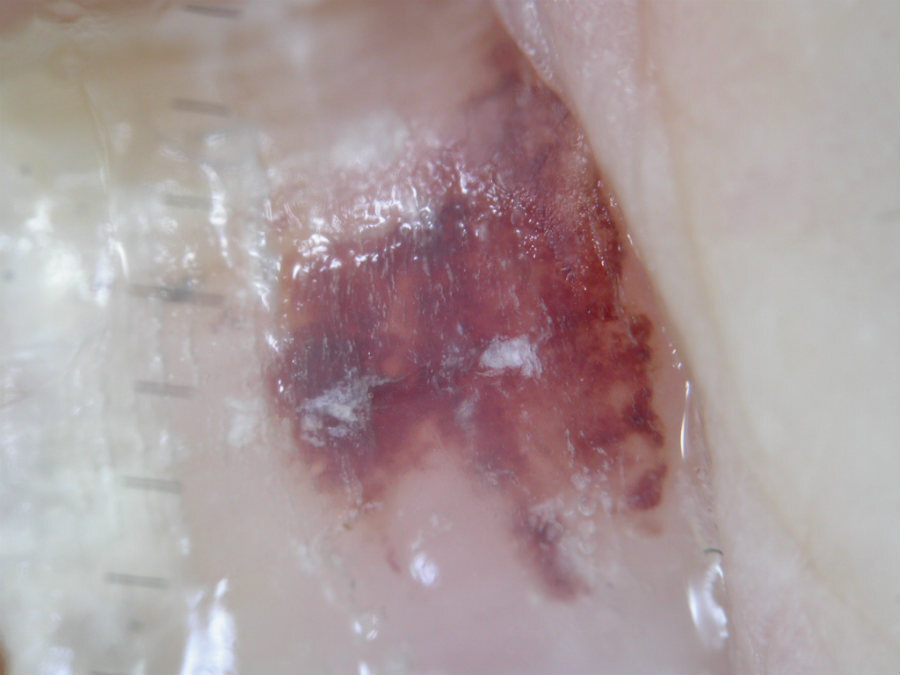
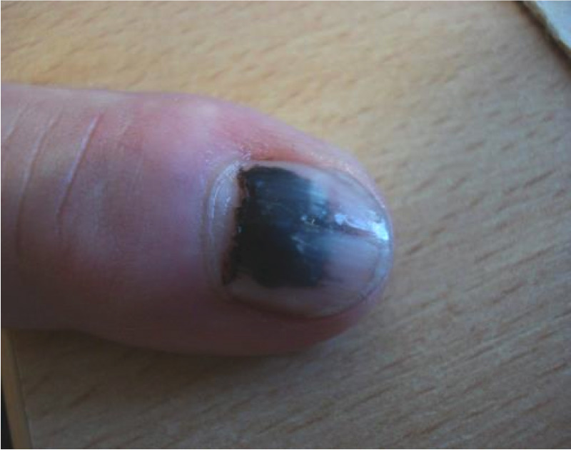
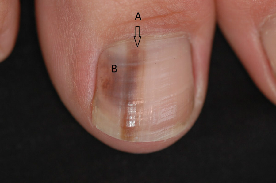
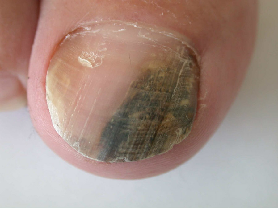
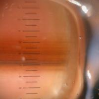
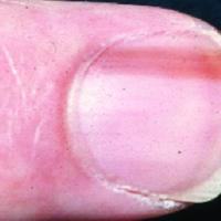
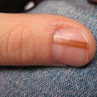
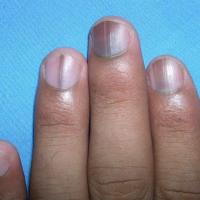
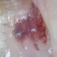
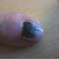
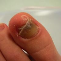
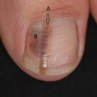
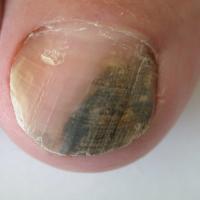
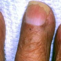
Red-brown-black discolouration (figures 2-22)
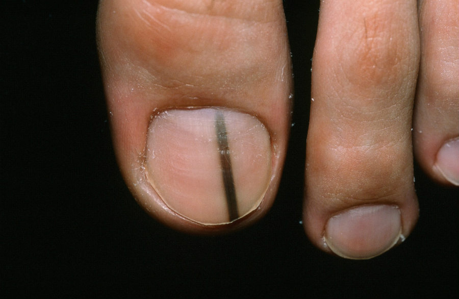
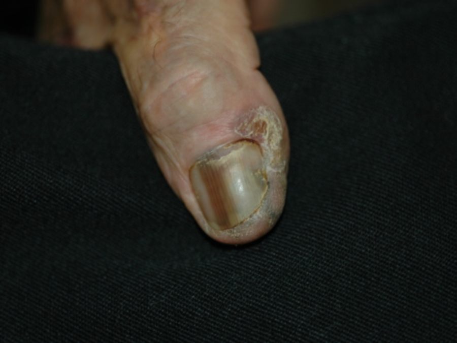
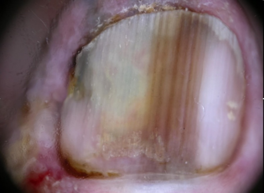
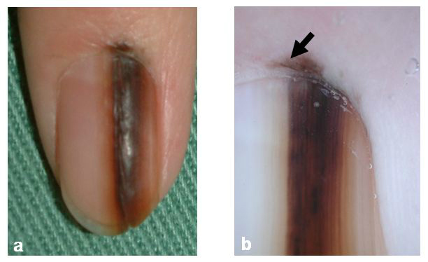
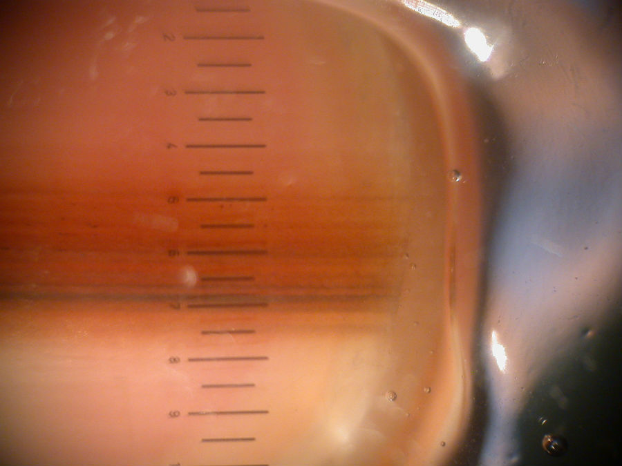
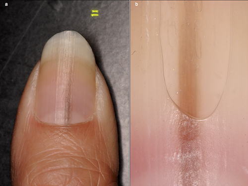
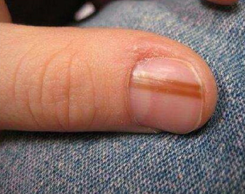
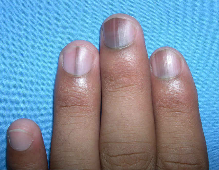
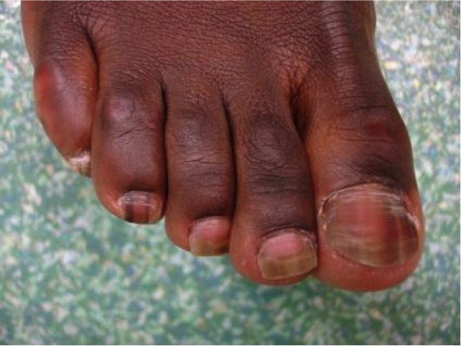
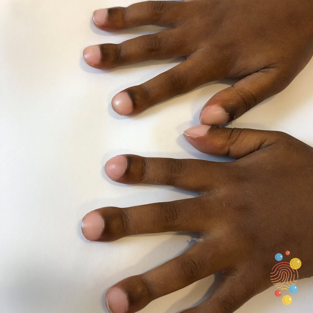
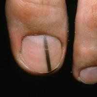
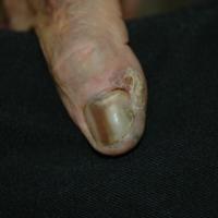
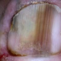
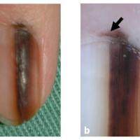
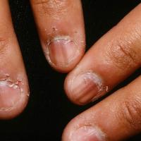
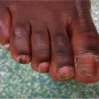
Longitudinal melanonychia is a term used to describe a hyperpigmented band that runs longitudinally from the proximal nail fold to the free end of the nail plate. The first diagnosis to consider is melanoma, although there are several benign causes.
- Subungual melanoma
- A differential diagnosis of subungual melanoma should be considered in the presence of any new or changing solitary black, brown, and occasionally red (in hypomelanotic lesions) line
- Hutchinson's sign - pigment affecting the nail fold, is very suspicious of melanoma
- Black-skinned individuals, Asians, Hispanics, and Middle Easterners frequently have benign longitudinal pigmented bands. The number and width of the bands increase with age. However, the presence of a new, solitary band in an adult could represent a melanoma
- Other causes of longitudinal melanonychia include benign melanocytic naevi, trauma, pregnancy, Addison's disease, post-inflammatory hyperpigmentation, a number of medications, and HIV positive patients in untreated cases as well as in those receiving zidovudine
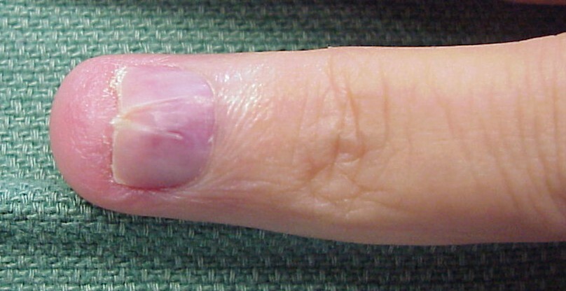
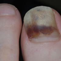
- Subungual haematoma - a collection of blood in the space between the nail bed and nail plate that may be associated with an episode of trauma, or more commonly, a period of vigorous activity where no trauma is recollected. Sometimes, especially with the toenails, there may be no history of trauma. The following are clinical clues to a subungual haematoma:
- Usually red or red-black colour depending on the age of the bleed. Brown is not a colour usually seen
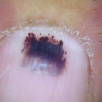
- Dermoscopy may reveal the following:
- That the haemorrhage is broken up into several zones, some of which are homogenous, and some which have droplets of blood that may be seen separated from the main zone of pigmentation
- The pigment may not be continuous in the longitudinal axis, with clear nail at either the proximal or distal margin
- The pigment may vary in any axis with much more irregularity at the lateral margins when compared to a subungual melanoma
- Blood may be seen as a discrete layer of material on the lower aspect of the nail plate at the free margin
- Over time (usually several months) there is normal proximal nail growth, and a gradual distal drift of the pigment
- An anecdotal clinical observation is that traumatic causes of subungual bleeding are associated with a proximal white transverse band in many instances. This is more common for trauma to digits of the hand than the foot
- Onychomycosis usually cause a yellow-white discolouration, but occasionally it can be black
- Unlike melanocytic pigment, which starts in the matrix, the pattern of onychomycosis usually extends from the distal free edge with proximal progression
- Early reassurance can be given if the pigmented change and dystrophic nail can all be trimmed away with no disturbance of surrounding skin and there is no sign of a more proximal origin to the pathology
- Suspicion of fungus should always be explored by mycological assessment and in particular culture. There is a wide variety of potential organisms. Some of the pigmented fungi are non-dermatophytes and may represent a therapeutic challenge likely to be surmounted only if the pathogen is known - some cases of Aspergillus do respond to treatment
Medications
- Chloroquine may produce blue‐black pigmentation of the nail bed, and other antimalarials may produce longitudinal bands of pigmentation on the nail bed or in the nail
- Hyperpigmentation due to increased melanin in the nail and nail bed has been noted after treatment with cytotoxic drugs
Glomus tumour
- A glomus tumour is a benign vascular neoplasm, which most commonly presents as a red-blue area of discoloration under the nail. Lesions are usually tender, particularly following change in temperature or pressure
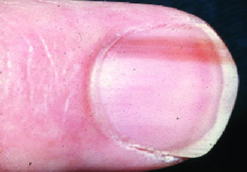
Splinter haemorrhages
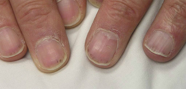
- Seen as 1-3 mm, red or brown-black, longitudinal bands of the nail bed (not plate)
- They are usually due to trauma, but can be secondary to a number of conditions including psoriasis, tinea, and most importantly subacute bacterial endocarditis
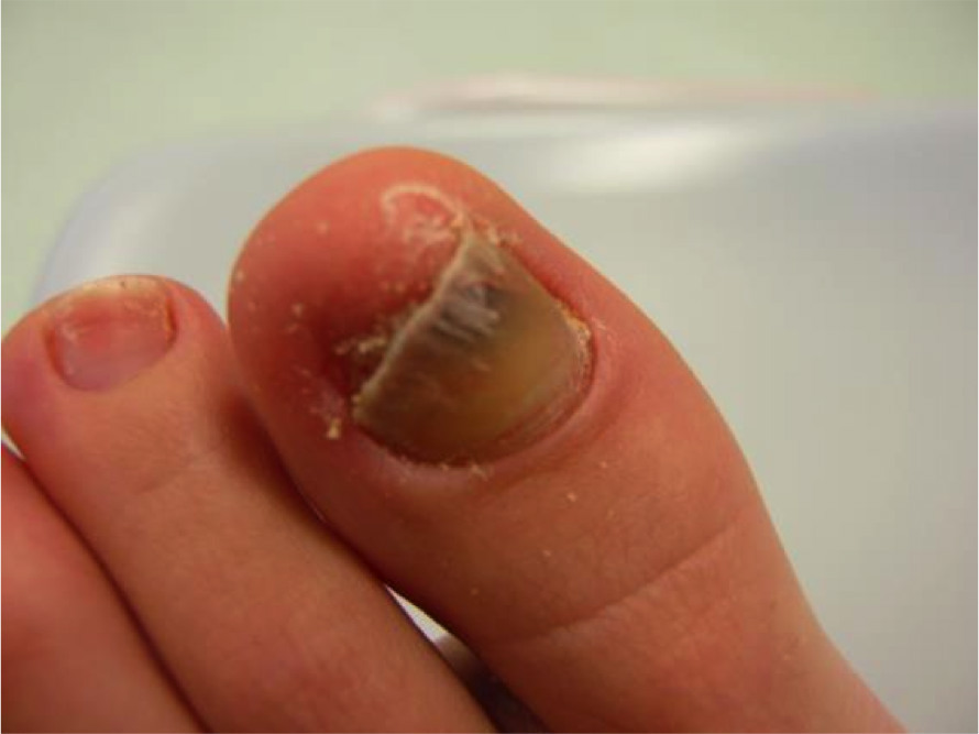
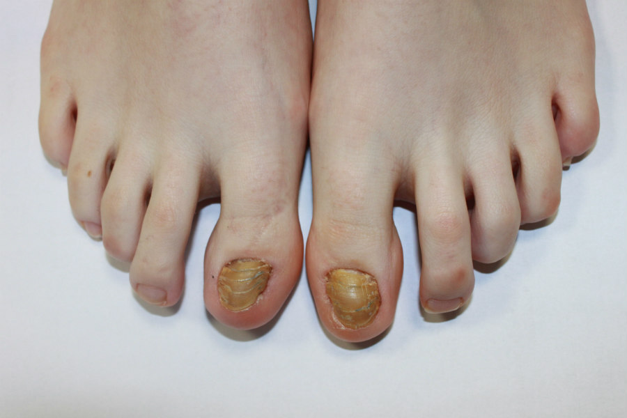
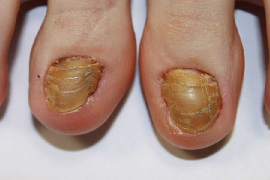
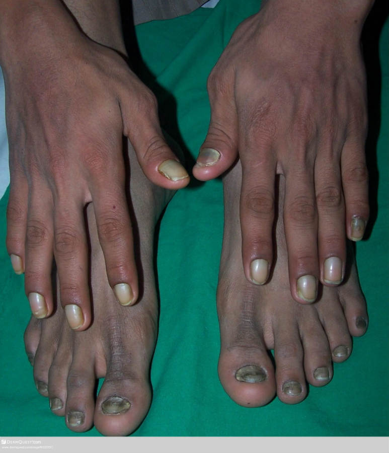
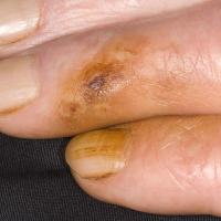
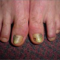
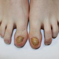
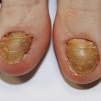
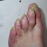
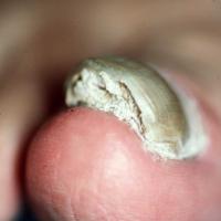
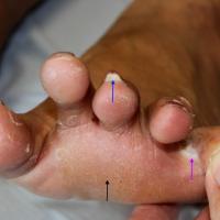
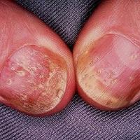
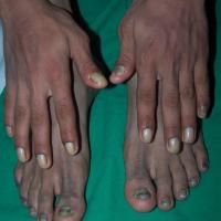
Yellow or white-yellow nails with or without onycholysis, thickening of the nail, or, subungual hyperkeratosis (figures 23-37)
Tobacco smoking
- Can result in yellow-brown discoloration of nails and fingertips
Trauma
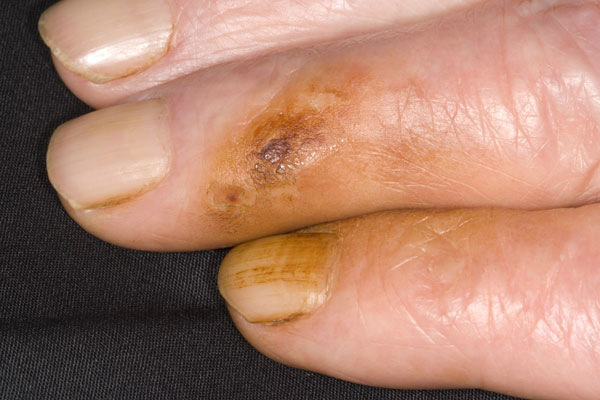
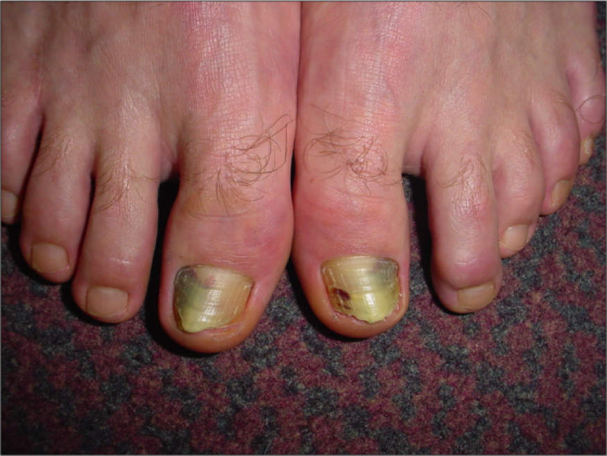
- Most commonly affects the big toenails, and is seen mainly in very active individuals, those with poor footwear, and in the elderly
- The nail becomes discoloured (yellow-brown), thickened, and then onycholitic
- The nails can start to grow out laterally, such changes can become gross when they are termed onychogryphosis
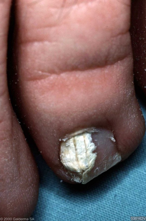
Tinea unguium
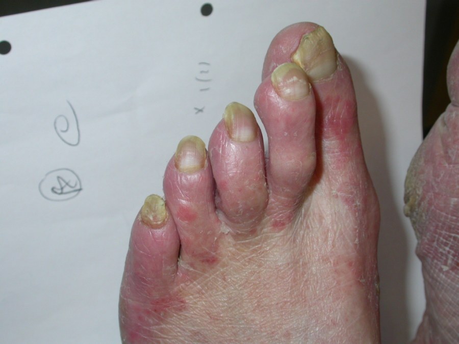
- Tinea unguium is a dermatophytic fungal infection
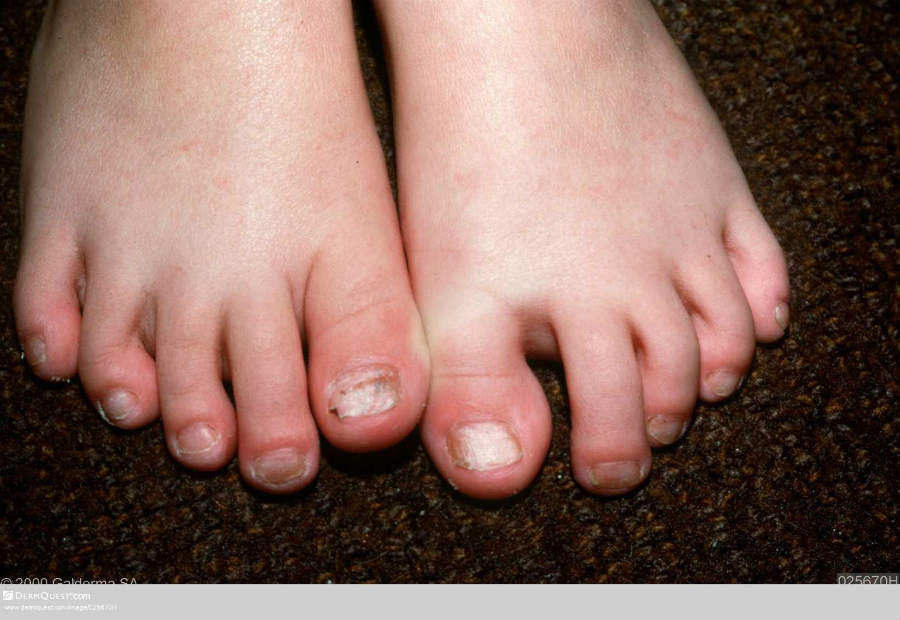
- The toenails are affected more often than the fingernails, indeed if there are no changes to the nails on the hands then tinea is much more likely than psoriasis
- The clinical features may be asymmetrical, but not always, and include onycholysis, subungual hyperkeratosis (which is often crumbly), and sometimes interdigital infection
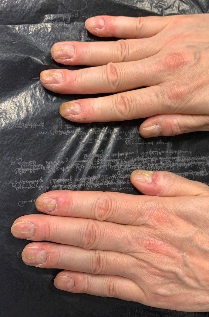
Psoriasis
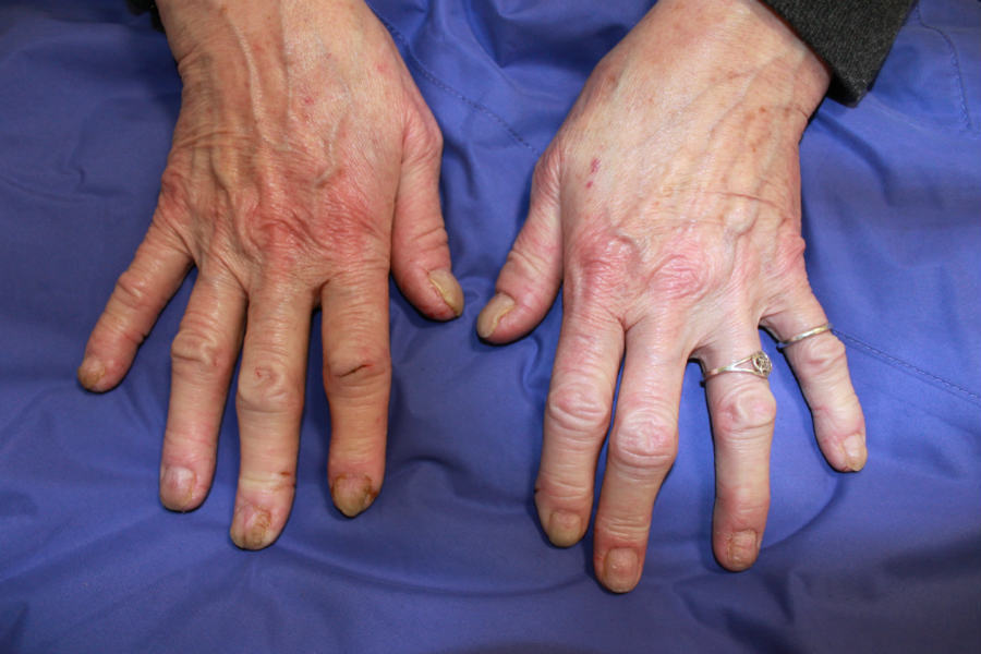
- Psoriasis tends to be more symmetrical then with tinea
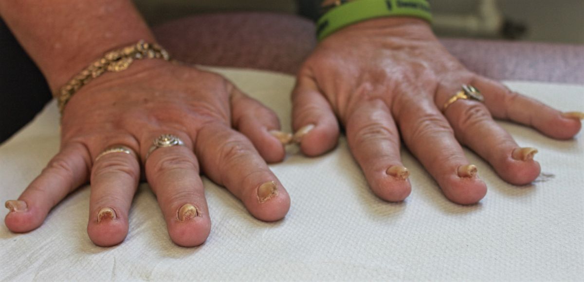
- Findings include pits, distal and lateral onycholysis, 'oil-drops', subungual hyperkeratosis, an uneven nail surface, paronychia, transverse midline depressions in the thumbnails, and occasionally splinter haemorrhages
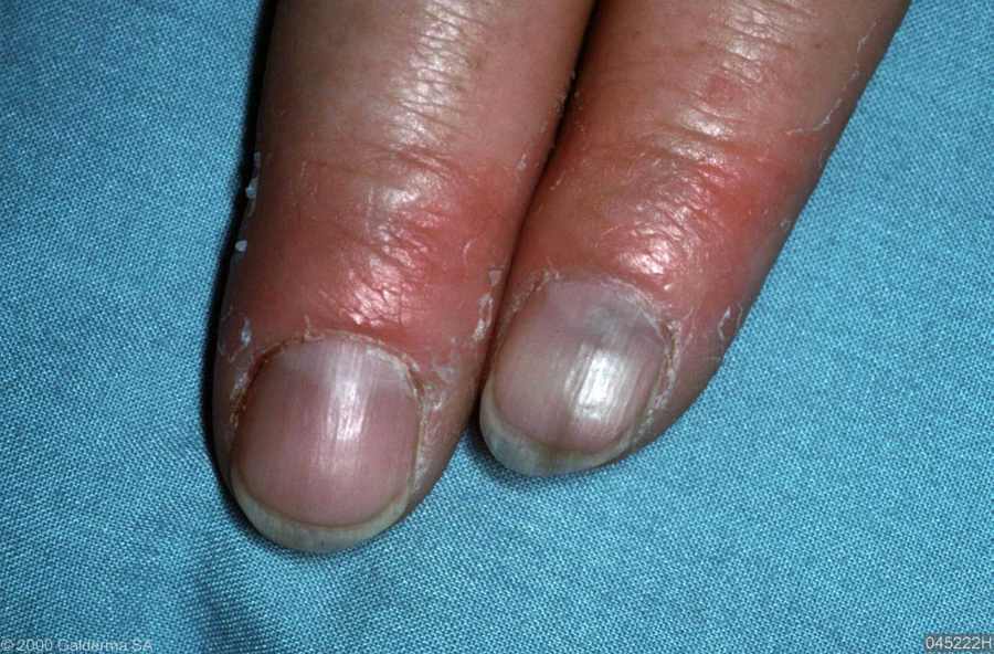
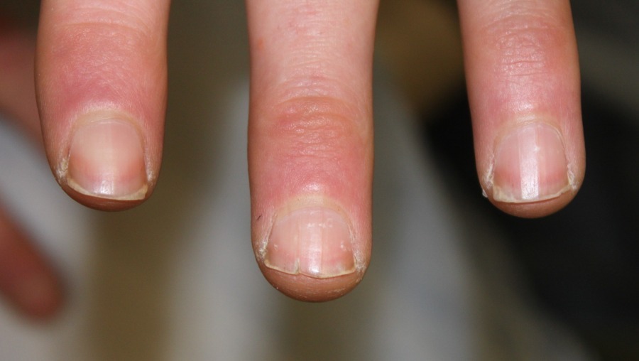
Paronychia
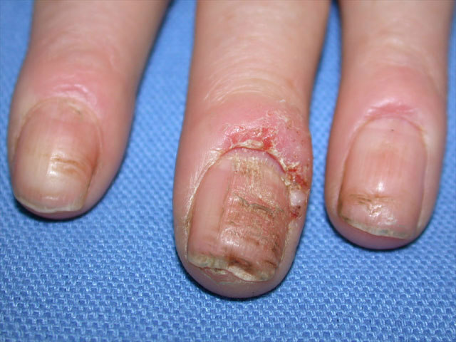
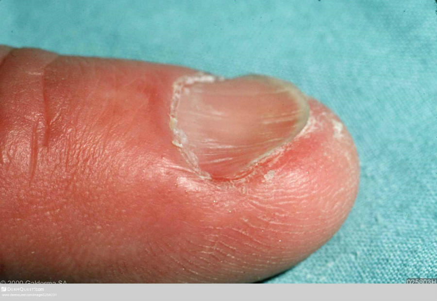
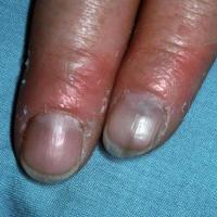
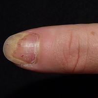
- Paronychia can lead to various nail changes such as discolouration, onycholysis and transverse ridging
Medications
- Onycholysis can occur secondary to a number of drugs including tetracyclines (photo-onycholysis), cardiovascular drugs (particularly practolol and captopril), trimethoprim/sulfamethoxazole, indomethacin, isotretinoin, isoniazid, and some cytotoxic drugs eg doxorubicin and 5-fluorouracil
Yellow Nail syndrome
- A very rare condition characterised by hypercurved, transversely thickened, smooth, yellow nails. It is associated with lymphoedema, pleural effusion, and (usually) ascites
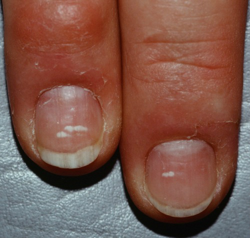
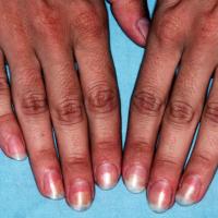
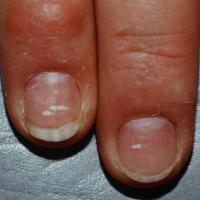
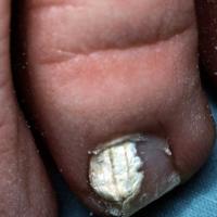
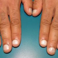
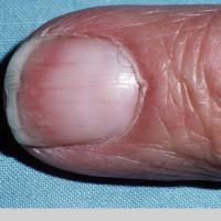
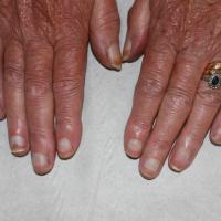
White nail discolouration - leukonychia (figures 38-44)
Minor trauma
- A common cause, and tends to cause focal areas of leukonychia
Tinea unguium
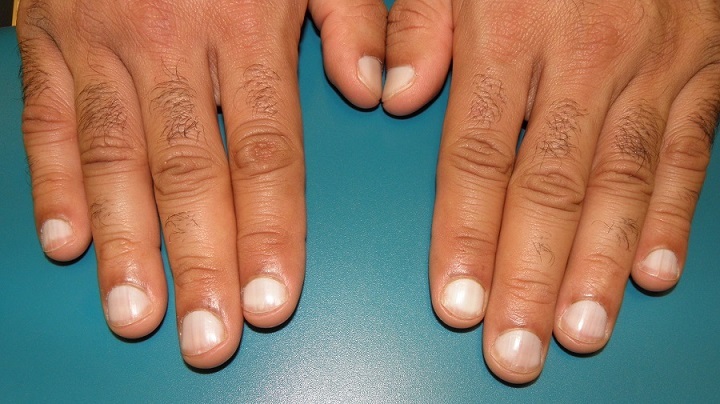
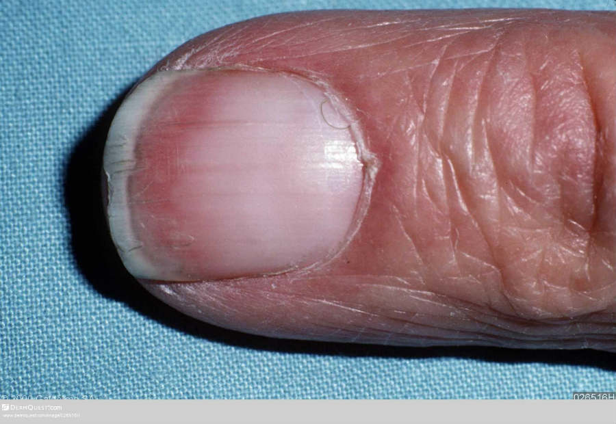
Terry's nails
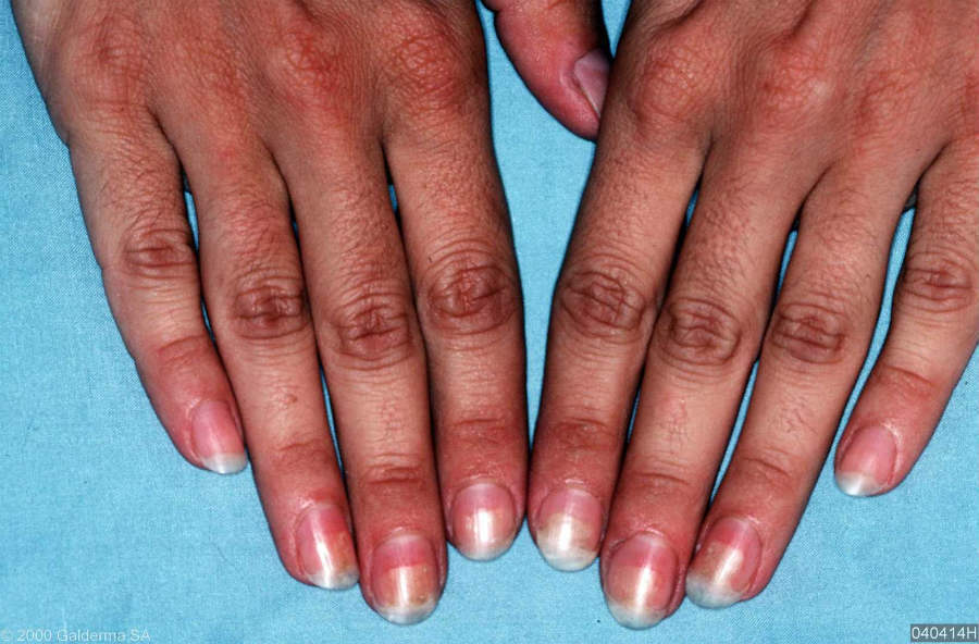
- A whiteness of most of the nail, except the distal edge
- Most commonly associated with cirrhosis, but also with a number of other systemic conditions including diabetes, cardiac failure, malabsorption and thyrotoxicosis
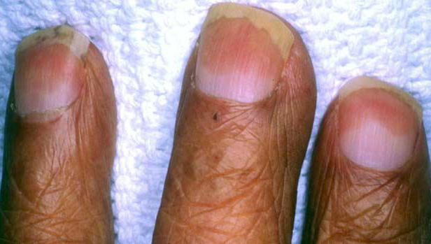
Half-and-half nails
- Describes nails where there is a proximal white zone and distal (20–60%) brownish sharp demarcation
- Associated with renal failure
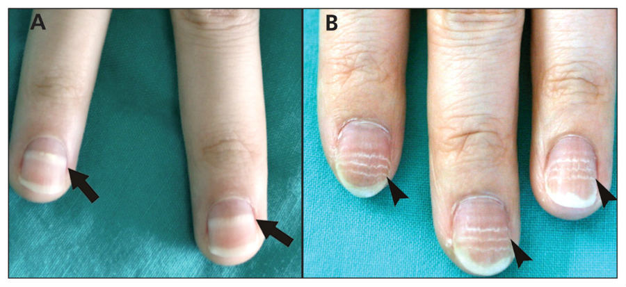
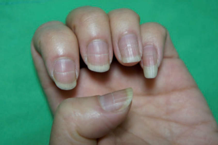
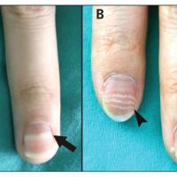
Transverse leukonychia (Mees’ lines)
- The 1–2 mm wide transverse band is in the arcuate form of the lunula
- Reflects a systemic disorder, drugs such as chemotherapy, or systemic infection that affect matrix function
Beau’s lines
- Narrow horizontal grooves in the nail plate, which often have a white colour, and occur when nail growth temporarily slows
- Beau's lines can follow infection, trauma, or systemic illness
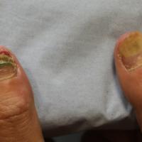
Other colour changes (figures 45-46)
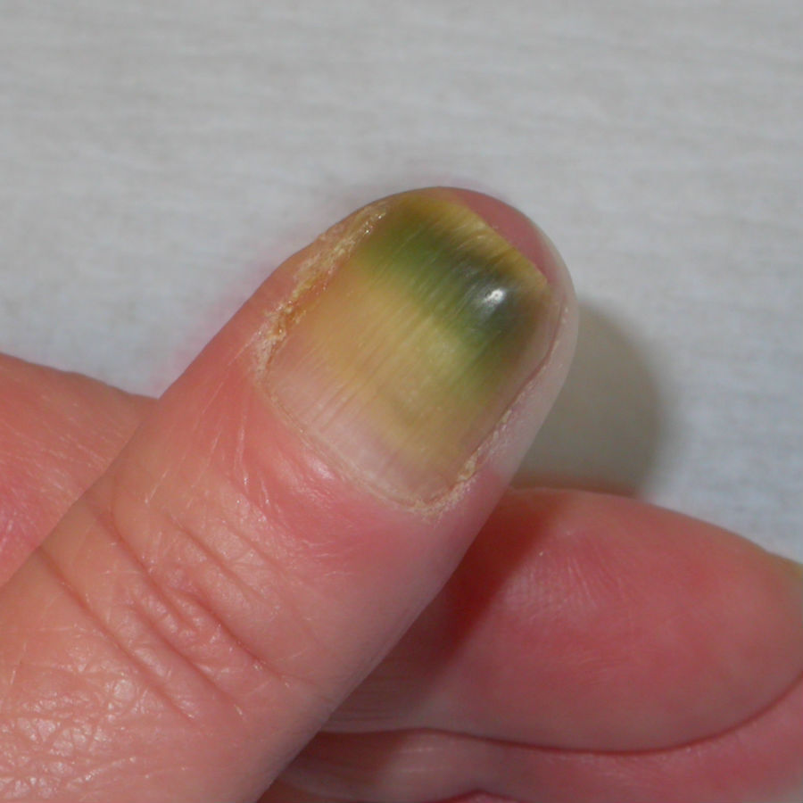
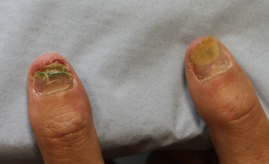
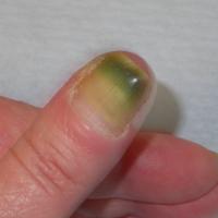
Pseudomonal nail infection
- Occurs most commonly secondary to other nail pathology
- Characterised by green-blue, or occasionally black, discolouration
Other drug-induced nail changes
- A bluish colour is seen with mepacrine, and hydroxyurea has been reported to result in blue lunulae
Nail pitting (figures 47-48)
- Can arise from psoriasis (small regular pits), eczema (larger and more irregular pits), and alopecia areata (causes small pits. Can be a bad prognostic sign for hair regrowth)
- Isolated pits can be a normal finding
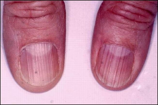
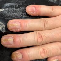
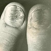
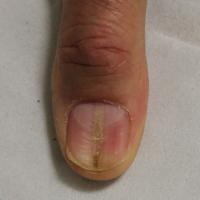
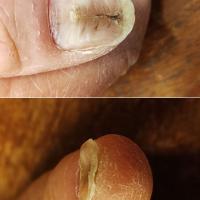
Longitudinal ridges (figures 49-62)
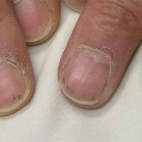
A few ridges are seen in normal nails.
Peripheral vascular disease can cause longitudinal ridges.
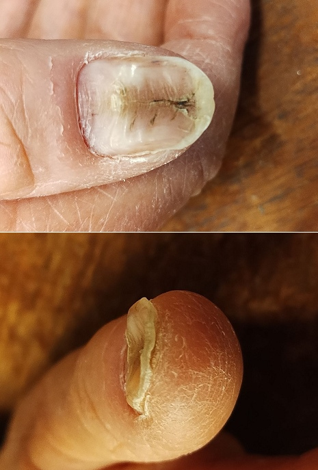
Myxoid cyst
- A myxoid cyst is seen around the proximal nail fold
- The cyst has a smooth shiny surface, and a clear jelly-like fluid may be expressed
- As a result of pressure the cyst often causes a lateral groove in the nail, a few mm across, which extends the length of the nail
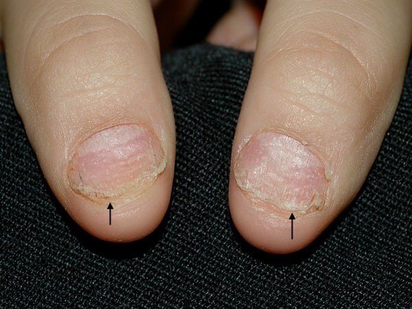
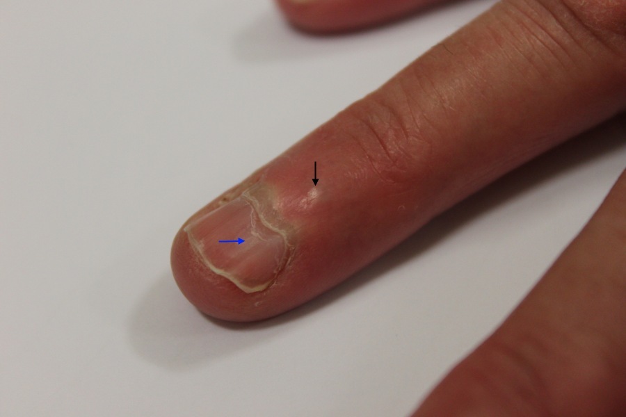
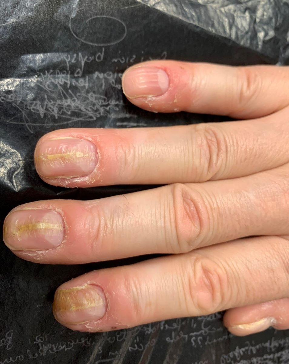
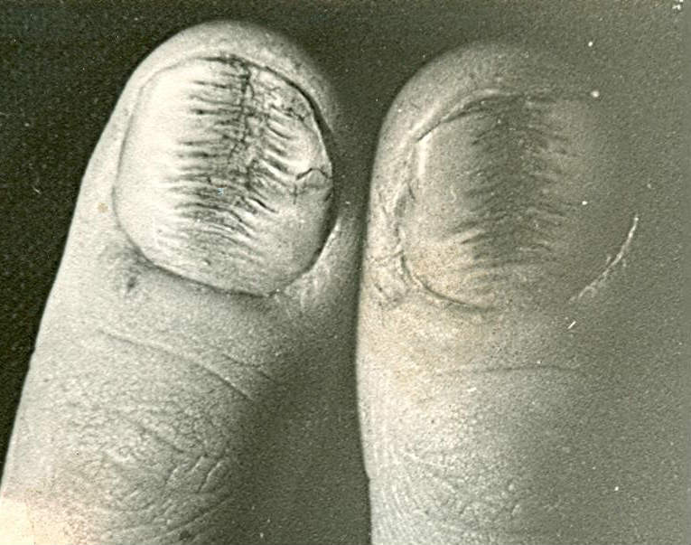
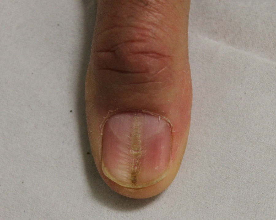
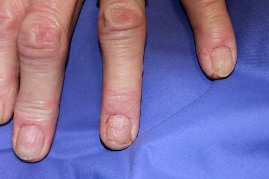
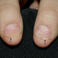
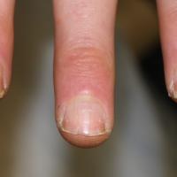
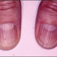
Median nail dystrophy (syn. median canaliform dystrophy) and habit-tic nail deformity (HTND)
- These conditions cause confusion in the literature as both have been described as having very similar appearances
- It is the author's view that habit-tic nail deformity should be the term used where there is the suggestion of repetitive trauma to the nail matrix, caused by rubbing or picking with another finger
- Both conditions tend to affect one or both thumbnails; other nails can be affected
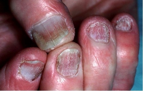
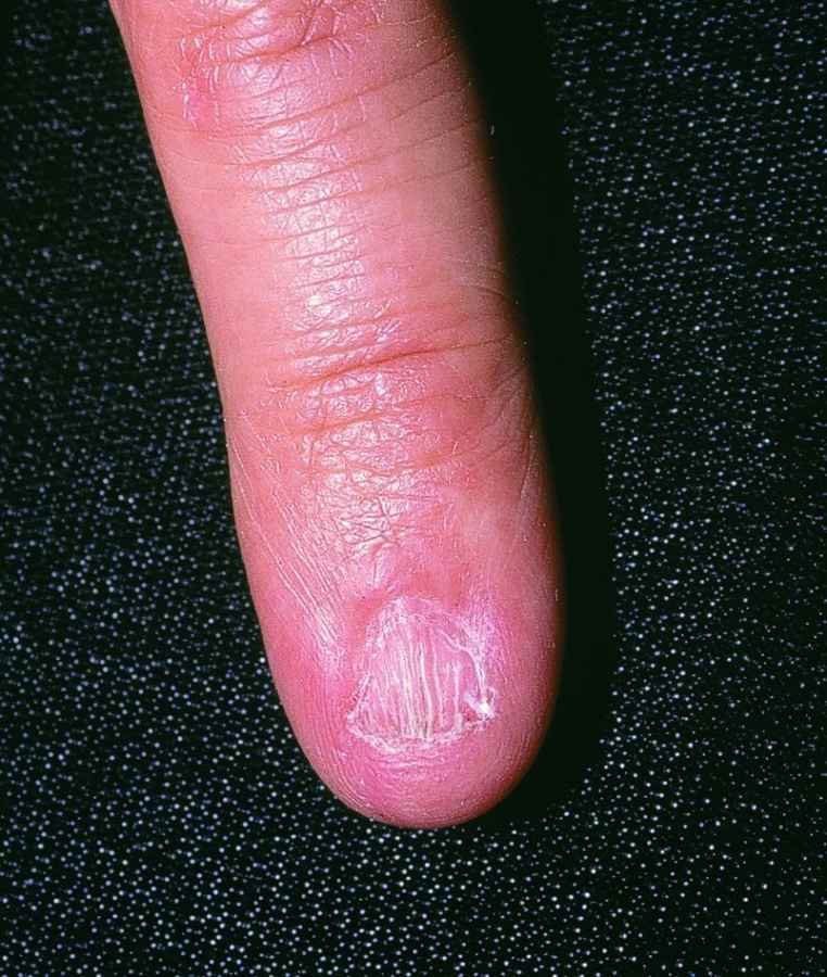
- A longitudinal depression with cross ridges giving a 'washboard' or 'fir tree' appearance
- In more severe cases, there can be paronychia, loss of the cuticle, and/or hypertrophy of the lunulae
- HTND generally responds well to habit reversal, although sometimes permanent nail dystrophy can result if the damage has been long-term. Median nail dystrophy tends to improve gradually
Darier's disease
- Darier's disease can cause longitudinal ridges with V-shaped nicks at the free end of the nail
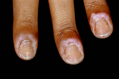
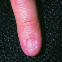
Lichen planus
- In lichen planus the fingernails are more frequently affected than toenails, with initial involvement of two or three fingernails before subsequent involvement of the remaining digits
- The most common changes are thinning of the nail plates and longitudinal ridges. Adhesion between the epidermis of the dorsal nail fold and the nail bed may cause partial destruction of the nail (pterygium unguis)
- Rarely, the nail is completely shed. One variant of nail lichen planus is associated with trachyonychia (see below)
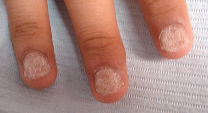
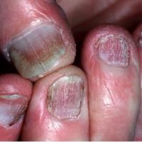
Trachyonychia
- Describes a rough surface affecting all of the nail plate, which is often associated with longitudinal ridging
- It can be idiopathic, or associated with alopecia areata, psoriasis, lichen planus, and ichthyosis vulgaris
- When the condition occurs on all the twenty nails of the fingers and toes, it is known as twenty-nail dystrophy
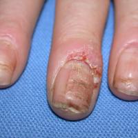
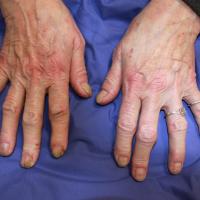
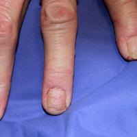
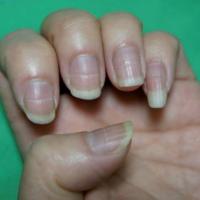
Transverse ridges (figures 63-68)
Eczema
- Eczema can cause irregular transverse ridging, with thickening and sometimes discolouration
Chronic paronychia
Beau’s lines
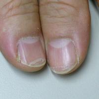
- Horizontal grooves in the nail plate that occur when nail growth temporarily slows
- Beau's lines can occur after infection, trauma, or systemic illness
Onychomadesis
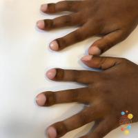
- Similarly results from temporary growth arrest of the nail plate, but differs from Beau's lines in that the full thickness of the nail is involved, causing a proximal separation of the nail plate from the nail bed
- It most frequently occurs several months after hand-foot-and-mouth disease but can occur after other viral infections
- Nails affected by Beau's lines or onychomadesis regrow normally with time
Various nail disorders (figures 69-73)
Nail cosmetic dystrophy
- From artificial nails, can cause onycholysis, ridging, sepsis and marked atrophy
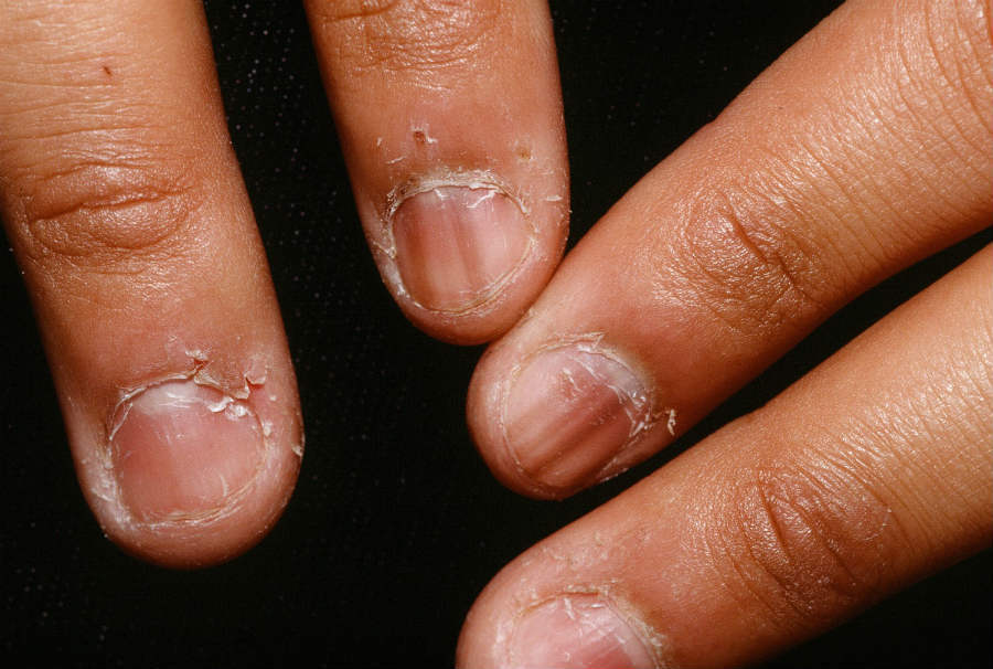
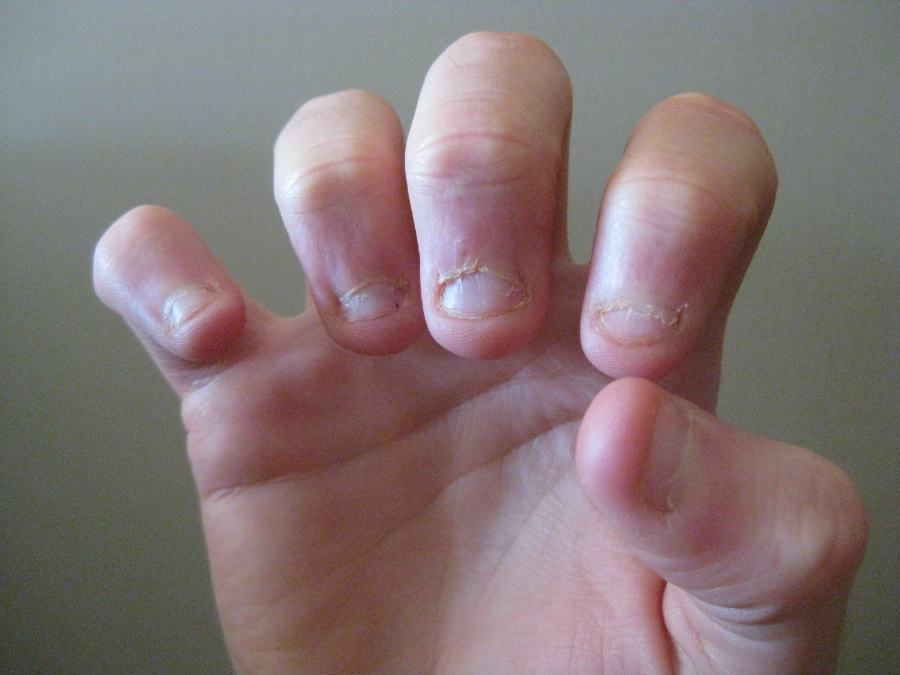
Nail biting
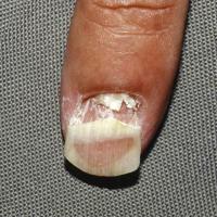
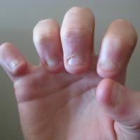
- Nails are typically short, with up to 50% of the nail bed exposed. The free edge may be even or ragged. Surface change may include splitting of the nail into layers
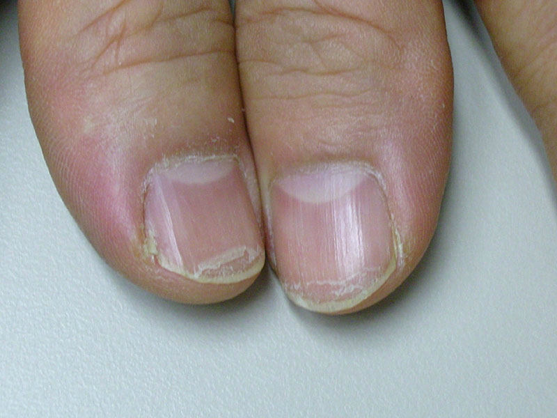
Lamellar nail dystrophy
- Characterised by the distal free end of the nail splitting into layers. It is seen more commonly in women, and is generally associated with frequent immersion in water
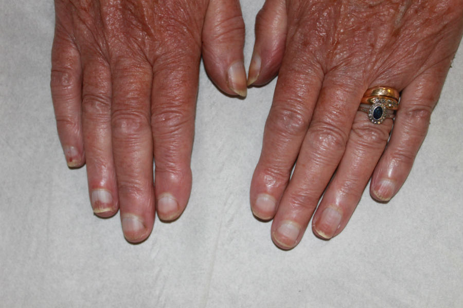
Clubbing
- Increased transverse and longitudinal nail curvature with hypertrophy of the soft‐tissue components of the digit pulp
Thyroid acropachy of Grave's disease
- Swelling of distal digits with overgrown nail plates that has a similar appearance to clubbing. Nails may lift off the nail bed
Koilonychia
- A concavity of the nailplate
- It is common in infancy as a benign feature of the great toenail
- In adults the most common systemic association is with iron deficiency and haemochromatosis, although the majority of adults with koilonychia demonstrate a familial pattern
Pincer nail deformity
- A transverse over-curvature of the nail plate
- Usually caused by ill-fitting footwear but it can occur in patients with psoriasis, SLE, Kawasaki disease, cancer, end-stage renal disease, and some genetic syndromes (eg paronychia congenita)
- Patients often have pain at the borders of the nail where the nail plate curves into the tips of the digits
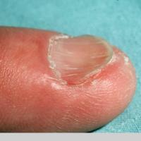
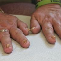
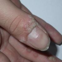
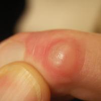
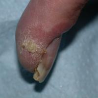
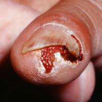
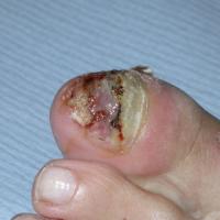
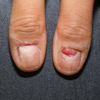
Skin lesions (figures 74-83)
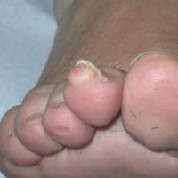
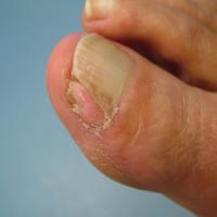
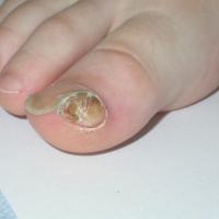
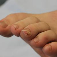
Many skin lesions can affect the nail including:
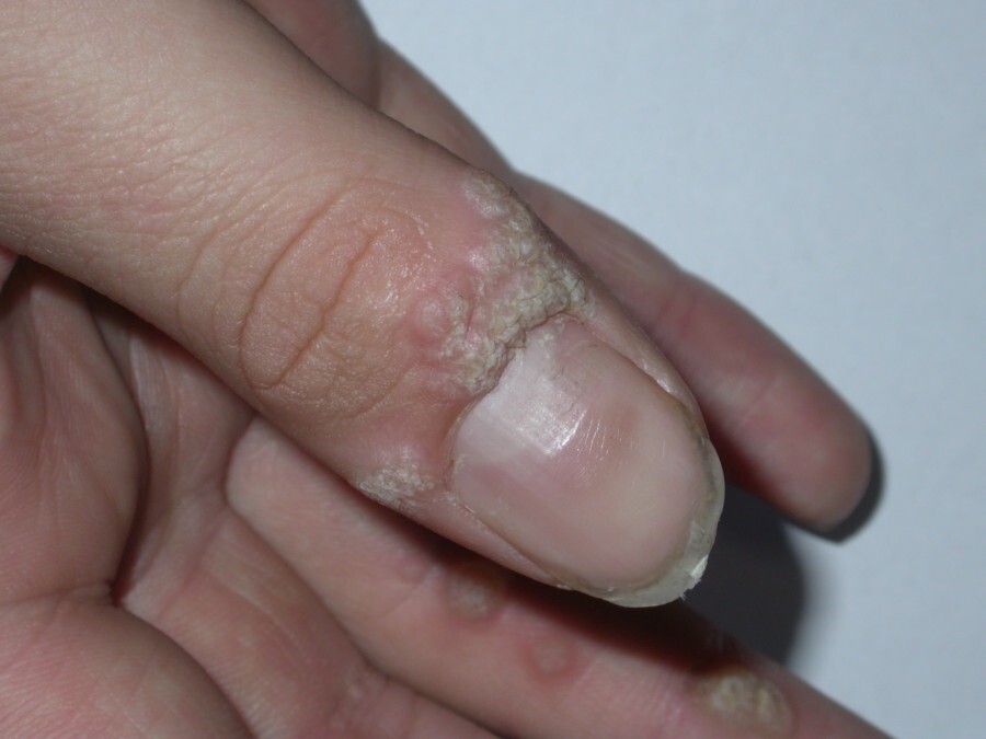
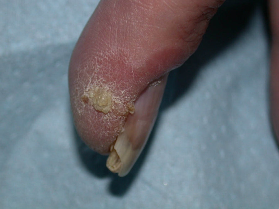
- Warts
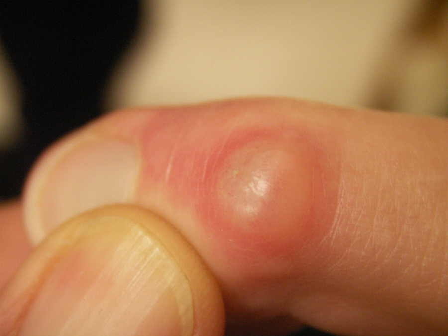
- Myxoid cyst - seen around the proximal nail fold. The cyst has a smooth shiny surface, and a clear jelly-like fluid may be expressed. As a result of pressure the cyst often causes a lateral groove in the nail, a few mm across, which extends the length of the nail
- Pyogenic granuloma - a red papule or nodule that bleeds very easily. Needs to be differentiated from malignancy
- Bowen's disease - a red or skin-coloured scaly lesion
- Squamous cell carcinoma - a nodule, ulcer, or occasionally a verrucous-like mass. Can cause partial or complete nail destruction
- Melanoma - in addition to longitudinal melanonychia, subungual melanoma can sometimes grow, destructively, under the nail
- Acquired fibrous nodule - presents as a smooth-edged lesion, commonly in the periungual tissue as opposed to within the nail unit
- Periungual fibroma - associated with tuberous sclerosis (Koenen tumour). They present as smooth, firm, flesh-coloured lesions that emerge, usually, from the nail folds
- Subungual exostosis - a firm nodule that develops below the nail bed. As it grows upwards the nail plate separates from the bed. The big toenail is the most commonly affected digit
Drug-induced nail disorders
Clinical Images
Please refer to notes on image rights at bottom of the page with regards to individual image
ownership.
1
2
3
4
5
6

7
8
9
10
11
12
13
14
15
16
17
18
19
20
21
22
23
24
25
26
27
28
29
30
31
32
33
34
35
36
37
38
39
40
41
42
43
44
45
46
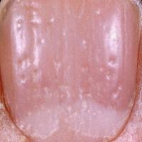
47
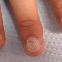
48
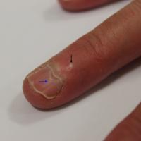
49
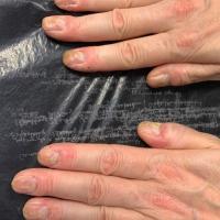
50
51
52
53
54
55
56
57
58
59
60
61
62
63
64
65

66
67
68
69
70
71
72
73
74
75
76
77
78
79
80
81
82
83
Investigations
Suspected fungal infection
-
In order to prevent high rates of false negative results it is important to the cut the nail back and remove the subungual debris by careful curettage or by using a scalpel blade, and sending all of the specimen for mycology. Simply asking the patient to provide a nail clipping will miss many cases of fungal infection
Management
-
General measures
-
Good hand care - protect hands from solvents and other chemicals by using gloves. Do not use soap, but use plenty of emollients. Avoid manicures
-
Patients should take good care of their feet by washing, frequent airing, and wearing sensible footwear
-
Nails should be cut straight (as opposed to curved) and not too short, in order to prevent ingrowing toenails
-
Patients with troublesome nails who are not able to manage them on their own should be seen regularly by a chiropodist
-
Patients with suspected melanoma or squamous cell carcinoma should be referred urgently to Secondary Care (two-week wait)
-
Patients with potentially destructive lichen planus of the nails should be referred urgently to dermatology, as early intervention may prevent long-term nail damage
-
For fungal infection of the nail refer to the chapter Tinea unguium
-
Pseudomonal nail infection - it is important to try and correct any underlying nail pathology. Treatments reported as helping include vinegar, 1% hydrogen peroxide cream, gentamicin drops, ciprofloxacin drops
-
Most other nail conditions, including psoriasis, provide a major therapeutic challenge
Disclaimer - the author PCDS cannot accept responsibility for any misleading or incorrect statements, and the management of individual patients remains the direct responsibility of the individual doctor. We do however hope that visitors to this site can contact us regarding comments that are considered misleading or incorrect so that we can continue to improve the site.
Image Rights - The PCDS would like to thank Dermatoweb, DermQuest (Galderma), and others who have contributed images. All named individuals and organisations maintain copyright for the relevant images.