Squamous cell carcinoma - cutaneous
LAST UPDATED: Jun 05, 2026
Introduction
Cutaneous squamous cell carcinoma (cSCC) is a malignant tumour arising from the keratinocytes of the epidermis. cSCC is the second most common skin cancer; the incidence is increasing by 5-6% per year in the UK with over 52,000 new cases each year.
Athough most cases of cSCC, if recognised and treated promptly, are very manageable, cSCC can metastasize. The incidence of cSCC and mortality risk is very much greater in immunosuppressed patients.
Prompt diagnosis and urgent referral to Secondary Care is essential for patients suspected as having cSCC.
This chapter is set out as follows:
Aetiology
- Much more common in white skin as opposed to skin of colour
- Men are more affected than women
- Excessive exposure to sunlight is the main aetiological factor (except in skin of colour) and so incidence increases with age
- The long-term use of phototherapy eg UVB and PUVA, used in the management of psoriasis and other dermatological conditions, as well as sunbed usage, increases the risk
- Genetic factors
- Individuals with fair skin, blue eyes and blonde hair are at higher risk
- Patients with xeroderma pigmentosum, albinism or epidermolysis bullosa can develop lesions at a very young age
- Patients on immunosuppressive therapy (and in particular following kidney or other organ transplant) are at a very much higher risk of developing cSCC and such lesions tend to behave more aggressively
- Chronic inflammation
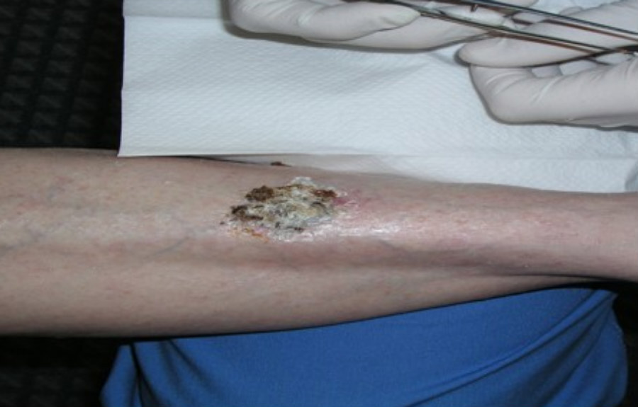
- SCC can arise in areas of chronic skin inflammation such as leg ulcers
- In pipe smokers the increased risk of developing SCC on the lips may be secondary to heat damage
History
- cSCC predominantly arise in older patients, although immunosuppressed patients and the other high risk groups referred to above eg xeroderma pigmentosum, can develop lesions at a much younger age
- Lesions may start de-novo or from pre-existing skin lesions such as AK and Bowen's disease. Transformation can also occur in areas of chronic inflammation such as leg ulcers
- cSCC grow more quickly than basal cell carcinoma. Patients may complain of pain/discomfort when the lesion is touched, bleeding, or sensory changes
Clinical findings
General features
- Distribution
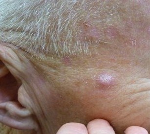
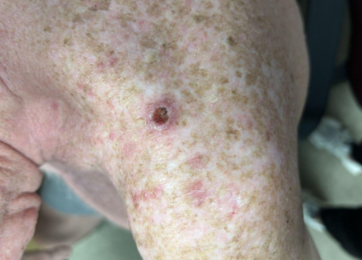
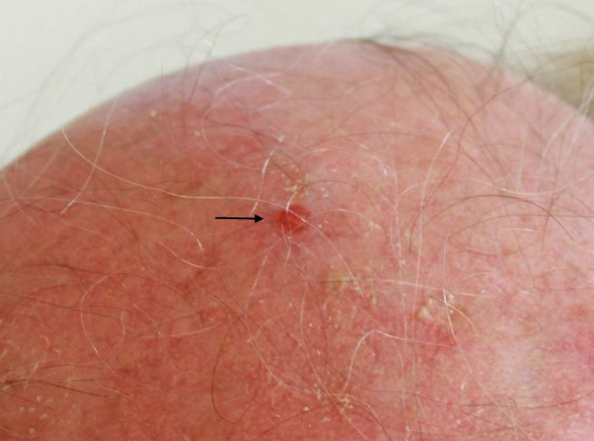
- SCC predominantly arise on sun-exposed sites and there are often other signs of sun-damaged skin
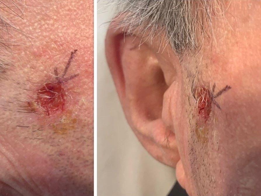
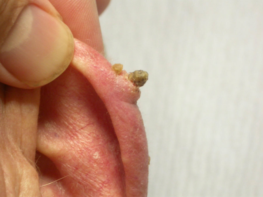
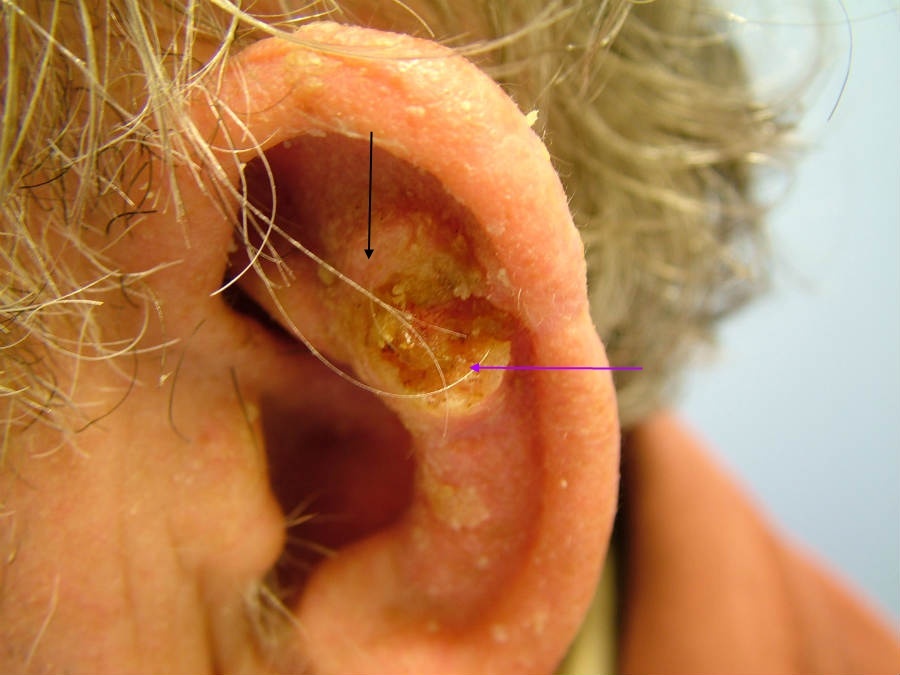
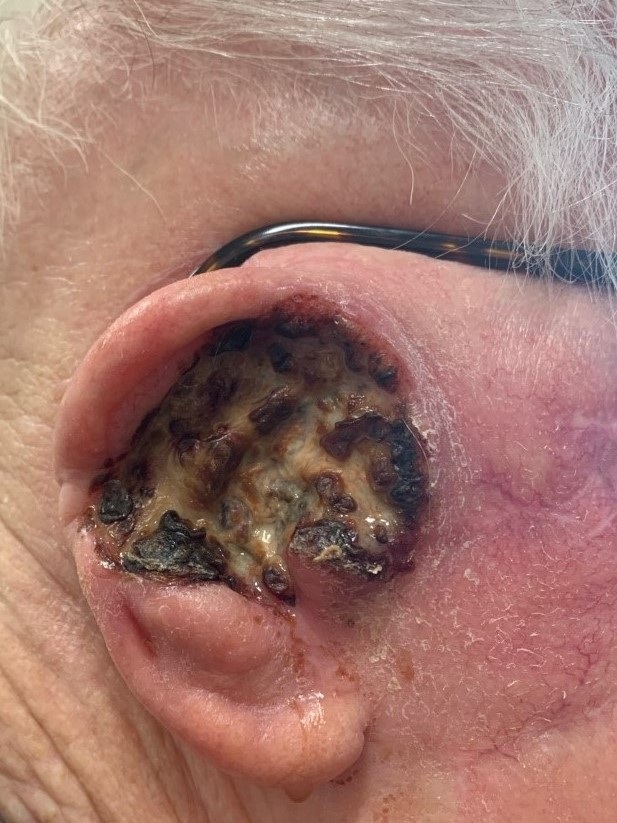
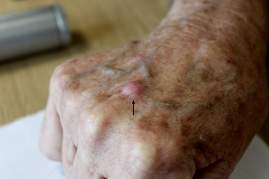
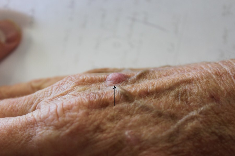
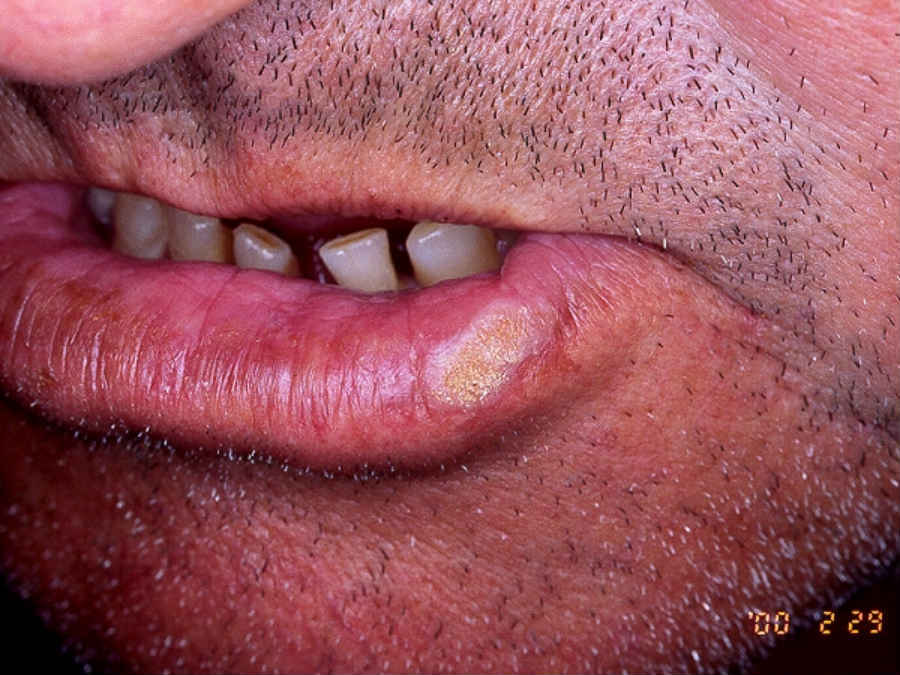
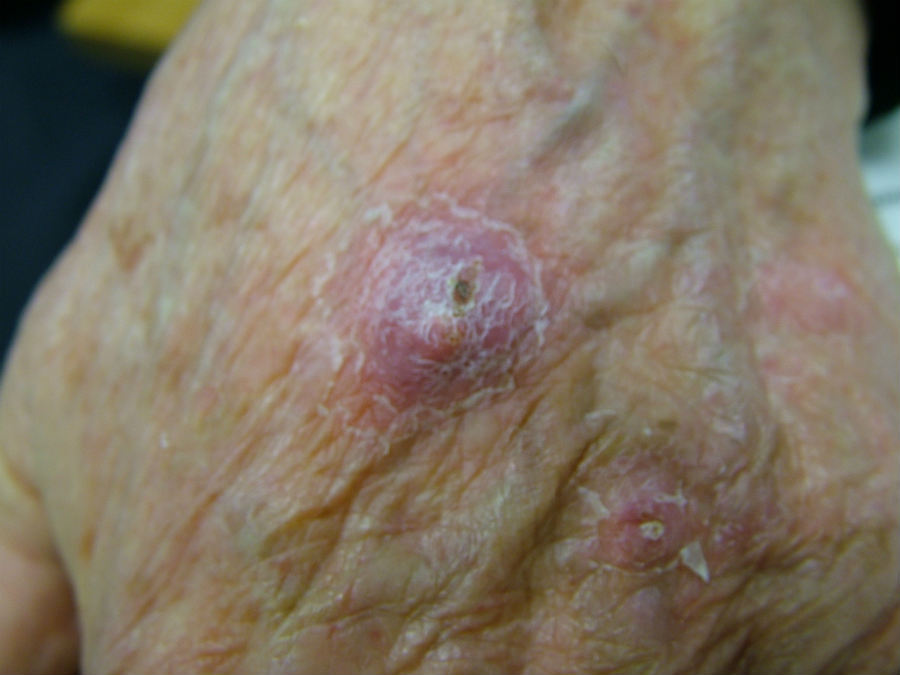
- The most commonly affected areas are the backs of hands and forearms, upper part of the face and, especially in males, the lower lip and pinna
- Clinical features
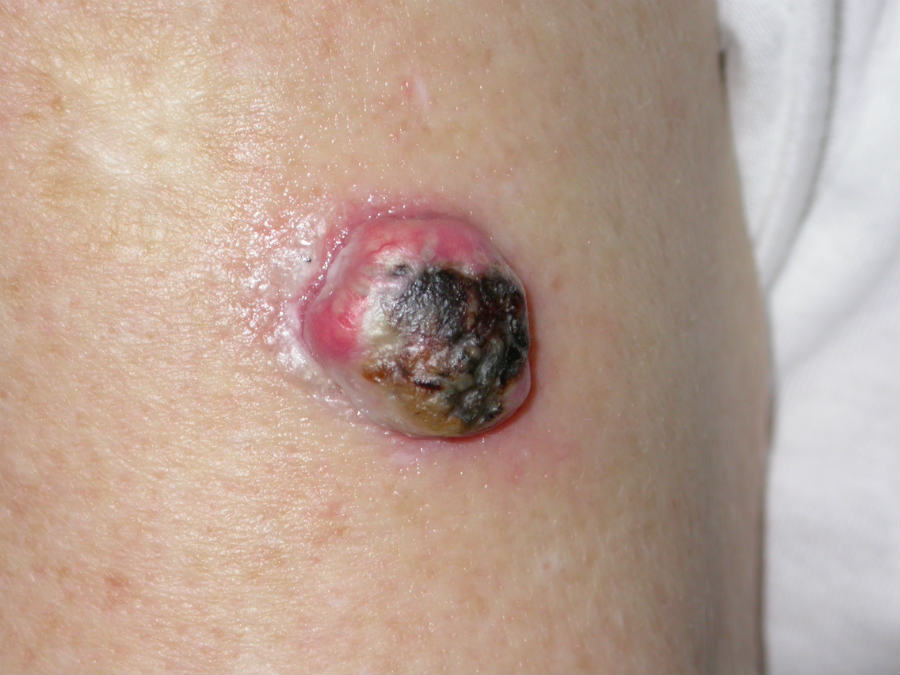
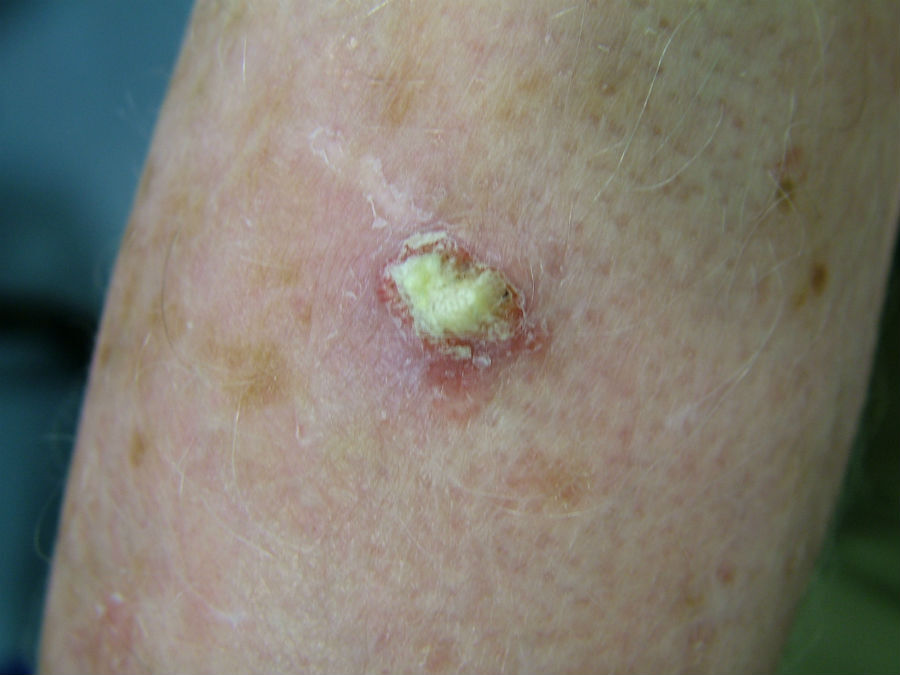
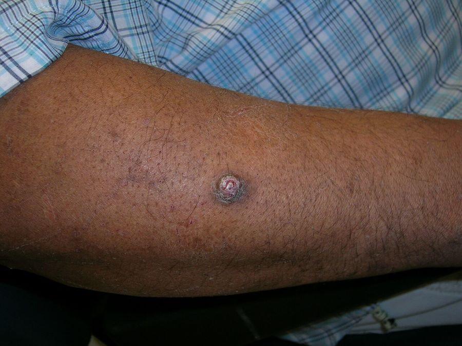
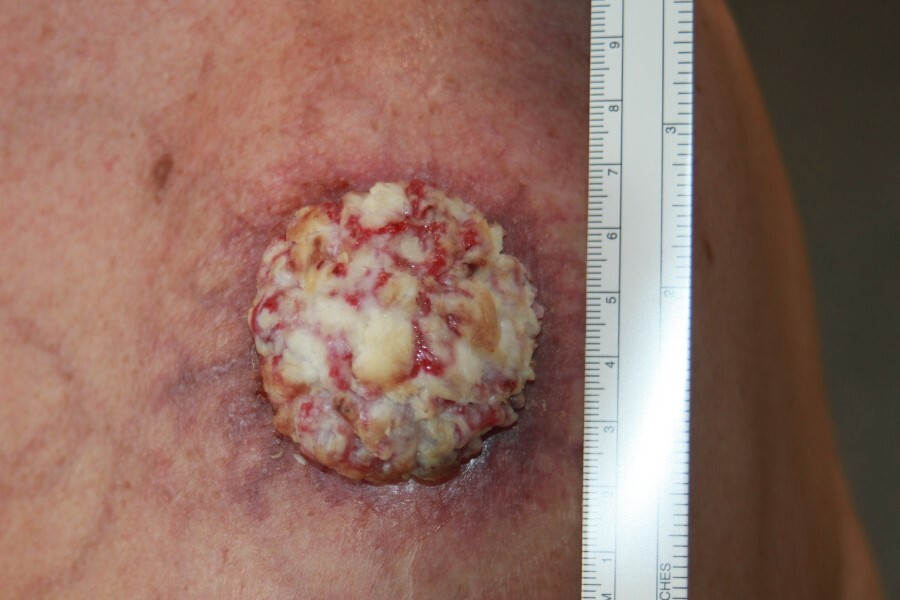
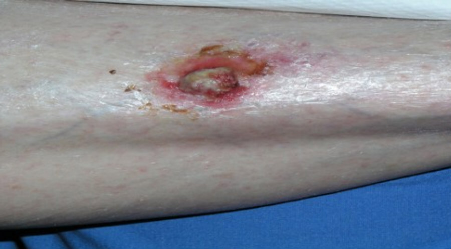
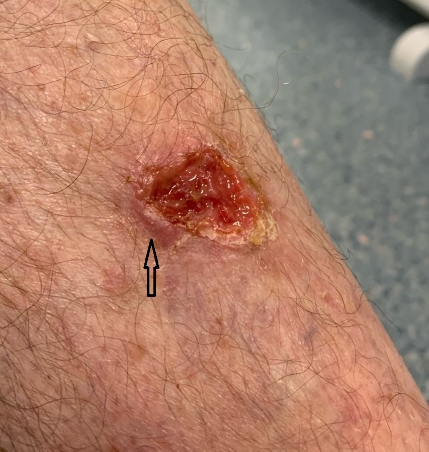
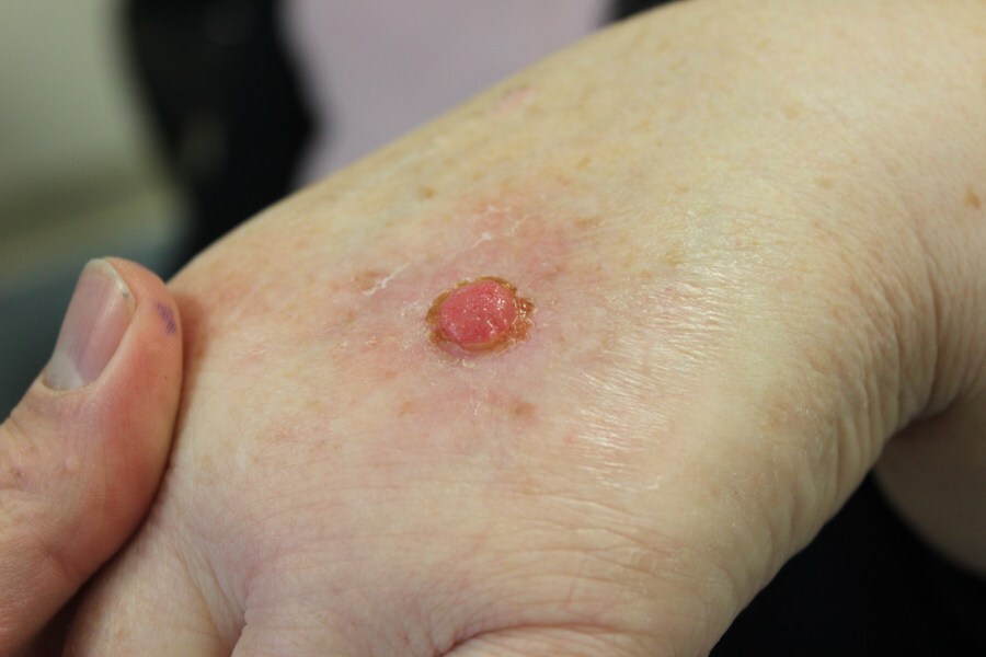
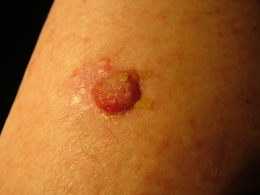
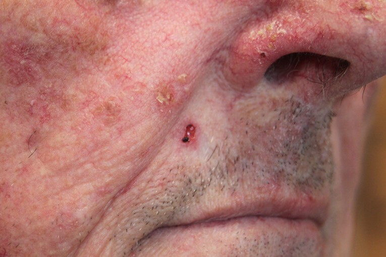
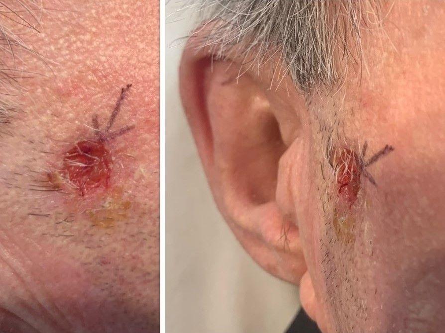
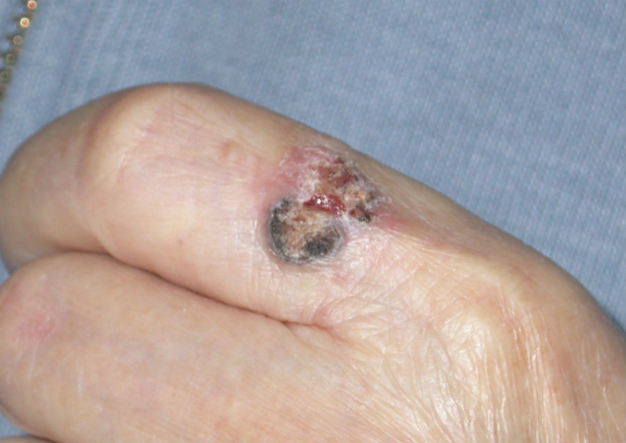
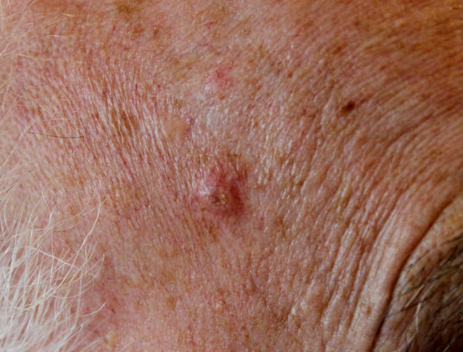
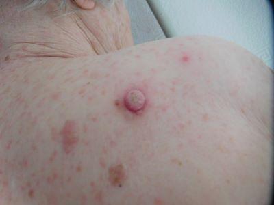
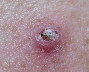
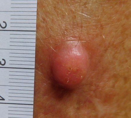
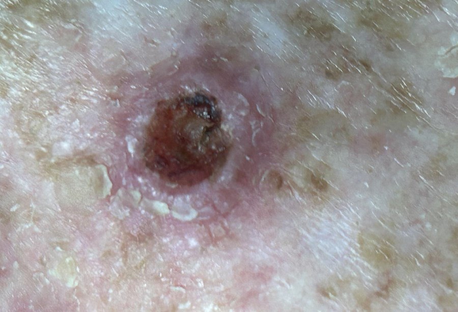
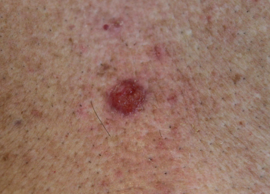
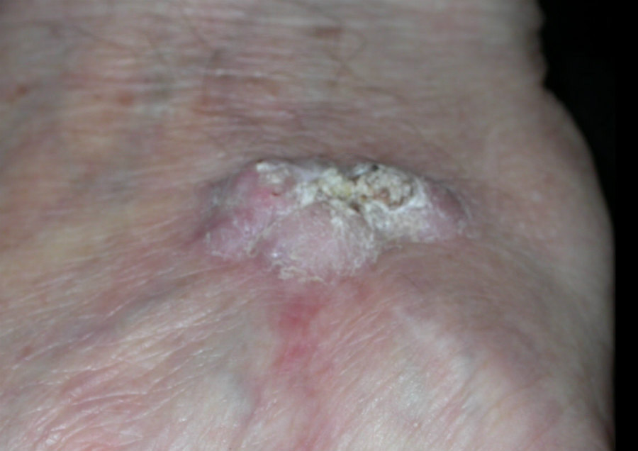
- Induration ie firm to palpate - in the vast majority of cases the first clinical sign is that of induration, which may take on the characteristics of nodular, plaque-like, verrucous, or ulcerated
- The limits of induration are not sharp and usually extend beyond the visible margins of the lesion
- The surrounding tissue is often inflamed
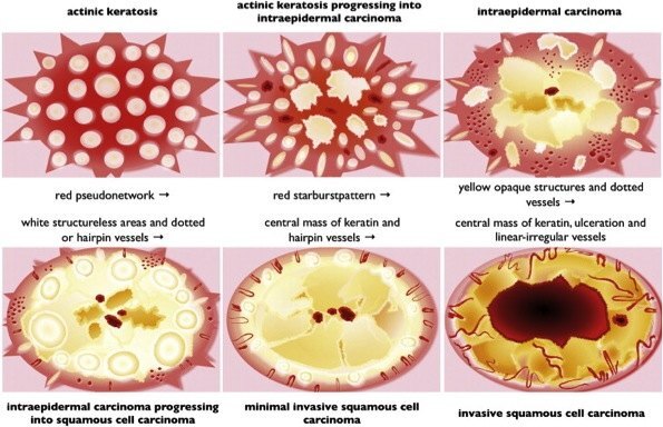
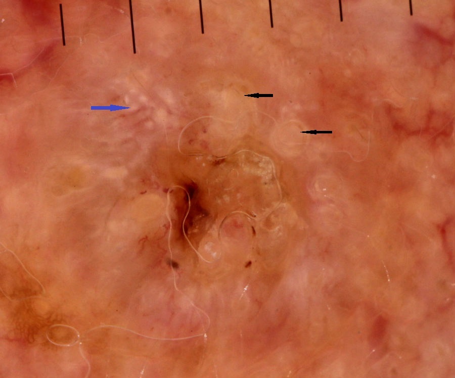
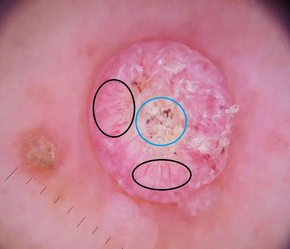
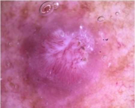
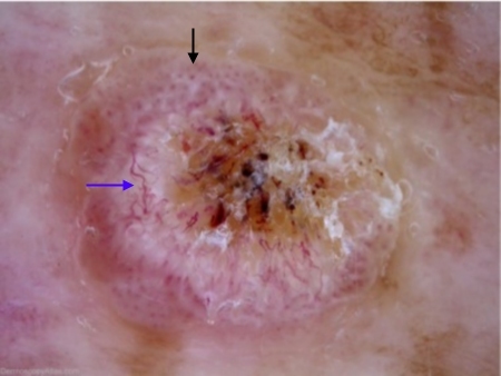
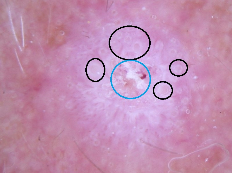
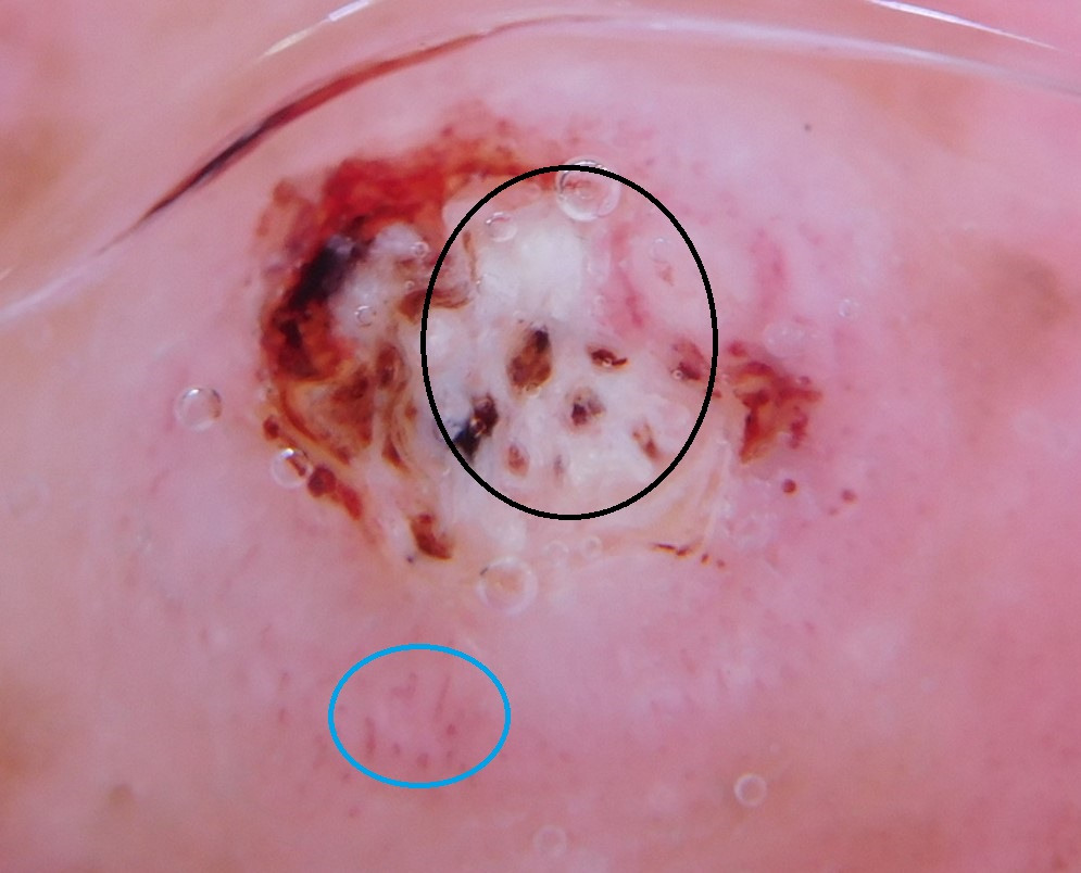
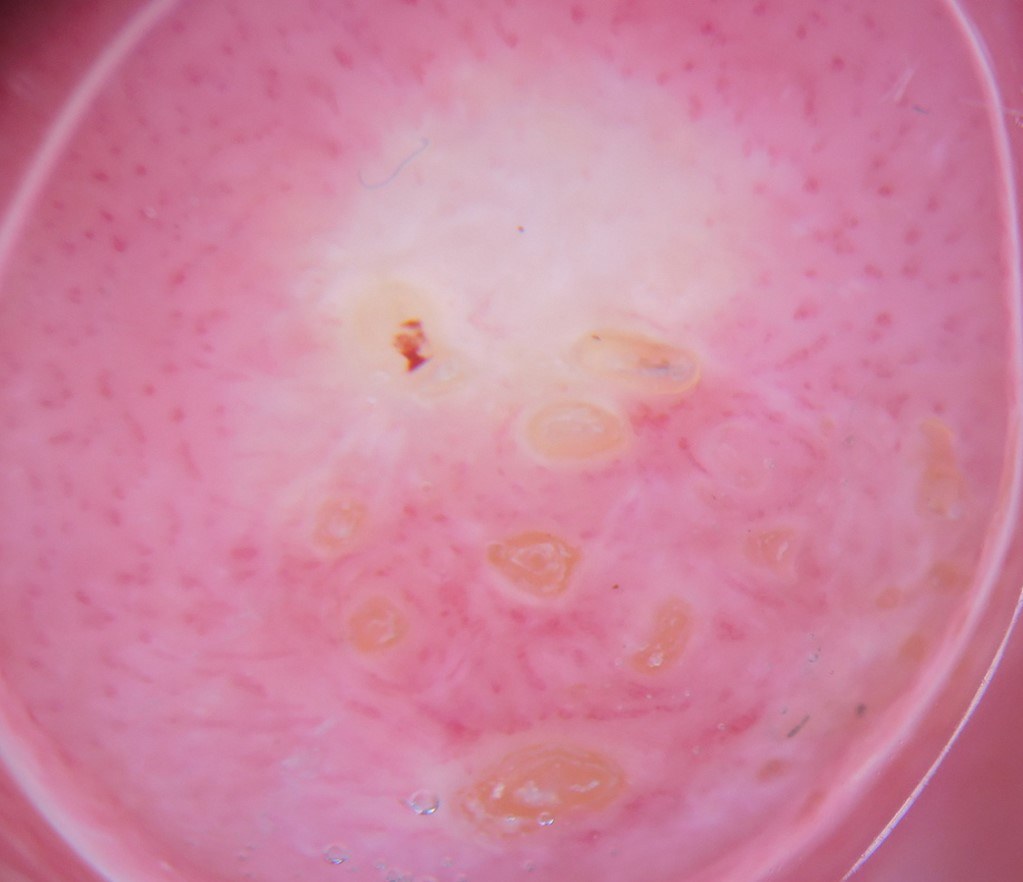
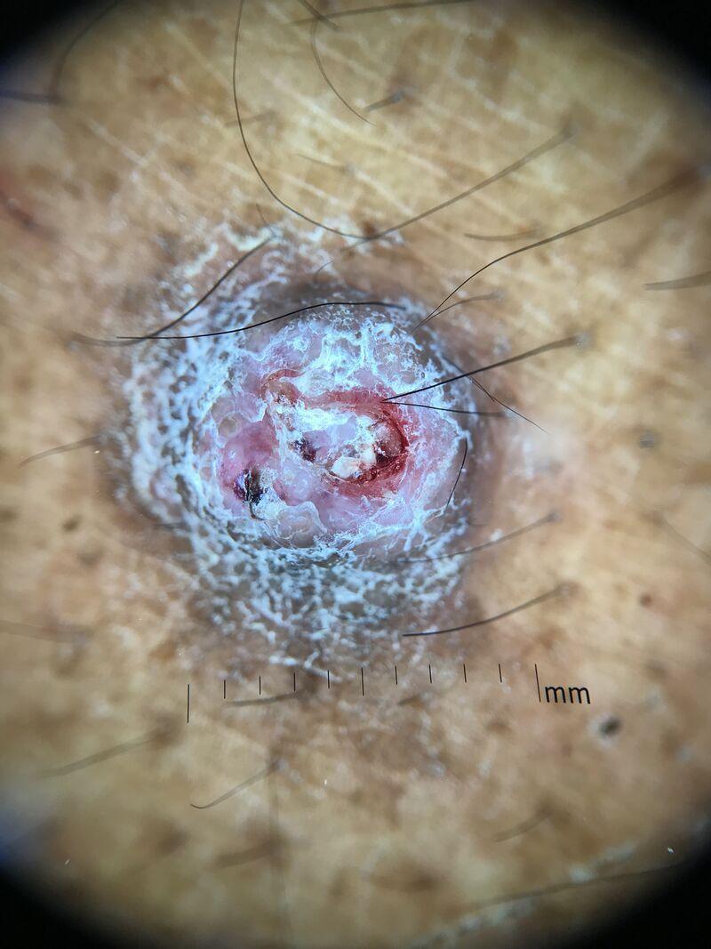
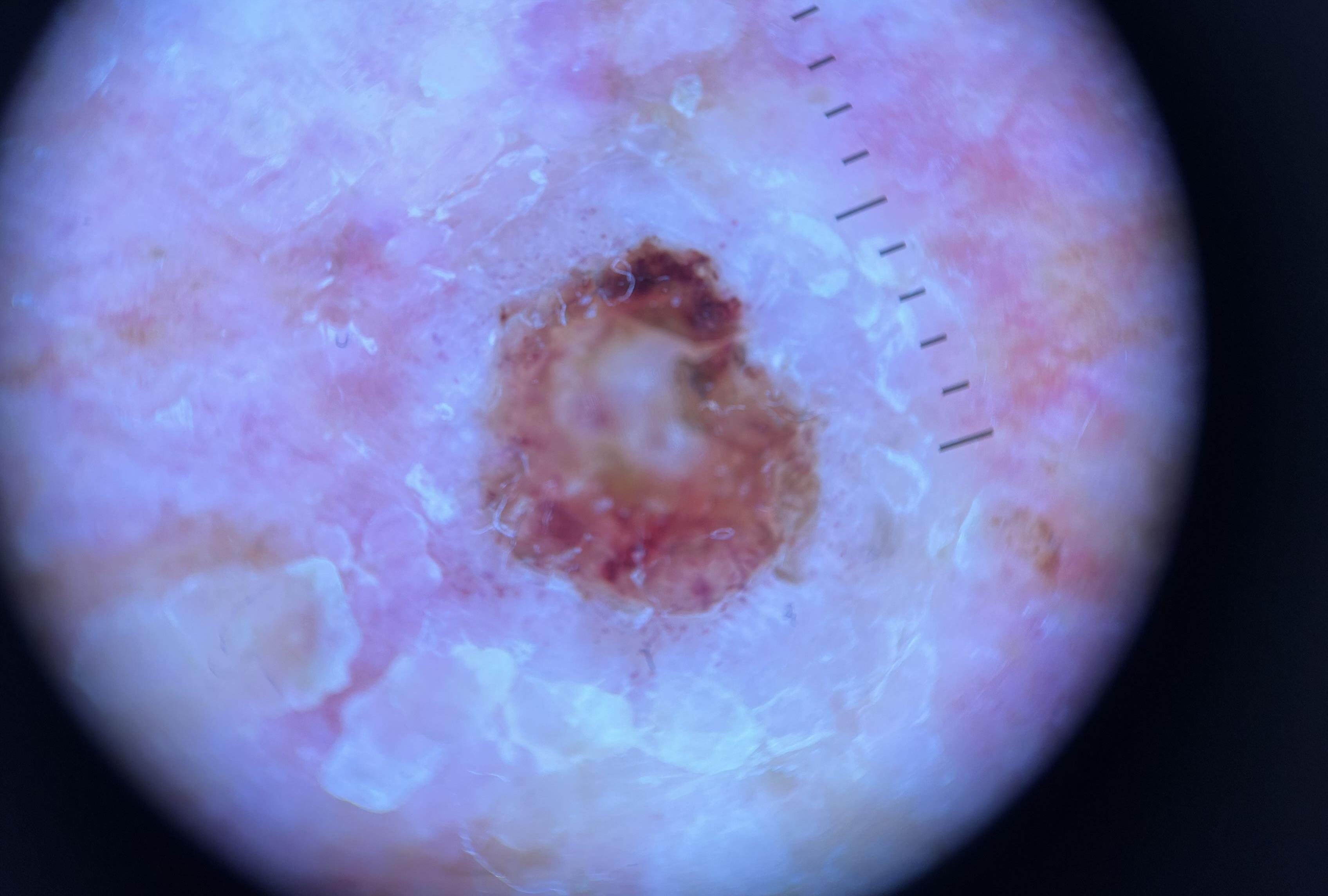
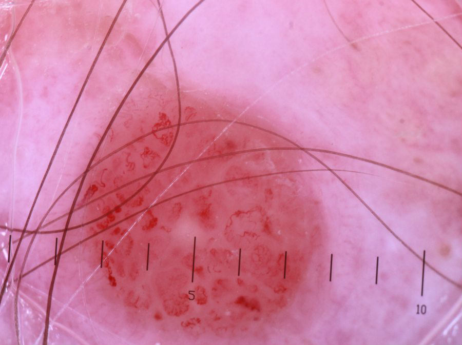
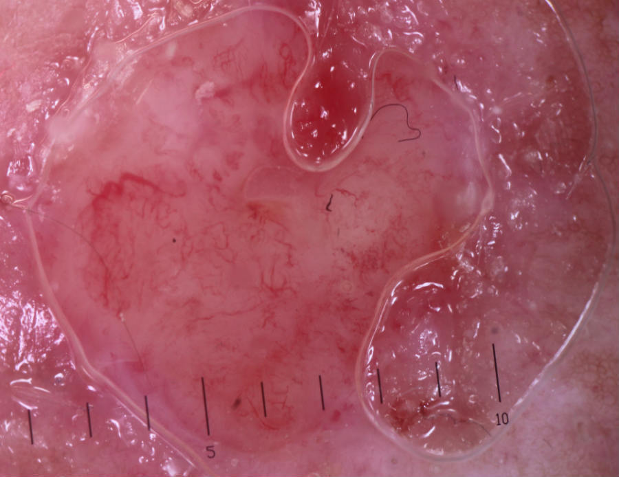
- Dermoscopic features (hover over terminologies for description) may include:
- A central white keratin mass, which may look yellow if keratin mixes with serum due to erosion or ulceration
- Other white structureless areas
- Targetoid hair follicles - these are variably large roundish structures composed of a yellow to light-brown structureless centre and a white outer rim (these structures are sometimes referred to as white or yellow clods or circles), and are found around the central keratin
- Peripheral radial vessels (ie pointing to the centre), which may be looped, and often irregular. Vessels are occasionally surrounded by white halos. In poorly differentiated SCC, vessels predominate and are usually polymorphous (ie more than one type of vessel). Click here for more information Vessel patterns in dermoscopy
A spectrum of presentations
Different types of SCC have differing morphological characteristics:
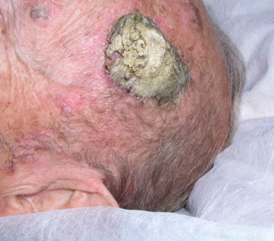
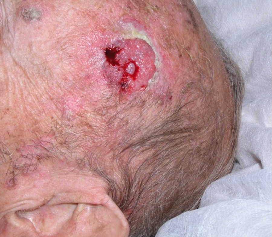
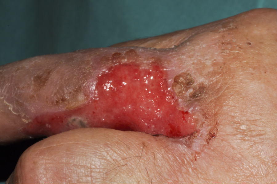
- Well-differentiated SCC (less aggressive) - grow slower and have a keratotic surface in the early stages. The keratin tends to be able to be removed quite easily. As the tumour enlarges the balance between keratin production and cellular mass shifts towards cellular proliferation - so even in a well-differentiated tumour the surface tends to ulcerate and growth takes place predominantly at the margins with an increasing area of granulation tissue occupying the centre. This may be shed later to reveal an ulcer or an indurated, eroded margin (as opposed to a BCC, which may have a more regular ulcer) and a purulent, exuding surface that bleeds rather easily. The outline is rounded but often irregular
- Moderate-poorly differentiated SCC (more aggressive) - the organisation of the keratin on the tumour diminishes causing keratin often to be quite sparse or even absent. Lesions may look like granulation tissue or a 'juicy' nodule. In an undifferentiated (anaplastic) tumour the stratum corneum may have been replaced by erosion of the surface or frank ulceration, sometimes presenting as a punched-out ulcer with no palpable component
Features of SCC in skin of colour
- Key risk factors include chronic wounds, HPV, immunosuppression, radiation sites, and chemical exposure, with UV less dominant than in lighter skin
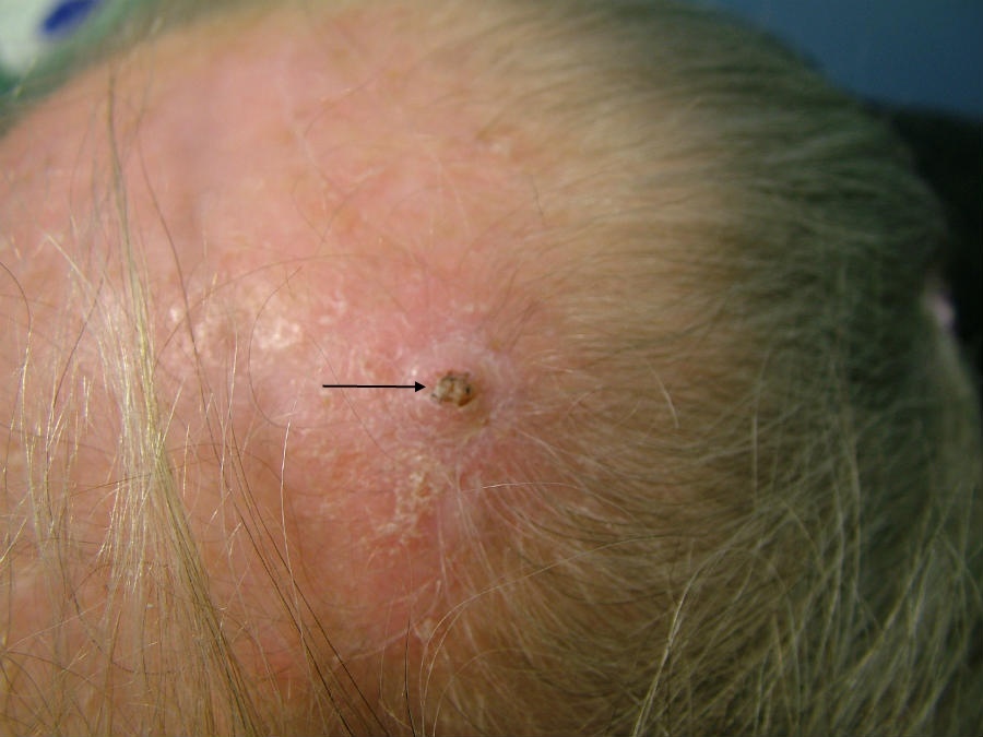
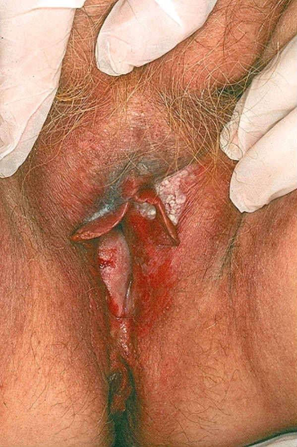
- SCC often presents differently than in lighter skin, appearing more frequently in less sun-exposed areas such as the legs, feet, anogenital skin, and sometimes arising from scars or inflammation. In terms of the head and neck, SCC can also affect hair-covered areas of the scalp
- Diagnosis can be delayed due to misconceptions, requiring better awareness and education for early detection
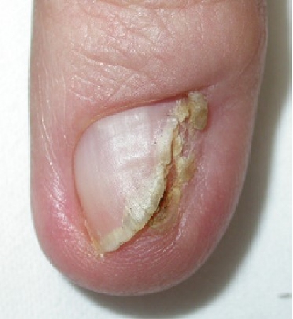
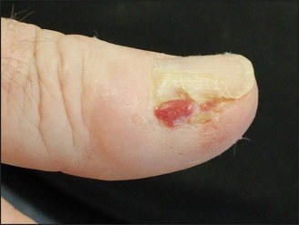
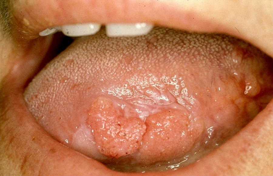
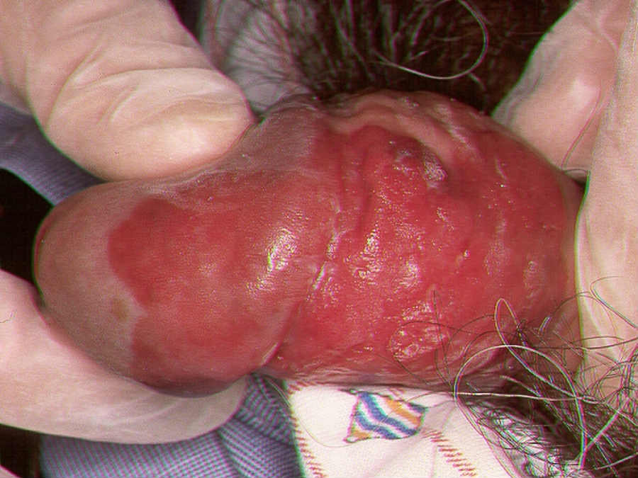
SCC at other sites
Clinical Images
Please refer to notes on image rights at bottom of the page with regards to individual image
ownership.
1
2
3
4
5
6
7
8
9
10
11
12
13
14
15
16
17
18
19
20
21
22
23
24
25
26
27
28
29
30
31
32
33
34
35
36
37
38
39
40
41
42
43
44
45
46
47
48
49
50
51
Management
Disclaimer - the author PCDS cannot accept responsibility for any misleading or incorrect statements, and the management of individual patients remains the direct responsibility of the individual doctor. We do however hope that visitors to this site can contact us regarding comments that are considered misleading or incorrect so that we can continue to improve the site.
Image Rights - The PCDS would like to thank Dermatoweb, DermQuest (Galderma), and others who have contributed images. All named individuals and organisations maintain copyright for the relevant images.