Keratoacanthoma
LAST UPDATED: Mar 23, 2026
Introduction
A keratoacanthoma (KA) is a rapidly evolving tumour of the skin, composed of keratinising squamous cells originating in pilosebaceous follicles and resolving spontaneously if untreated.
This chapter is set out as follows:
Aetiology
-
Sun exposure plays a role
-
In some cases the lesion may follow an injury to the skin
History
-
Males are more commonly affected than females
-
Lesions grow rapidly
Clinical findings
Distribution
Clinical features
- Lesions look very similar to well-differentiated SCC, although their behaviour may differ in that:
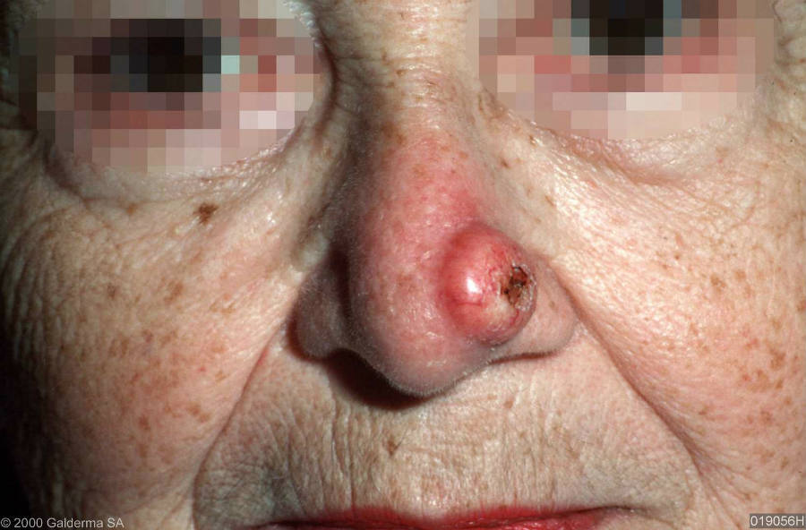
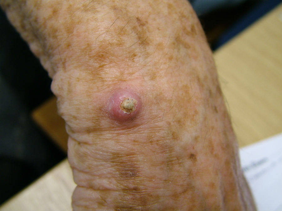
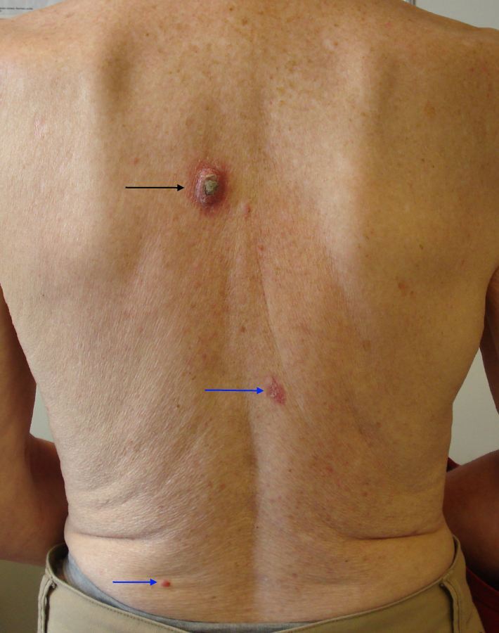
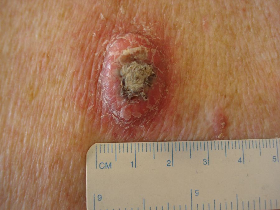
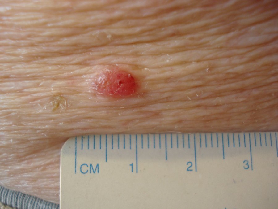
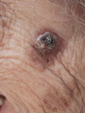
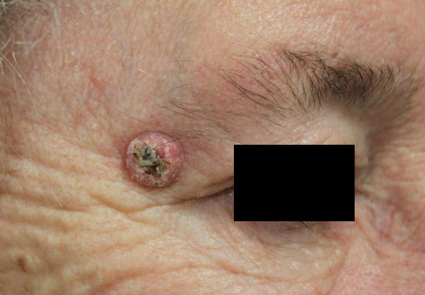
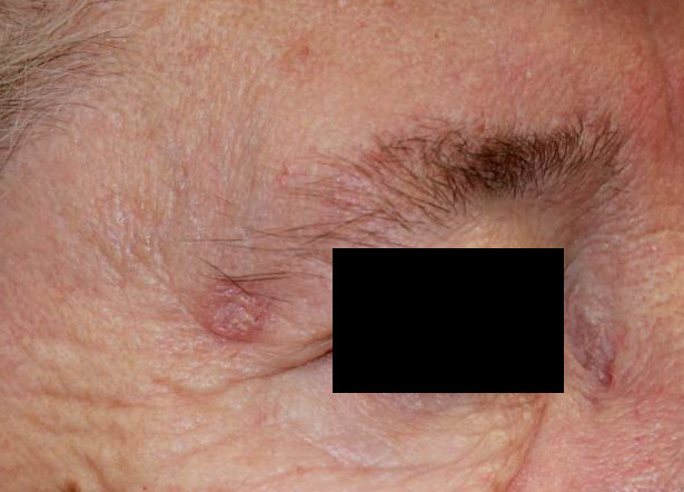
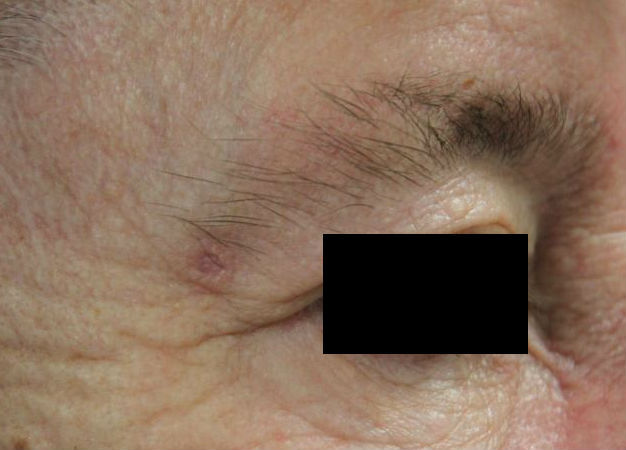
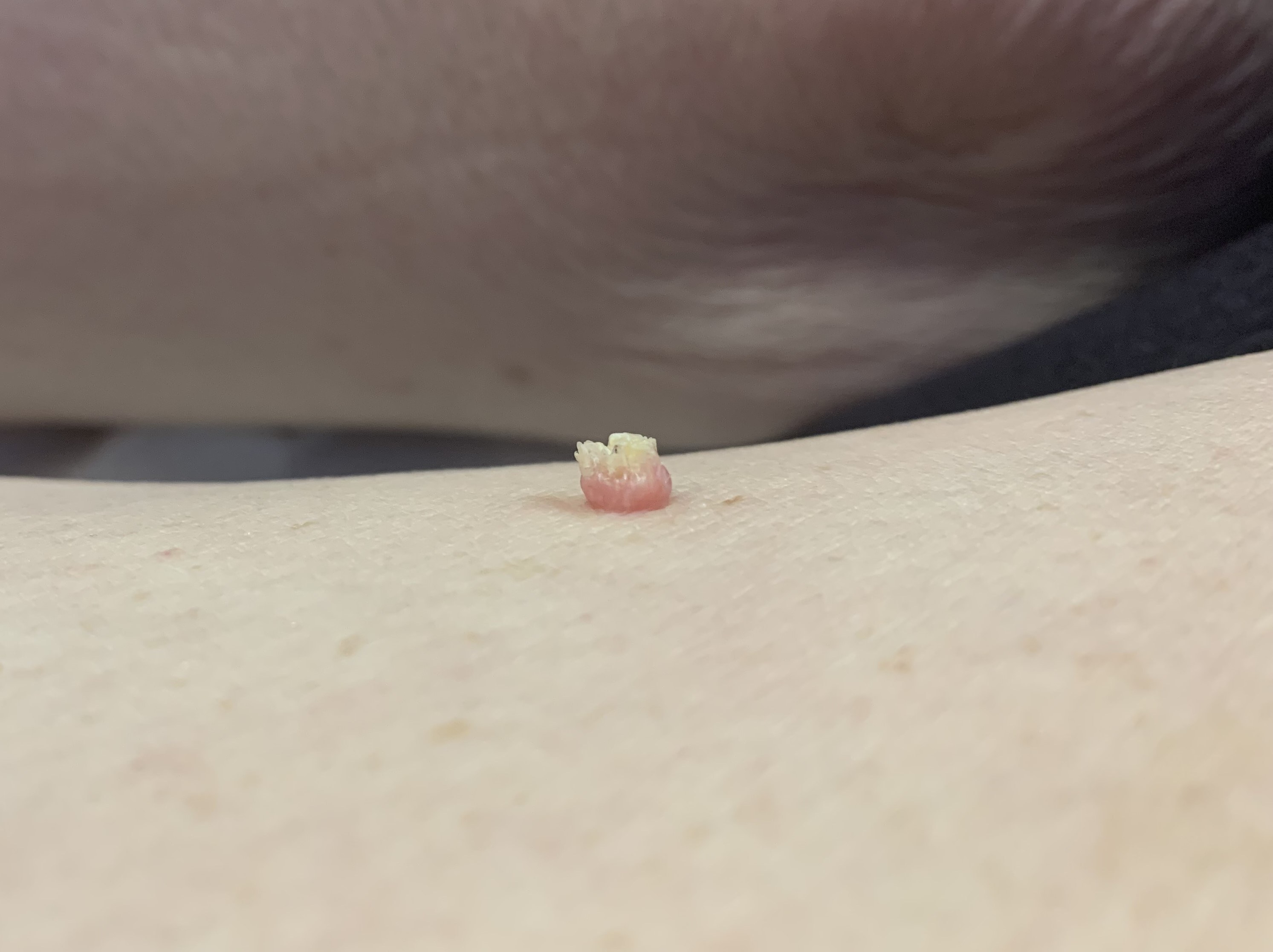
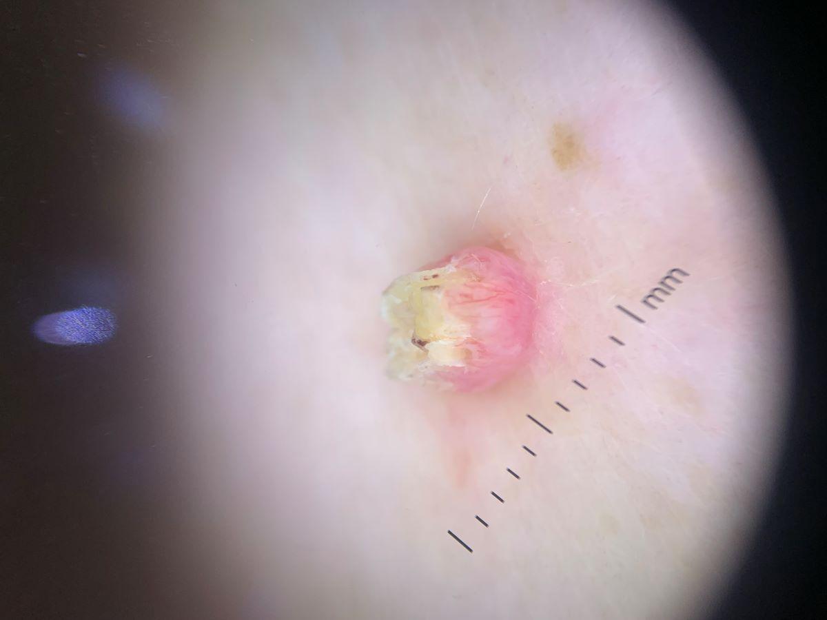
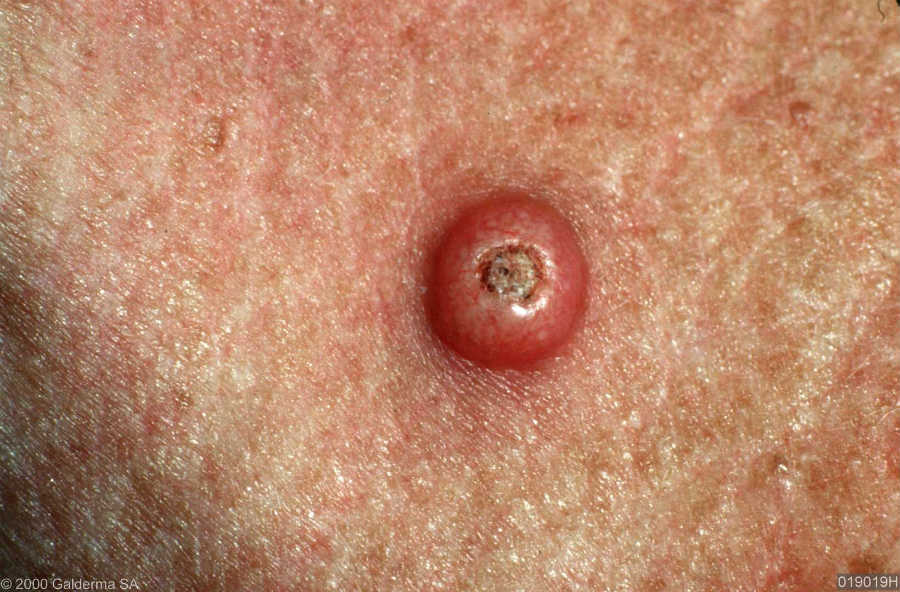
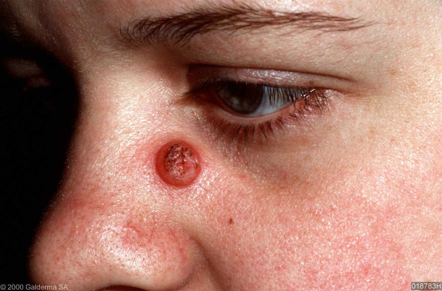
- KA grow rapidly over a period of about 12 weeks from a small, firm, rounded, skin-coloured to red papule, in to a very symmetrical dome-shaped nodule with a smooth shoulder of skin and a central keratin core, giving it a volcano-like appearance
- Lesions start to resolve after approximately three months
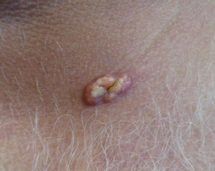
- On rare occasions a keratoacanthoma can appear yellow, looking almost sebaceous in nature
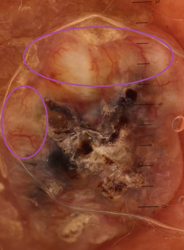
Dermoscopic features (hover over terminologies for description) are often similar to those of an SCC, and may include:
- A central white keratin mass, which may look yellow if keratin mixes with serum due to erosion or ulceration
- Other white//yellow structureless areas
- Targetoid hair follicles - these are variably large roundish structures composed of a yellow to light-brown structureless centre and a white outer rim (these structures are sometimes referred to as white or yellow clods or circles), and are found around the central keratin
- Peripheral and often radial vessels (ie pointing to the centre), which may be serpentine, arborising, or looped; occasionally glomerular. Click here for more information on Vessel patterns in dermoscopy
Clinical Images
Please refer to notes on image rights at bottom of the page with regards to individual image
ownership.
1
2
3
4
5
6
7
8
9
10
11
12
13
14
15
Management
-
It can be difficult to distinguish clinically and histologically between a KA and an SCC and so patients should be referred urgently (two-week wait) to Secondary Care - dermatology or plastic surgery (check local pathways)
Disclaimer - the author PCDS cannot accept responsibility for any misleading or incorrect statements, and the management of individual patients remains the direct responsibility of the individual doctor. We do however hope that visitors to this site can contact us regarding comments that are considered misleading or incorrect so that we can continue to improve the site.
Image Rights - The PCDS would like to thank Dermatoweb, DermQuest (Galderma), and others who have contributed images. All named individuals and organisations maintain copyright for the relevant images.