Tinea manuum (hands), pedis (feet), and unguium (nails)
LAST UPDATED: Jul 25, 2023
Acknowledgements: This chapter was kindly updated by Ivan Bristow PhD FRCPodM Fellow, Royal College of Podiatry.
Introduction
This chapter discusses tinea manuum, tinea pedis and tinea unguium and is set out as follows:
Aetiology
Clinical findings
Tinea manuum (hands)
-
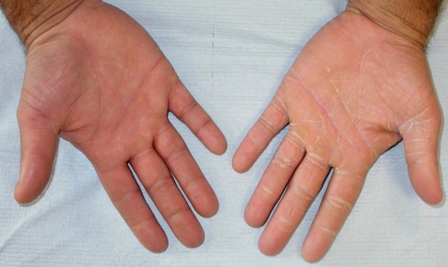
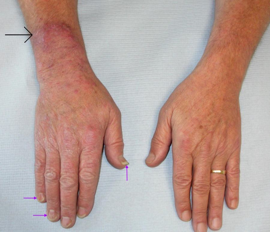
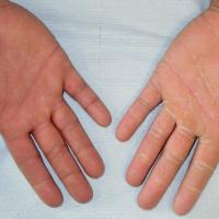
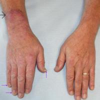
Red and / or scaly areas. Involvement of the creases gives the impression that the patient has been decorating with white paint
-
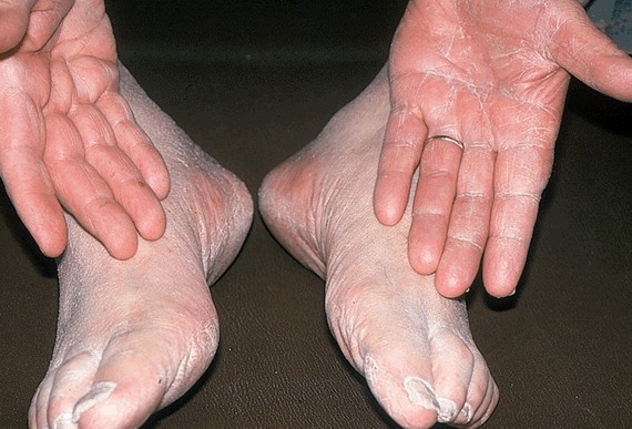
‘Two foot, one hand’ disease is the name given to a common pattern of infection where both feet and only one hand is infected. Always examine the hands, feet and nails
Tinea pedis (feet)
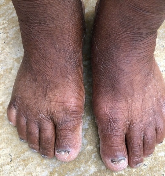
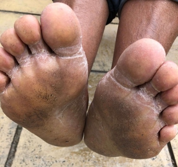
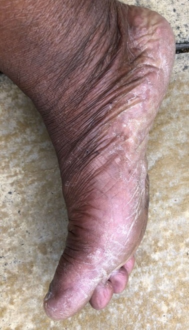
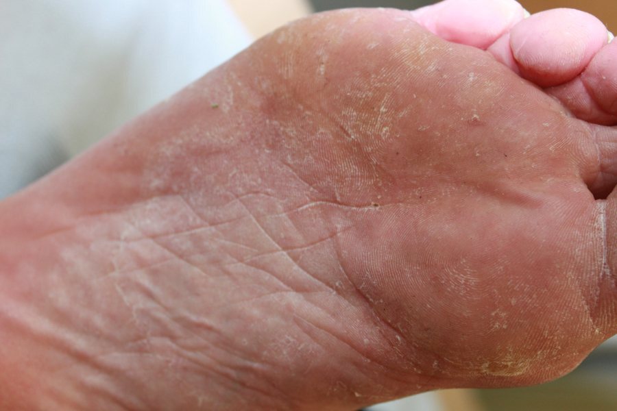
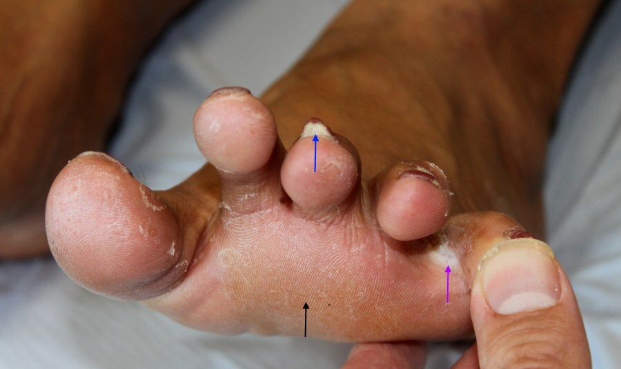
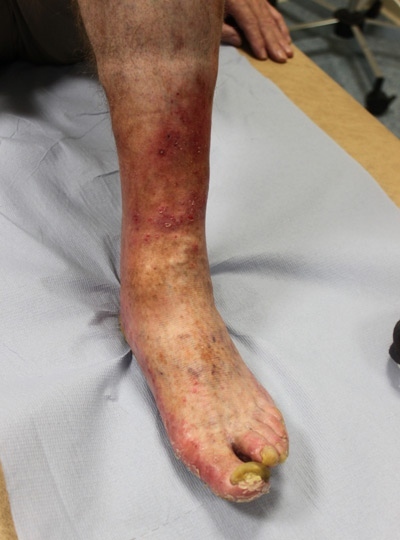
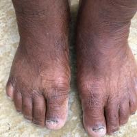
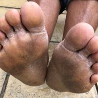
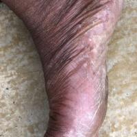
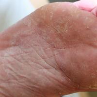
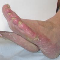
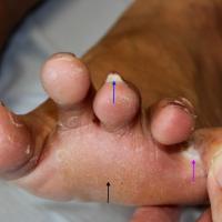
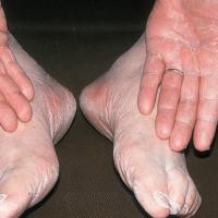
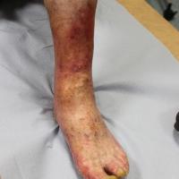
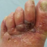
- Plantar skin involvement is the most common presenting as a dry, dusty appearance on a mildly erythemic background. The chalky white appearance is accentuated in the plantar skin creases and there can be a moccasin distribution. The features are often mistaken for dry skin
-
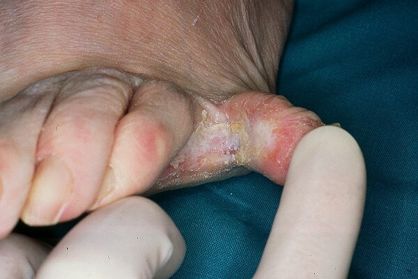
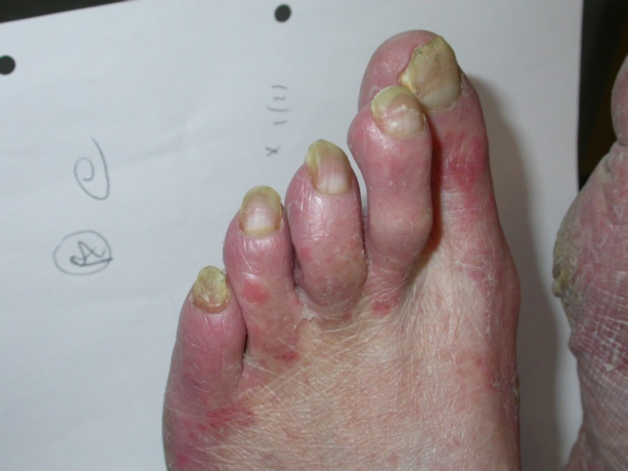
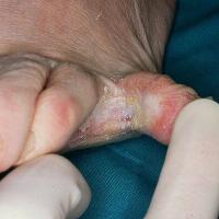
Interdigital involvement can affect any web space, especially the fourth web space. The skin can be dry and scaly or macerated and possibly malodorous
-
The dorsal foot can have very fine scale in the skin creases, which may affect one foot more than the other
-
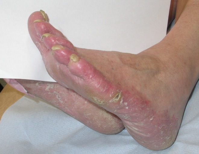
Erythema may be present with a leading scaly edge
-
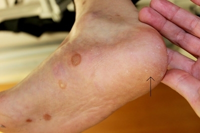
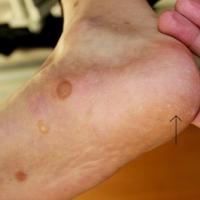
On occasions tinea pedis can cause a bullous reaction
Tinea unguium (nails) - syn. onycomycosis
-
The toenails are affected more often than the fingernails
-
May be asymmetrical, but not always
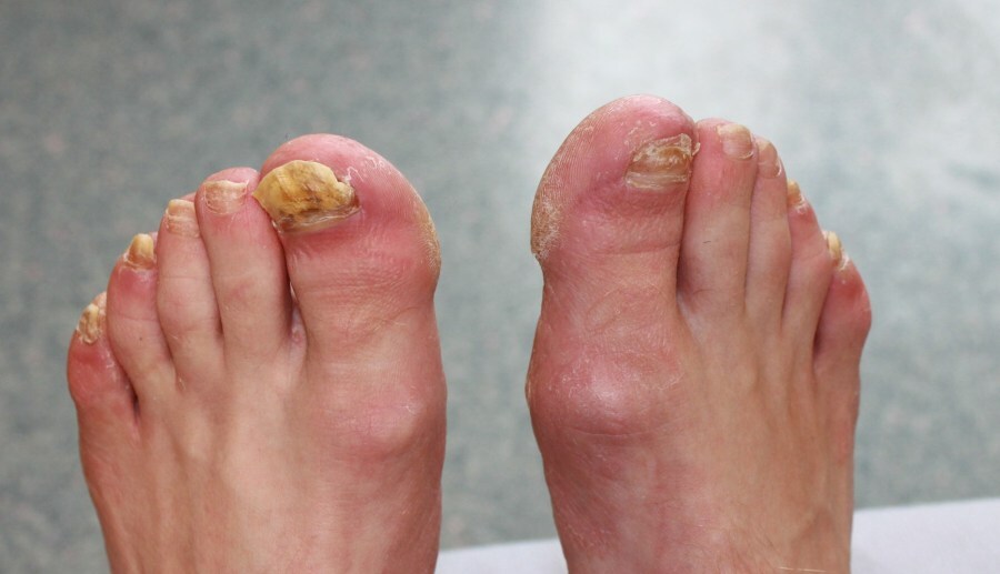
- Typically, the 1st, 2nd and 5th toenails are most frequently affected
-
Can be associated with interdigital infection (plantar and interdigital)
-
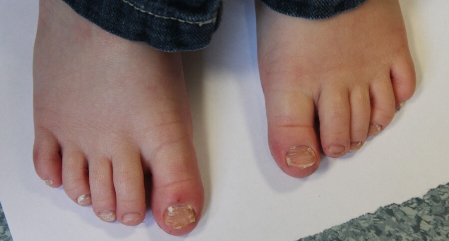
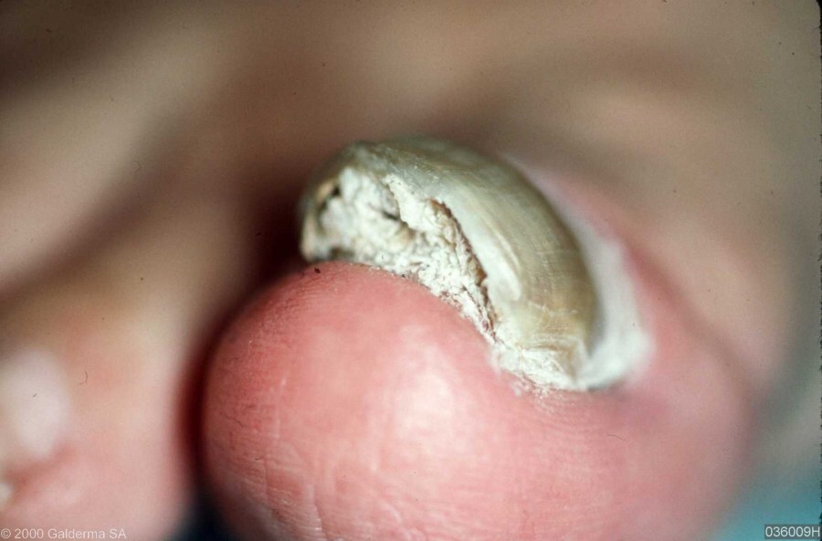
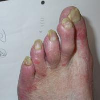
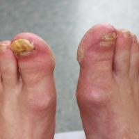
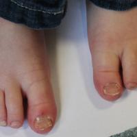
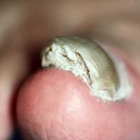
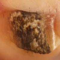
In most cases the nail thickens and turns yellow. If the infection starts distally the nail plate can be seen to crumble
-
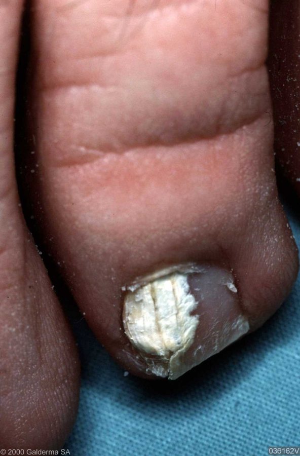
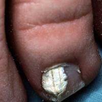
With superficial involvement the nail surface becomes white and powdery
-
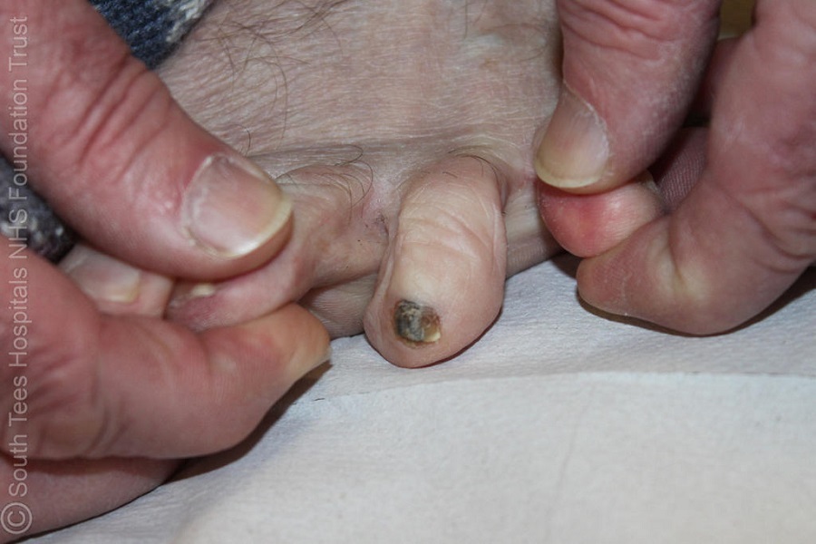
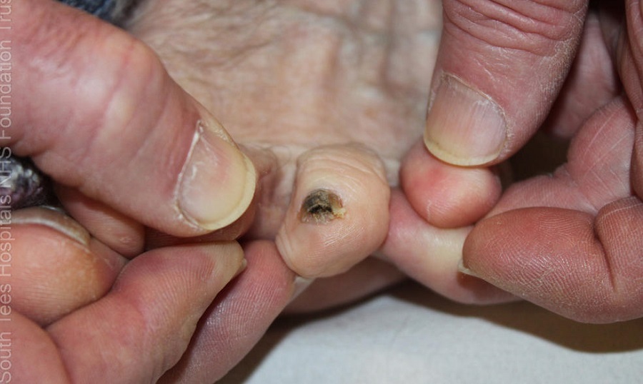
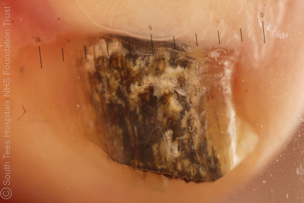
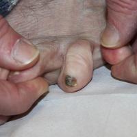
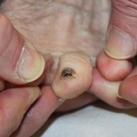
Very occasionally a fungal nail infection can appear brown-black, such cases must be differentiated from melanoma - in fungal infection the discolouration tends to start at the distal nail, whereas the most common presentation of melanoma is a line growing continuously from the proximal nail
-
Exclude skin diseases that may cause similar nail changes eg psoriasis by taking a good history and examining the whole of skin
-
Psoriasis may co-exist with fungal infection, as may any dystrophic condition
Clinical Images
Please refer to notes on image rights at bottom of the page with regards to individual image
ownership.
1
2
3
4
5
6
7
8
9
10
11
12
13
14
15
16
17
18
19
20
21
Investigations
-
The skin
-
Adequate scrapings should be taken with the back of a scalpel blade. The scrapings should be sent to microbiology either in a commercial sample pack or in folded black paper held by a paper clip. Adhesive tape (eg sellotape) stripping can be useful if scale is limited
-
The nails
-
Infection should be confirmed prior to treatment
- In order to prevent high rates of false negative results it is important to include samples of the diseased part of the nail from as proximal as possible, including any sub-ungual debris by careful curettage or by using a scalpel blade and sending all of the specimen for mycology
- This must be done by a health professional and not the patient
Management
Tinea manuum
-
Treatment is with a topical antifungal agent eg terbinafine (Lamisil ®) cream OD for 2 weeks, or one of the imidazole creams such as miconazole (Daktarin ®) BD for 2-4 weeks. Terbinafine is slightly more expensive but faster to cure
-
Systemic treatment should be used if there is co-existent nail involvement in which case treat as per tinea unguium
Tinea pedis
-
Treatment - for interdigital involvement or fine scaling treat as above
- Alternatively, patients may purchase and apply a single dose of terbinafine film forming solution (Lamisil Once®), which shows similar cures rates to terbinafine cream when applied OD for 2 weeks
- Topical treatments need to involve the soles and heels of the feet as well as the interdigital spaces. Systemic treatment should be used if there is co-existent nail involvement in which case treat as per tinea unguium
-
Recurrence is common and patients need to be advised to keep feet well aerated by wearing breathable footwear and leaving shoes off around the home. Prophylactic treatment with topical antifungals used once to twice a week can help
Tinea unguium
-
Topical treatments - these have a low cure rate but may be suitable for treating distal nail infection (as opposed to involvement of the nail matrix) or superficial white infection. Options include Tioconazole ® nail solution BD for 6-12 months or Amorolfine (Loceryl®, Curanail®) nail lacquer twice per week for 3-6 months for fingernails, and 6-12 months for toenails. Nails should be filed / cut back as much as possible prior to applying the treatment
-
Systemic treatment in adults
-
Terbinafine (Lamisil ®) is the most effective treatment (with eradication rates of 69% against 48% for itraconazole), except in cases of Microsporum canis when itraconazole is more effective. Terbinafine 250 mg OD should be given for 6 weeks for fingernails and 3-4 months for toenails
-
If the patient is unable to take terbinafine or the tinea appears resistant to treatment then itraconazole can be used as pulse therapy - 400 mg OD for 1 week out of 4 (one cycle), two cycles are needed for fingernails, three to four cycles for toenails
- Clear nail growth from the proximal edge indicates no further oral medication is needed, it can still takes a number of months for any previously affected nail to fully grow out
-
Systemic treatment in children
-
Terbinafine for 6-12 weeks (weight > 40 kg use 250 mg OD; weight 20-40 kg use 125 mg OD; weight up to 20 kg use 62.5 mg OD)
-
Griseofulvin 10 mg/kg up to a dose of 1000 mg daily, for a year or more, with food
-
Recurrent episodes - may be due to tinea pedis, in which case once the infection been eradicated Terbinafine ® cream should be applied once a week to the feet (including interdigital spaces) to try and prevent recurrence
Disclaimer - the author PCDS cannot accept responsibility for any misleading or incorrect statements, and the management of individual patients remains the direct responsibility of the individual doctor. We do however hope that visitors to this site can contact us regarding comments that are considered misleading or incorrect so that we can continue to improve the site.
Image Rights - The PCDS would like to thank Dermatoweb, DermQuest (Galderma), and others who have contributed images. All named individuals and organisations maintain copyright for the relevant images.