Melanoma: lentigo maligna melanoma (including lentigo maligna)

LAST UPDATED: May 15, 2026
Introduction

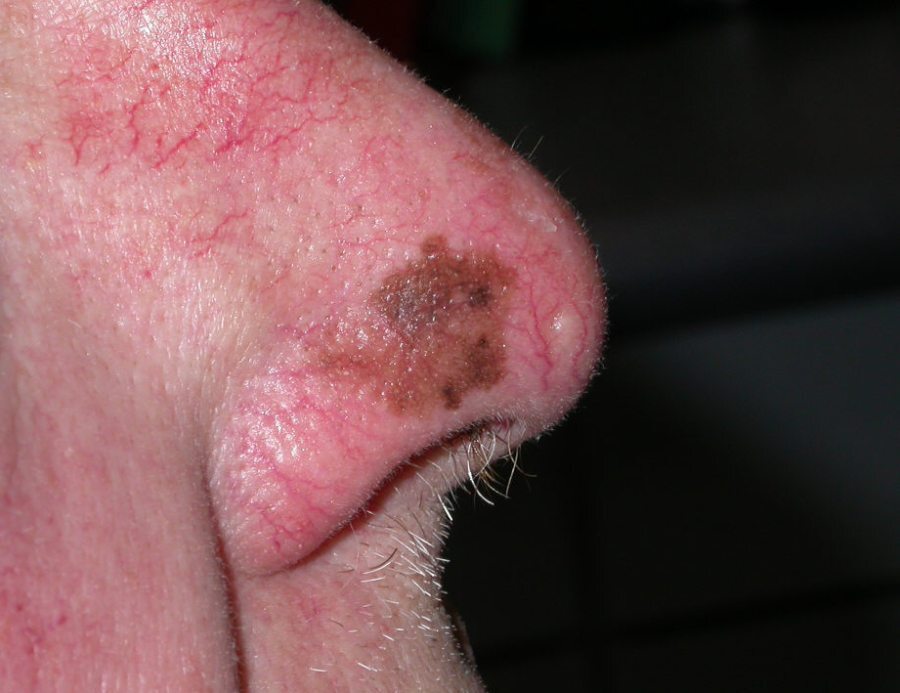
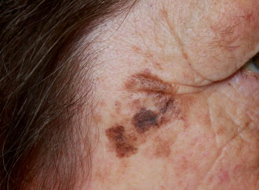
Lentigo maligna (LM) is a slow growing melanoma in situ that occurs around hair follicles, most commonly affecting UV-damaged skin of the head and neck. Lentigo maligna melanoma (LMM) is diagnosed when the melanoma cells invade into the dermis. It is the second most common form of melanoma. 

This chapter is set out as follows:


Aetiology

  • LM usually occurs in older people, with a peak incidence between 65 and 80 years
  • The incidence of LM appears to be increasing in younger age groups
  • The risk of LM and LMM is higher in individuals with lightly pigmented skin, markers of actinic skin damage (solar lentigos and actinic keratoses), and a history of non-melanoma skin cancer
  • Unlike with superficial spreading melanoma the risk is not associated with the total number of melanocytic lesions

History

  • LM grows very slowly over many years before progressing into LMM 

Clinical findings

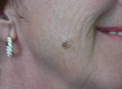
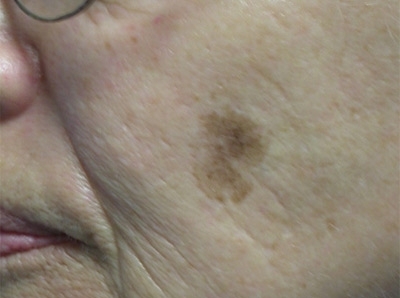
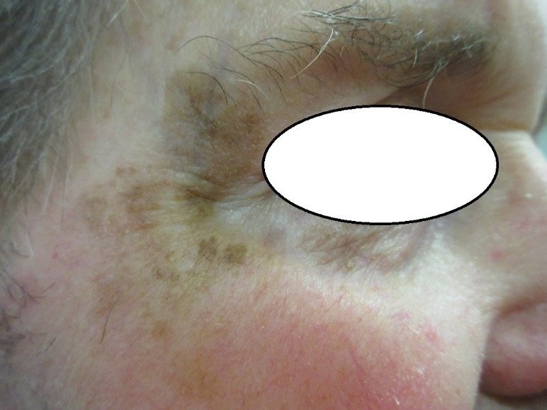
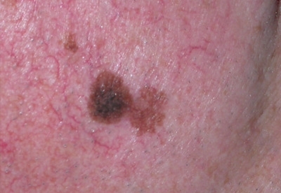
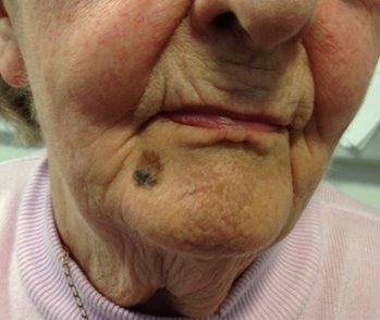
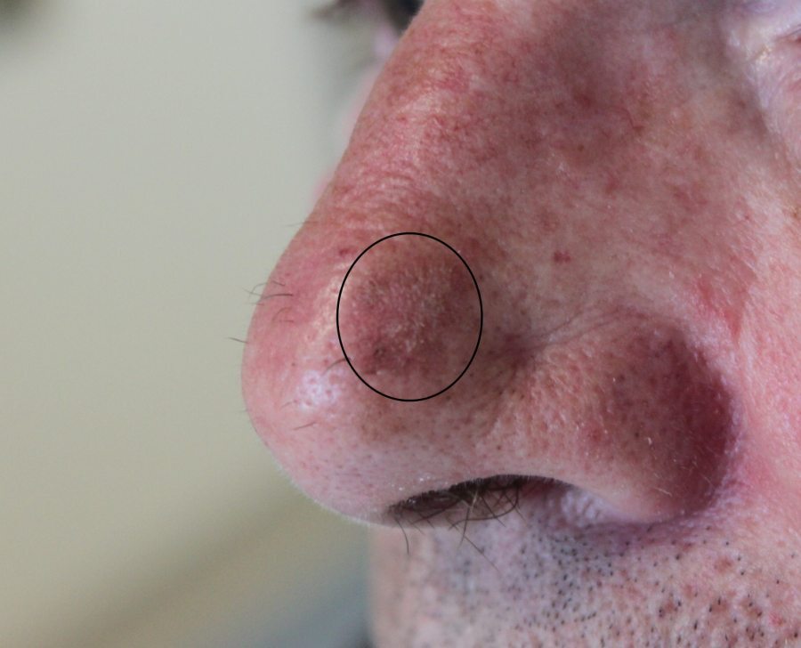
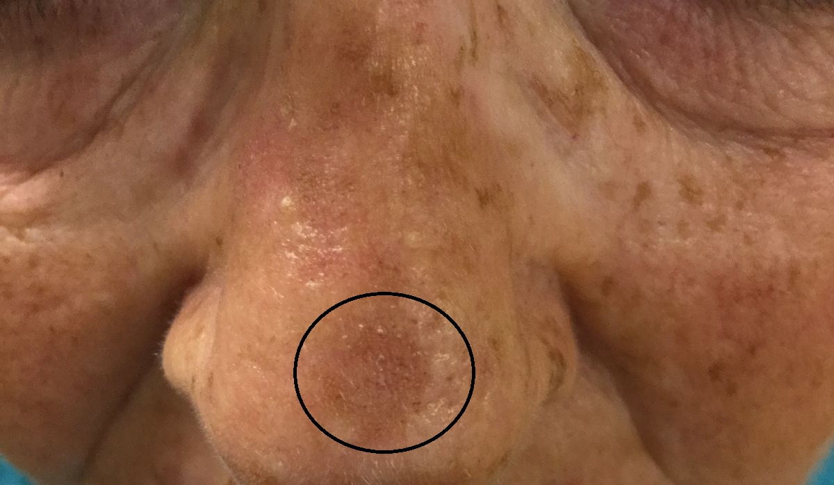
Clinical features of lentigo maligna 

  • Distribution
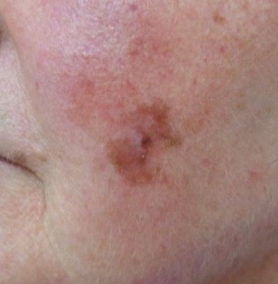
    • Sun-exposed areas, especially the face
       
  • Morphology - the ABCDE rule are key clinical features
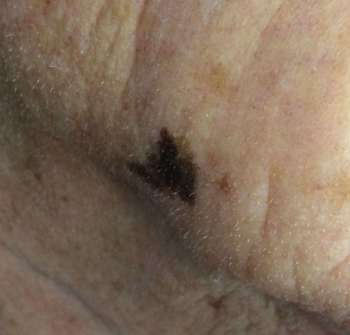
    • A = asymmetrical in shape and/or colour, the lesion becomes more asymmetrical as it enlarges
    • B = border irregular (jagged or notched)
    • C = colours can include different shades of brown and black; occasionally pink and white. Often variegated
    • D = dimensions greater than 6mm in diameter
    • E = evolving
       
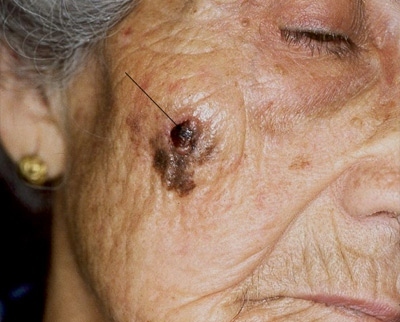
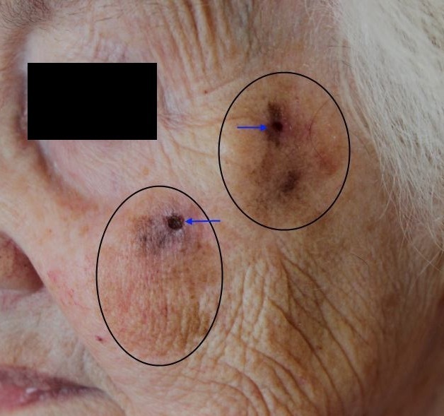
  • Transformation into LMM can be suggested by:
    • Development of a nodule
    • Increasing number of colours, especially blue or black
    • Ulceration or bleeding

Dermoscopic features of flat pigmented facial lesions

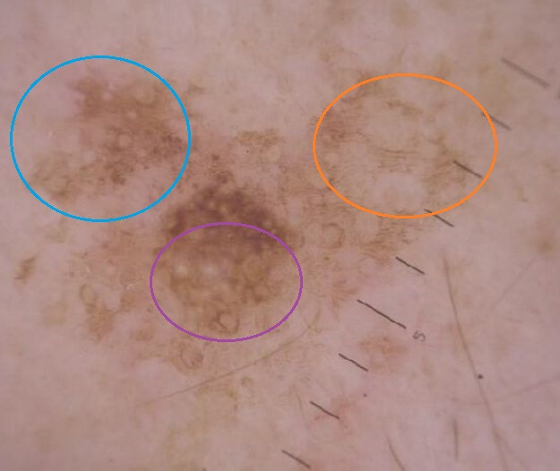
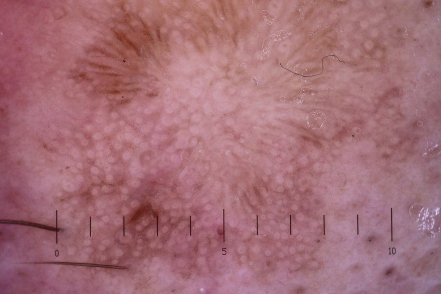
The structures of the facial epidermis differ to those on the trunk as the rete ridges (epidermal projections in to the dermis) are flat or absent, and there is an increased number of hair follicle units. As a result, lesions tend to lack a conventional pigment network found at other sites. When examining flatter facial lesions three of the key areas to focus on are:

  • The follicular openings - are these unaffected, is there pigmentation lining the openings, are they encircled, or are they obliterated?
     
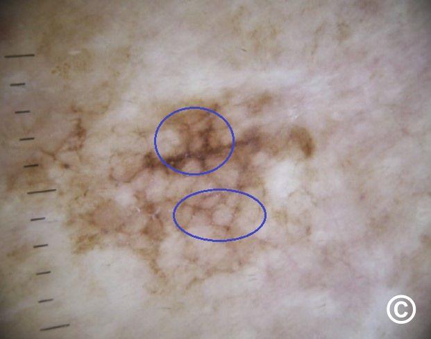
  • Structures in-between the follicular openings - these can be structureless (containing no structures), contain fine parallel lines ('fingerprinting'), have the appearance of a broad rounded 'network' (not a true network; sometimes referred to as a 'pseudonetwork'), or there can be angulated structures, sometimes intersecting to form a rhomboid-like pattern
     
  • The border - is the border well-demarcated or ill-defined?

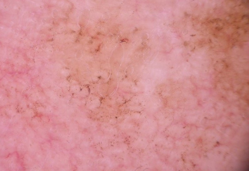
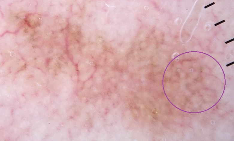
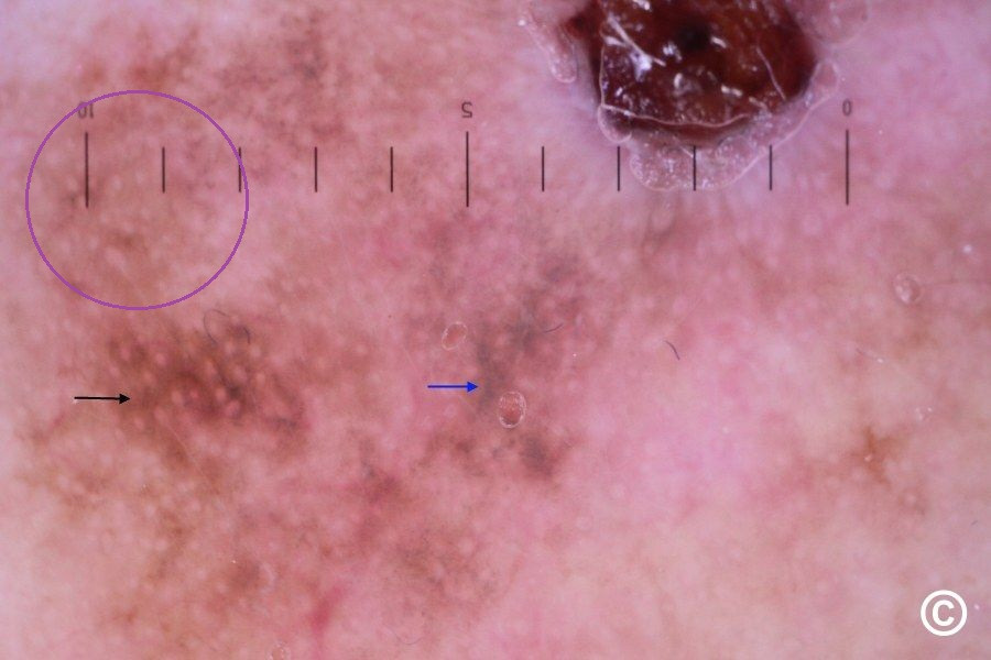
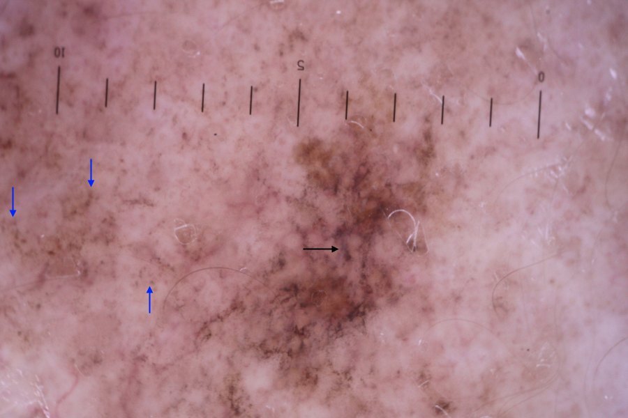
Dermoscopic features of lentigo maligna 

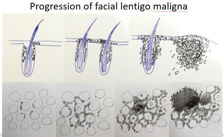
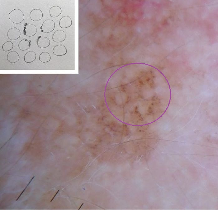
LM develops progressive features as follows:

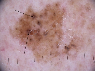
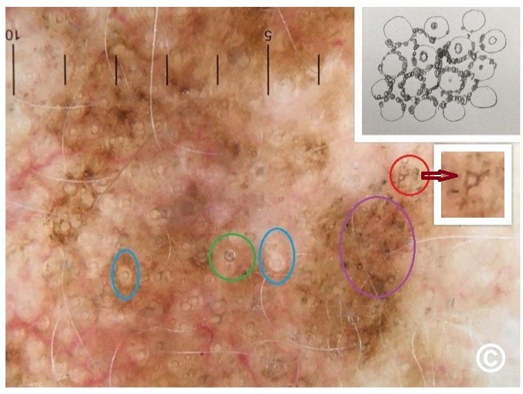
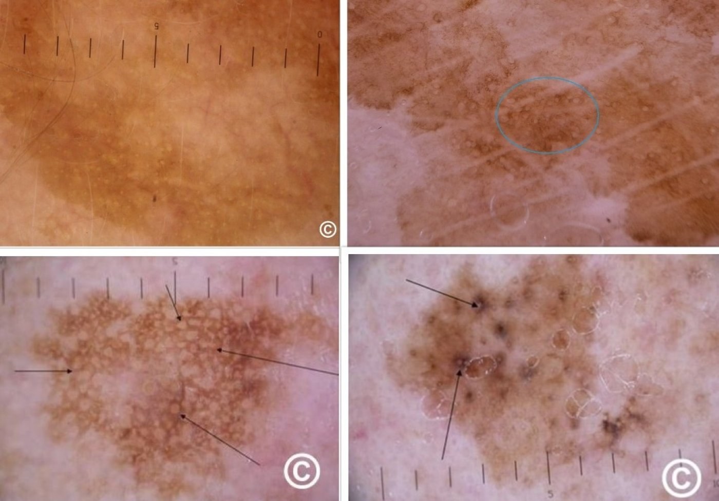
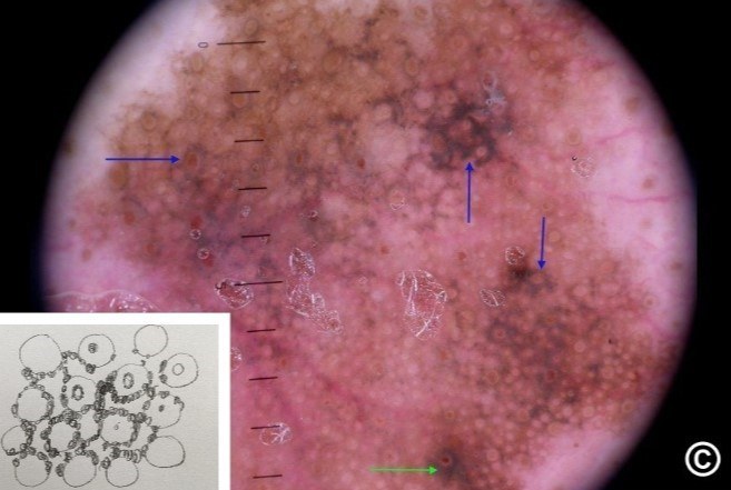
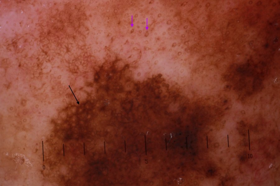
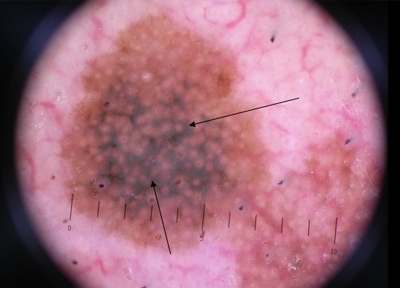
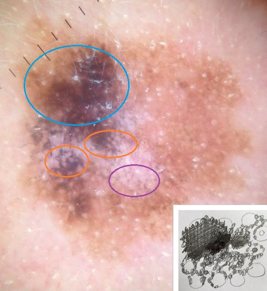
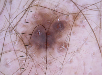
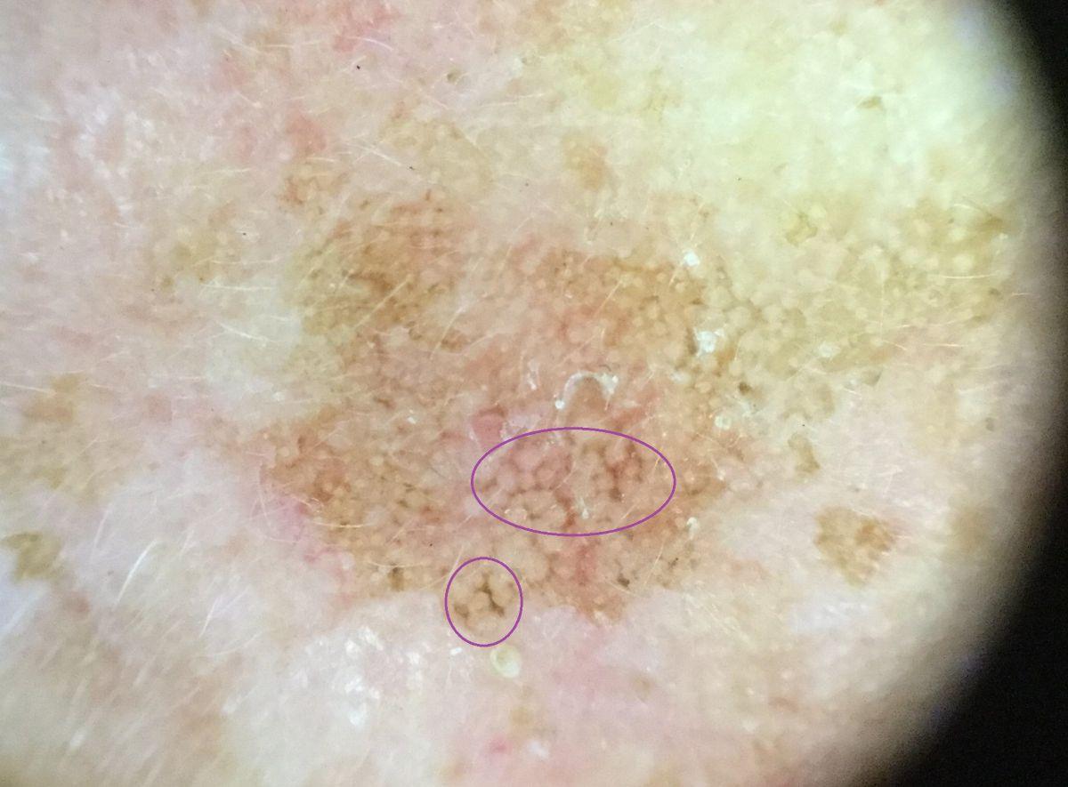
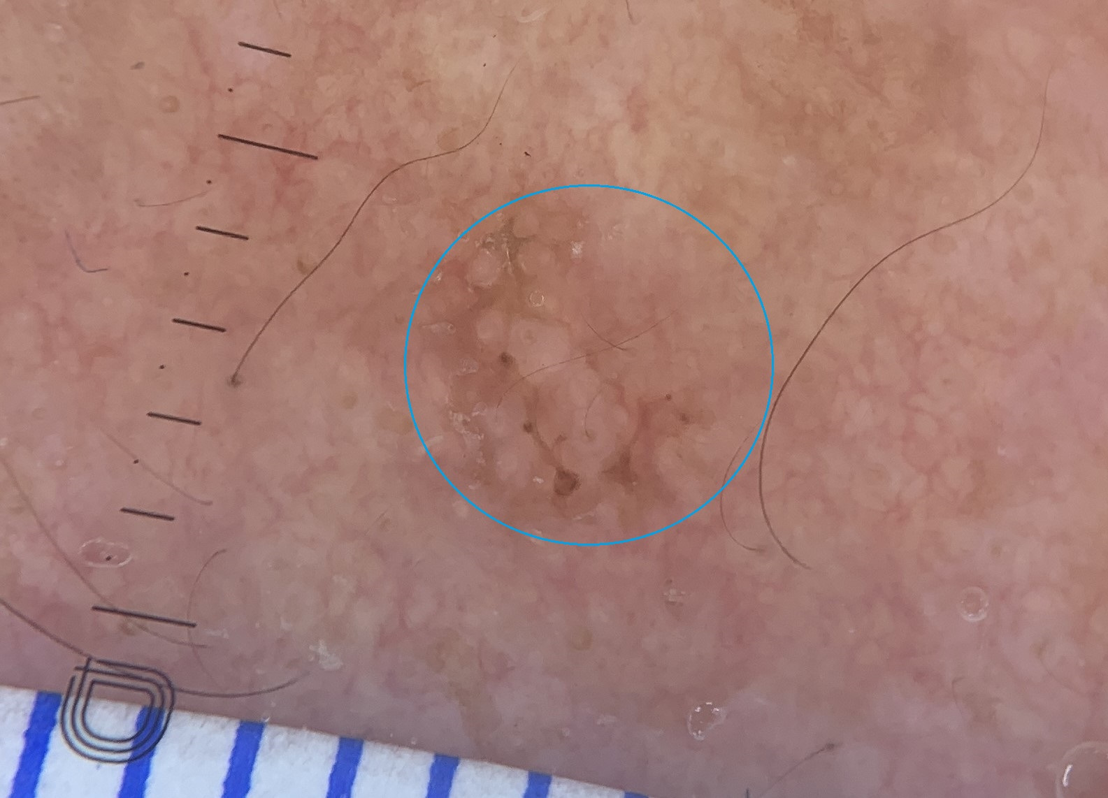
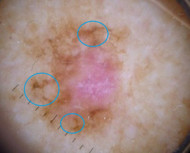
  • Hyperpigmented follicular openings - this can be regular or irregular peripheral pigmentation (eg crescent shaped), circles within circles (sometimes referred to as concentric circles) or dots within circles. Occasionally circles have linear projections (perifollicular projections). Although solar lentigos can have irregular follicular pigmentation, the colour in LM is often darker, often with a greyish hue, whereas in solar lentigo the colour is similar to that of the surrounding pigment. In LM these features correlate histologically with atypical melanocytes as single units or small nests extending down hair follicles
     
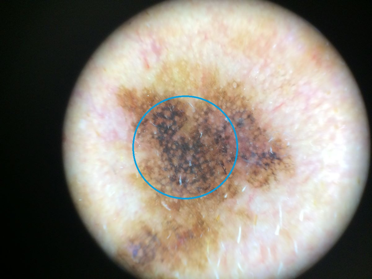
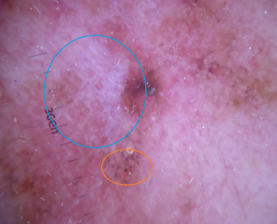
  • Annular-granular pattern with dots and short or polygonal lines - in terms of dots, the findings range from brown dots to blue-grey granularity scattered throughout the lesion; often clustering around follicular openings. Histologically these findings are explained by aggregates of melanocytes and small nests at the dermoepidermal junction between the follicles (brown dots), and by melanophages in the dermis (blue-grey granularity). The lines, which run around and between the follicles, reflect confluent junctional nests and aggregates of melanocytes
     
  • Rhomboid-like structures develop as the lines described above enlarge and intersect, often forming angulated structures 
     
  • Dark blotches - these appear as dark brown to black blotches, initially with sparing of follicular openings, and eventually as a homogenous black blotch with obliteration of the follicles 

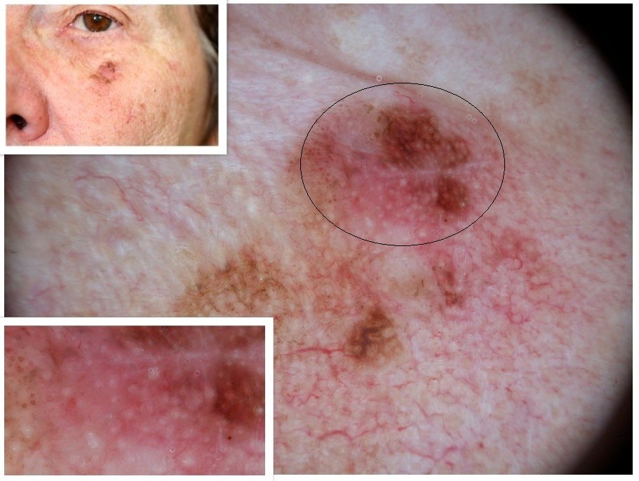
Lentigo maligna melanoma will share features of the above along with features of invasive melanoma elsewhere on the body. It is important to note that facial melanoma can also arise as a distinct entity from lentigo maligna. 

Additional notes on facial lesions

In the earlier stages of lentigo maligna it can be difficult to differentiate from non-malignant lesions, as such it is important to have a good understanding of the dermoscopic features of such lesions:

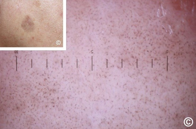
  • Solar lentigos - yellow to brown patches that dermoscopically have a well-demarcated, continuous, 'moth-eaten' border. Follicular openings are often unaffected; occasionally irregular and hyperpigmented at the periphery although the colour is similar to that of the surrounding pigment. Lesions may be structureless or have short parallel lines ('fingerprinting'). Some may evolve into seborrhoeic keratoses 
     
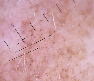
  • Lichenoid keratoses - grey patches, which dermoscopically show grey granules uniformly distributed throughout the lesion, sometimes referred to as 'peppering'. Lesions may have a preceding inflammatory component lasting 6-8 weeks with erythema and scale
     
  • Pigmented actinic keratoses - clinically the lesion should feel rough, and may have visible surface scale. Dermoscopically the hair follicle openings are often larger than normal, sometimes with white ring-like structures within or 'rosette-like' structures comprised of four grouped dots / globules best seen under polarised light. Other features can include grey confluent dots arranged in lines or grey to brown linear structures located between follicles, sometimes with small brown triangular structures  
     
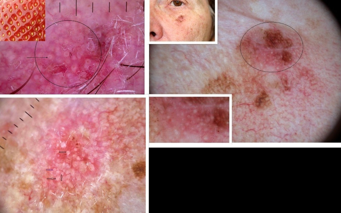
  • Dermoscopic erythema in facial lesions - erythema is a common feature of actinic keratoses (strawberry-like pattern). A pink-to-red hue or erythematous pseudo-network can also be found in some cases of lentigo maligna and melanoma. In facial lesions with erythema, melanoma should be considered a differential when:
    • The lesion lacks features of an actinic keratosis such as surface roughness and/or enlarged hair follicles
    • Additional features of lentigo maligna / melanoma such as an annular granular pattern, angulated structures, or atypical vessels are present

Dermoscopic features of cutaneous non-facial non-acral lentiginous growth pattern melanomas

One of the clues for lentiginous melanoma away from the head and neck (and palms and soles) are broad angulated grey or light brown lines that may form polygonal shapes, which are larger than the perifollicular rhomboidal structures previously described for facial LM. Other features can include chaos (asymmetry of colour or structures, or variable border demarcation), lentigo-like pigment pattern lacking a lentigo-like border, asymmetrically pigmented follicular openings, grey blue structures, eccentric structureless area, and bright white lines.


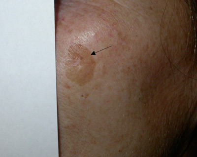
Clinical Images

Please refer to notes on image rights at bottom of the page with regards to individual image ownership.
Management

  • Any lesion suspicious for LM or LMM should be referred urgently to Secondary Care (2ww/USC pathway)
     
  • A skin biopsy can miss LM, especially in the early stages. Dermoscopy can help target the most atypical looking areas highlighting from where to take the biopsy
     
  • First-line treatment for LM and LMM is surgical excision
     
  • Topical imiquimod is a viable alternative for treating LM, particularly in patients who are not surgical candidates, and occasionally in difficult-to-treat locations. Such a decision should be taken in a skin cancer MDT. Complete clinical and histopathological clearance is achievable with imiquimod, but long-term follow-up is crucial due to potential for recurrence
     
  • For more information refer to the related chapter Melanoma - an overview

Disclaimer - the author PCDS cannot accept responsibility for any misleading or incorrect statements, and the management of individual patients remains the direct responsibility of the individual doctor. We do however hope that visitors to this site can contact us regarding comments that are considered misleading or incorrect so that we can continue to improve the site.

Image Rights - The PCDS would like to thank Dermatoweb, DermQuest (Galderma), and others who have contributed images. All named individuals and organisations maintain copyright for the relevant images.

Quick Links

The following pharmaceutical companies have had no involvement in the content of this website or in our conference programmes


Almirall
Galderma
Glenmark
Johnson & Johnson
La Roche-Posay
LEO Pharma
Pierre Fabre
Schuco