Drug rashes: mild to moderate
LAST UPDATED: Nov 16, 2021
Introduction
This chapter focuses on mild-moderate ACDR (adverse cutaneous drug reactions) and is set out as follows:
History
Working though this section can help determine whether a medication (drug) is responsible for the rash or other skin change. The next section (clinical findings) provides more detail on the different types of ACDR (adverse cutaneous drug reactions) that can result from medications.
This section is set out as follows:
- How soon after a drug has been started will ACDR arise?
- How soon after stopping the drug will the skin improve?
- Which drugs most commonly cause an ACDR?
- Serious ACDR
- Mild-moderate ACDR
How soon after a drug has been started will ACDR arise?
- Most ACDR start around 4-14 days after commencing the drug
-
However, some ACDR can take months or years to evolve such as those causing a drug-induced subacute cutaneous lupus erythematosus or a lichenoid eruption
-
It is important to ask about both prescribed drugs and over the counter medications
-
It is also important to be aware that an ACDR can occasionally arise if the patient is given a different brand of the same drug (the patient may for example notice that the new drug brand has a different colour)
How soon after stopping the drug will the skin improve?
-
Most skin changes settle within a few weeks of the drug being discontinued
-
Occasionally the skin changes can take several months to settle eg dapsone, and drugs causing pseudoporphyria
Which drugs most commonly cause an ACDR?
-
Antibiotics - especially the penicillins (eg amoxicillin), cephalosporins, sulfa antibiotics (see below) and the fluoroquinolones (eg ciprofloxacin)
-
The sulfa (sulpha) drugs - the term 'sulfa' refers to drugs that contain a sulphonamide group in their chemical structure, including the following:
-
Sulphonamide antibiotics - sulfamethoxazole, trimethoprim and co-trimoxazole (Septrin ®), which is a combination of the two. Co-trimoxazole tends to be associated with the most common and serious adverse effects of the group
-
Non-antibiotic sulphonamides - these are less likely to cause severe ACDR. Relevant drugs include:
-
Sulfasalazine and dapsone (these are the two most closely related to the sulphonamide antibiotics)
-
The sulfonylureas (eg gliclazide, glipizide and glibenclamide)
-
Furosemide, hydrochlorothiazide and sumatriptan
-
The aromatic anti-epileptic drugs - carbamazepine, phenytoin and lamotrigine
-
Allopurinol
-
Non-steroid anti-inflammatory pain killers (NSAIDs)
-
Some diuretics - furosemide, bumetanide and thiazides
-
Some angiotensin-converting enzyme (ACE) inhibitors
-
Gold
-
Penicillamine
-
Certain anti-fungal medications such as terbinafine
-
Blood
Which medications are unlikely to cause an ACDR?
-
The list of drugs includes digoxin, aspirin, paracetamol, codeine, tetracycline antibiotics, SSRI antidepressants and prochlorperazine
Serious ACDR
There are four main groups of potentially serious ACDR (for detailed information on the list below refer to the chapter Drug rashes: severe):
-
Urticaria (looks like a nettle rash), angioedema and sometimes anaphylaxis. Angioedema causes swellings of deeper tissues, often affecting the face (lips, tongue and eyelids) or other areas such as the genitalia (especially in men). The Drug‐induced serum sickness‐like reaction (SSLR) has migratory and pruritic urticarial wheals along with fever and arthralgias/arthritis, facial and periorbital oedema may coexist. Serum sickness-like reaction is most commonly seen in young children following the use of cephalosporins, however, other antibiotics, NSAIDs and a range of other medications have been associated
-
Stevens-Johnson syndrome / Toxic epidermal necrolysis - painful red areas of skin with local erosions and blisters, which quickly progresses resulting in sheet-like skin loss. Mucosal involvement includes the eyes, lips and mouth
-
Drug Hypersensitivity syndrome (also known as the DRESS syndrome) - systemic upset such as malaise and fever, a rash of variable appearance (widespread papules and plaques is the most common presentation followed by a measles type rash), haematological changes (especially eosinophilia) and usually solid‐organ disturbances (most commonly the liver). Lymphadenopathy is common, as is oedema of the head and neck
-
Erythroderma (syn. generalised exfoliative dermatitis) - a red scaly rash affecting 90% or more of the skin surface. It can also be part of the Drug Hypersensitivity syndrome. Most cases of erythroderma are not caused by an ACDR
Mild-moderate ACDR
There are many possible such reactions. For more detail on each category please refer to the section below on clinical findings. The list below provides a logical approach to this subject:
-
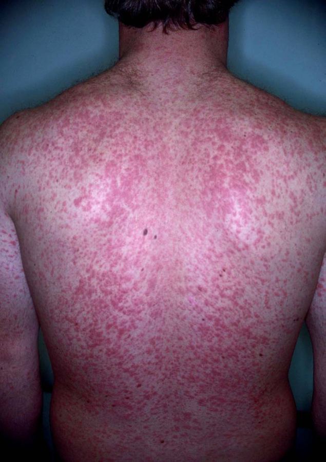
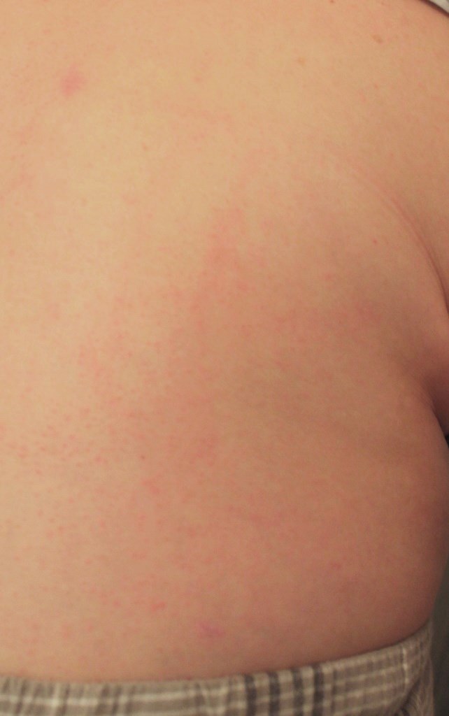
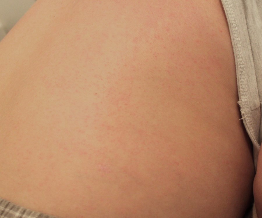
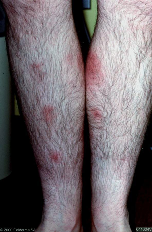
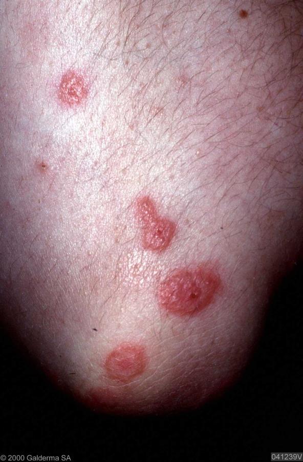
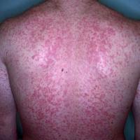
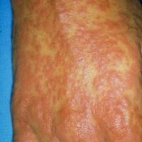
Exanthematic eruptions: these account for up to 95% of all ACDR. They usually resemble in appearance the classical rash of a viral infection (many pink-to-red macules usually first appearing on the trunk and then spreading to the limbs and neck. Lesions tend to blanch with pressure). Unlike with viral infections there is no fever
-
Several types of drug eruptions can itch:
-
Exanthematic eruptions can be itchy (above)
-
Urticaria - a nettle-like rash (see above under serious ACDR)
-
Drug-induced eczema is rare (see below under clinical findings)
-
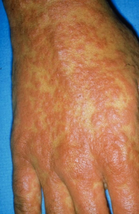
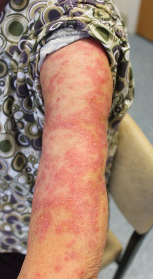
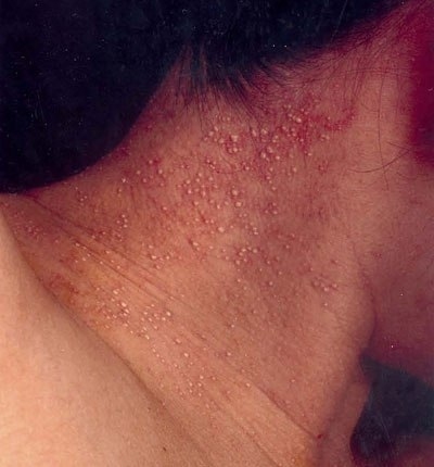
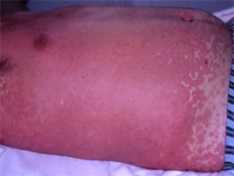
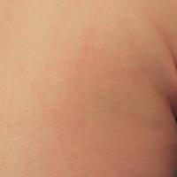
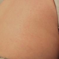
An urticated papular erythema (figures 3-4) - widespread itchy papules and plaques. Occasionally associated with the Drug Hypersensitivity syndrome (see above under serious ACDR)
-
No rash - drugs can occasionally cause an itch without a rash. Some of the drugs most commonly implicated include morphine and other opioids, statins, ACEI, digoxin, chloroquine and the sulphonamides. Confirming whether or not a particular drug is the prime cause for a patient’s itch is not always straightforward
-
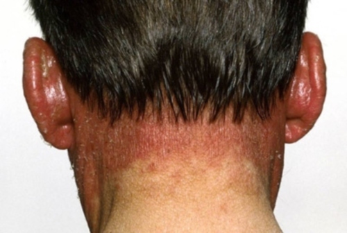
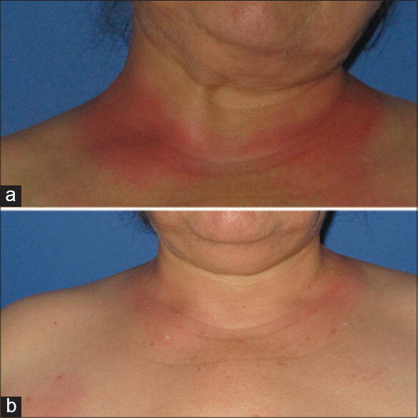
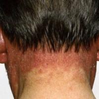
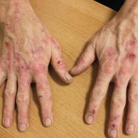
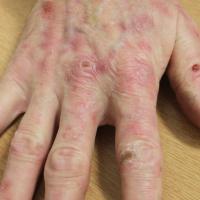
Does the rash mainly affect sunlight exposed sites? Several drugs can cause skin changes on areas exposed to sunlight, presenting in a number of ways:
-
Acute sunburn-like reactions
-
Eczematous changes with erythema and scaling
-
Hyperpigmentation
-
Lichenoid reactions
-
Pseudoporphyria - presenting as blistering / skin fragility, often on the backs of hands
-
Photo-onycholysis (nail changes)
- F or more detail on this group refer to the chapter Photodermatoses: drug / chemical-induced
-
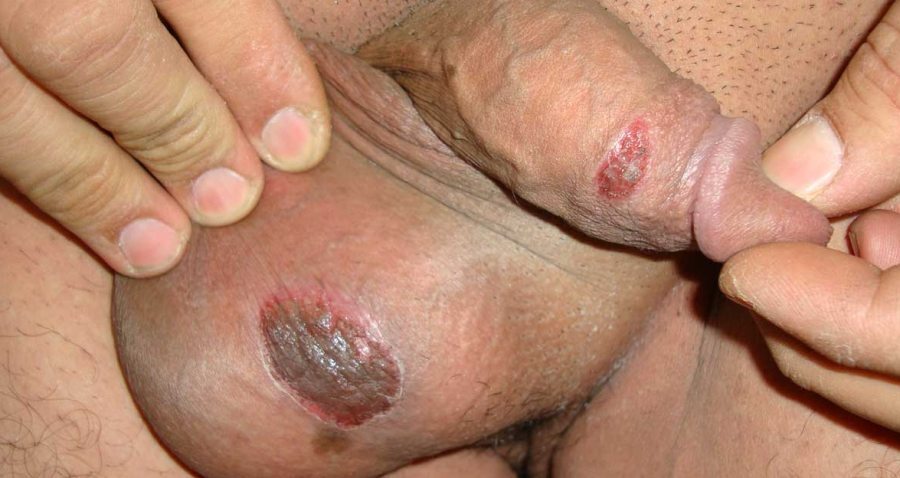
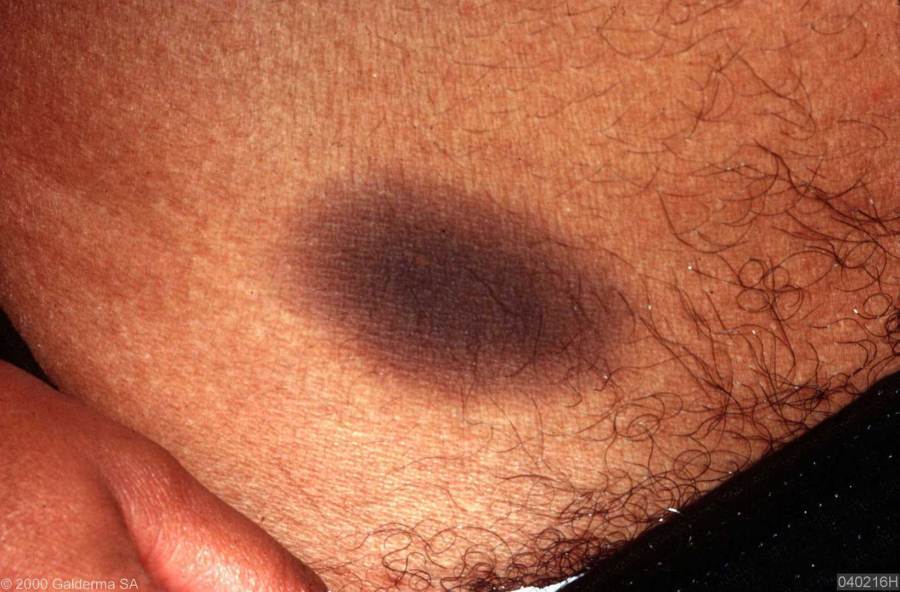
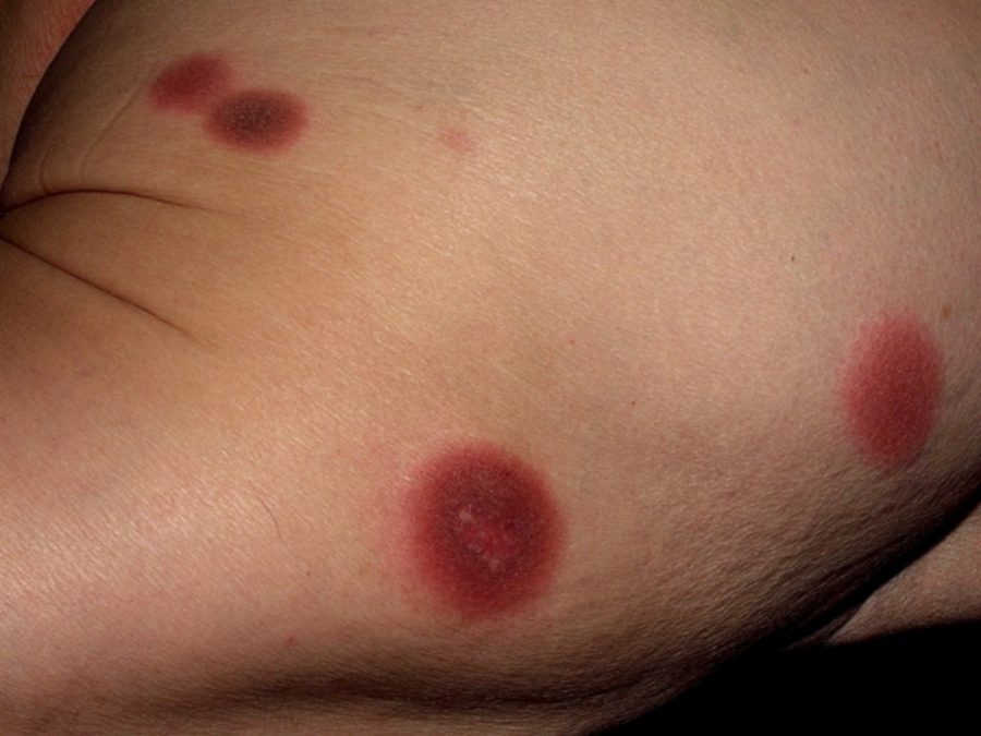
A fixed drug eruption: the rash comes and goes on exposure to the drug, the same body site is affected every time. The lesion(s) is a well-demarcated erythematous plaque that may blister, tending to heal with post-inflammatory hyperpigmentation
-
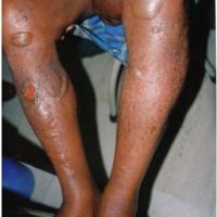
Are there any blisters? ACDR can cause a number of blistering eruptions including:
-
A fixed drug eruption (above)
-
Stevens-Johnson syndrome / Toxic epidermal necrolysis (refer above under serious ACDR)
-
Immunobullous reactions - very uncommon
-
Other possible presentations of ACDR:
-
Acneiform ie acne-like
-
Pustular ie white-yellow pustules - pustules can be localised but are more often generalised and known as acute generalized exanthematous pustulosis (AGEP)
-
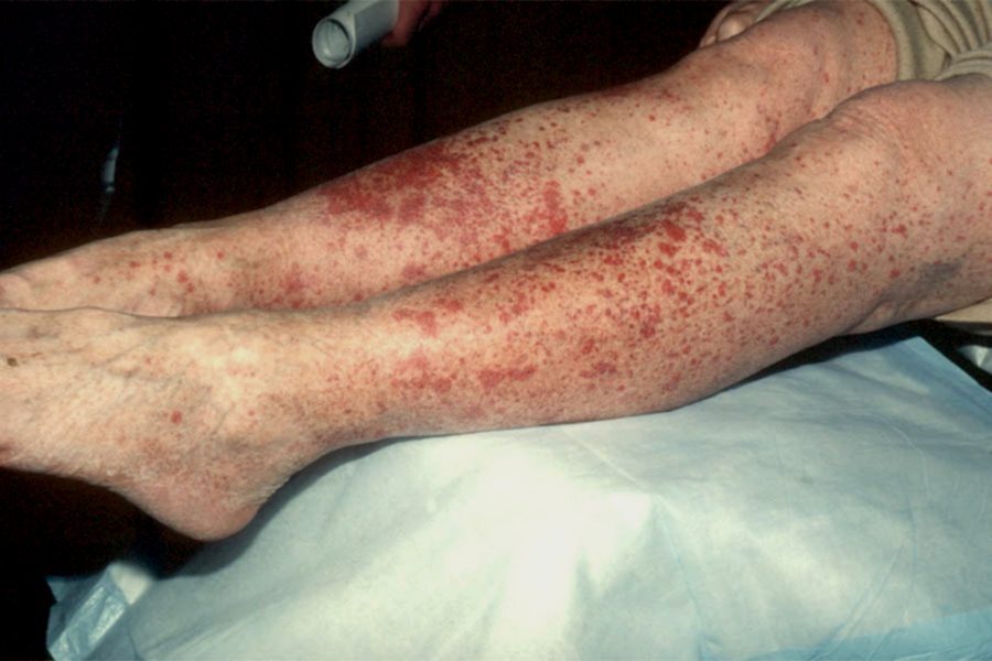
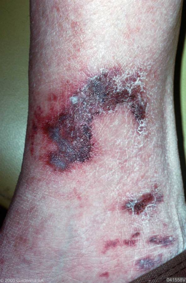
Purpura / cutaneous vasculitis - red-purple palpable lesions most commonly on lower legs, which do not blanch on pressure
-
Erythema nodosum (EN) - painful nodules on the shins
-
Erythema multiforme (EM) - those that are secondary to medication tend to cause more varied and less well-defined target lesions than when due to herpes simplex (antibiotics can be an exception)
-
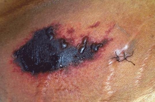
Skin ulceration and infarction - two of the most commonly associated medications that cause ulceration are hydroxyurea and nicorandil. Warfarin can cause infarction
-
Symmetrical drug related intertriginous and flexural exanthema (SDRIFE) - a symmetrical erythematous rash affecting the buttocks/natal cleft, peri-anal region or groins, usually along with one or more other flexural sites
-
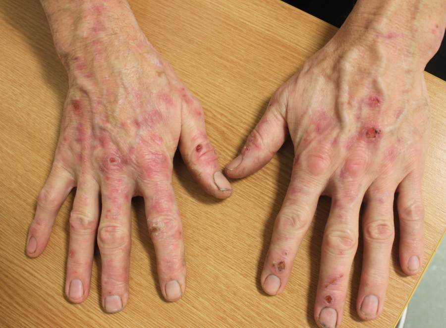
ACDR that sometimes present a long time after the drug was commenced (sometimes months or years) include:
-
Pigmentary changes ie developing darker areas of skin, either localised or more widespread
-
Subacute cutaneous lupus erythematosus (SCLE) - erythema and scale that is usually more widespread than compared to non-drug induced SCLE
-
Lichenoid reactions - can be clinically indistinguishable from normal lichen planus although is frequently more severe. Occasionally only affects sun-exposed sites
Clinical findings
Exanthematous (figures 1-2)
- The most common presentation of ACDR causing 95% of all such rashes
-
More common if inter-current viral infection
-
Chronology - symptoms most commonly occur at 7-14 days, but can be earlier if been exposed to the medication before
-
Clinical features
-
Distribution - tends to start on the trunk and move to extremities, spares mucosal membranes
-
Morphology - maculopapular / morbilliform
-
If drug not stopped it may progress to an exfoliative dermatitis
-
Large numbers of drugs implicated. The most commonly associated drugs are antibiotics (ampicillin, penicillin, cephalosporins), the sulphonamides, isoniazid, phenytoin and carbamazepine, NSAID, gold, thiazides and allopurinol
Photosensitive eruptions (figure 5)
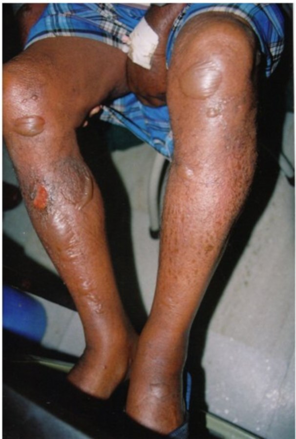
Bullous (blistering) drug reactions (figures 6-8)
- Can present in a number of ways:
- Fixed drug eruption
- SJS / TEN - refer to the related chapter Drug rashes - severe
- Immunobullous reactions - very uncommon
-
Immunobullous reactions include:
-
Pemphigus - penicillamine, captopril, piroxicam and rifampicin
-
Pemphigoid - psoralens, furosemide, clonidine, ibuprofen and the sulfa drugs
-
Linear IgA - vancomycin
-
Pseudoporphyria - refer to the chapter Pseudoporphyria. The most commonly associated drugs are NSAIDs, diuretics and nalidix acid
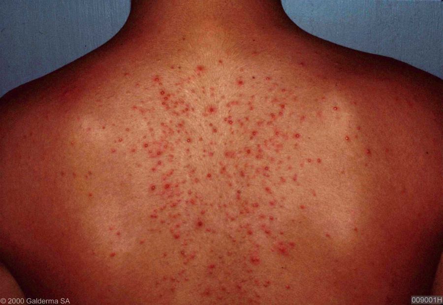
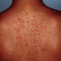
Acneiform (figure 9)
- Clinical features - tends to cause monomorphic papules and pustules without comedones
-
Most commonly associated drugs - hormonal treatments (eg progestagens in contraceptive pills), steroids, lithium, phenytoin and isoniazid
-
Always ask patients with very muscular physiques if they use anabolic steroids - patients may not come forward with this information at the first time of asking
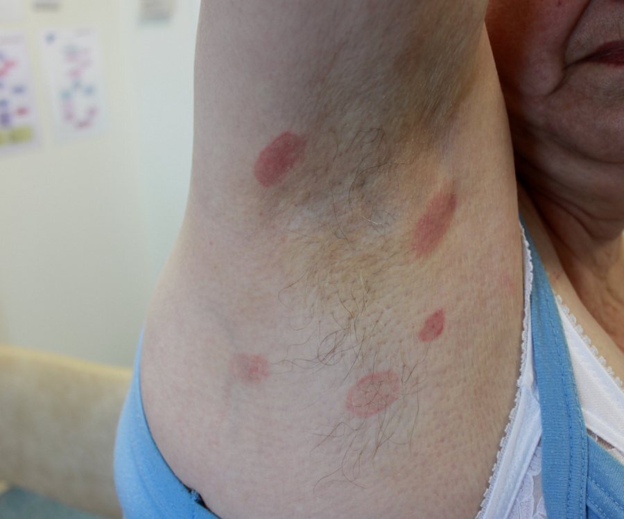
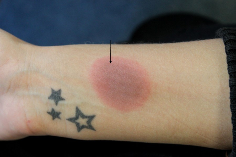
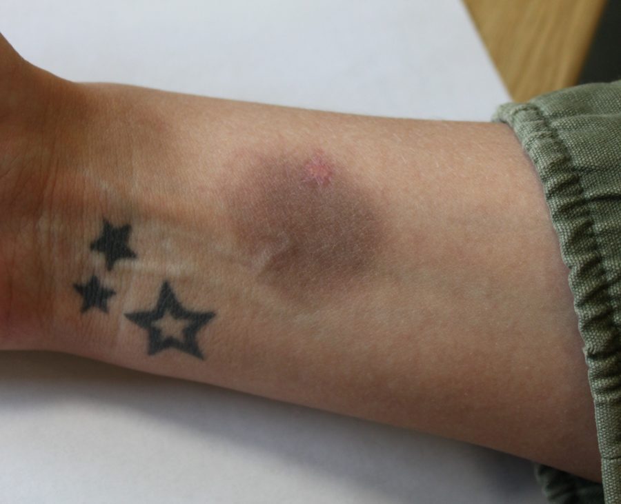
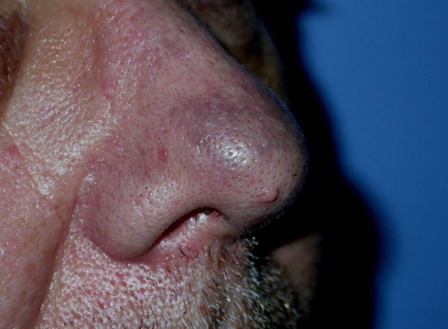
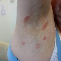
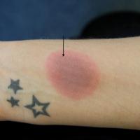
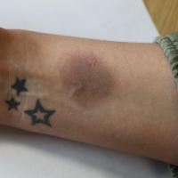
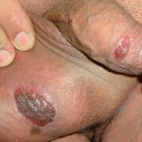
Fixed drug eruption (figures 10-15)
- Chronology - symptoms occur within 48 hours of commencing the medication
-
Clinical features:
-
Distribution - lips, face, distal limbs, hands & feet, glans penis and perianal skin. Occurs at same site on re-exposure
-
Morphology - well-demarcated erythematous plaque that may blister
-
Tends to heal with post-inflammatory hyperpigmentation
-
Large numbers of drugs implicated. The most commonly associated drugs - tetracyclines, sulphonamides, NSAID and quinine. Other drugs listed include co‐trimoxazole, penicillins, metronidazole, rifampicin, erythromycin, paracetamol, carbamazepine, sulphasalazine, calcium‐channel blockers, Angiotensin‐converting enzyme inhibitors, omeprazole, azole systemic antifungals, complementary medicines
-
Occasionally the eruption is secondary to food (eg cashew nuts, asparagus), or a chemical in food, agents have included sodium benzoate (a preservative in acidic foods eg carbonated drinks, fruit juice and jam), Splenda ® (an artificial sweetener), and tartrazine (an orange colouring in food and drinks - now seldom used). Green tea and Cannabis can also cause a fixed drug eruption
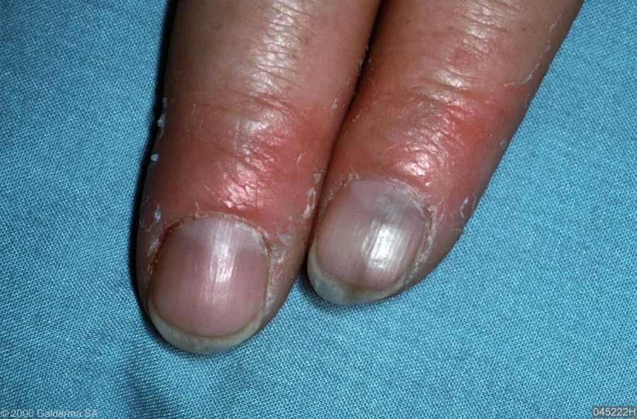
Hair and nails
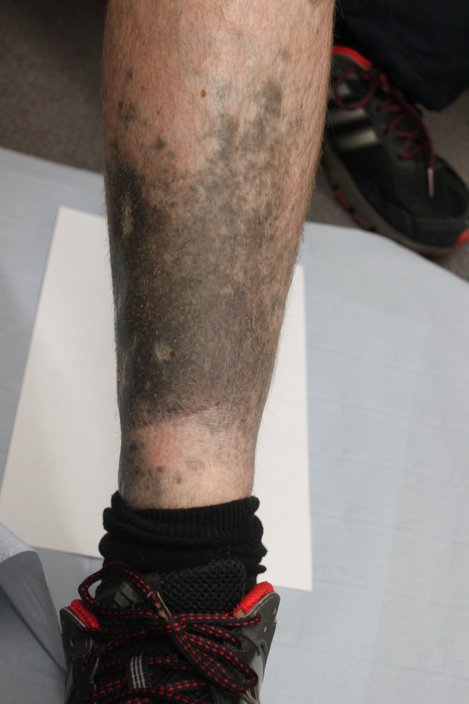
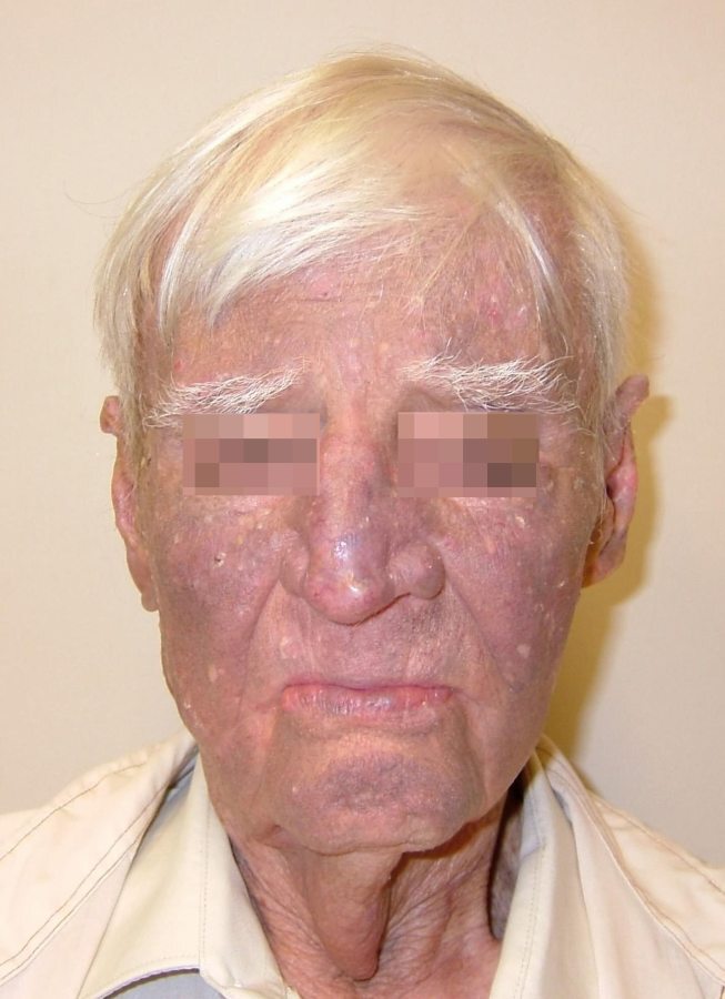
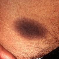
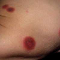
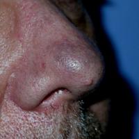
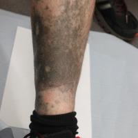
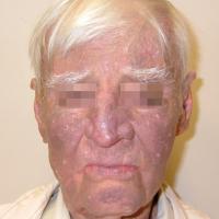
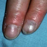
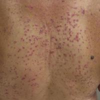
Pigmentary (figures 16-19)
- Chronology - very variable, can be a long time after the medication was commenced
-
Most commonly associated drugs - minocycline, amiodarone, anti-malarials, co-danthramer (can stain peri-anal and surrounding skin in patients with incontinence)
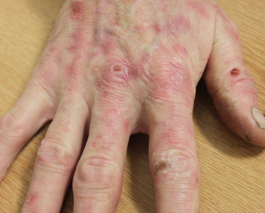
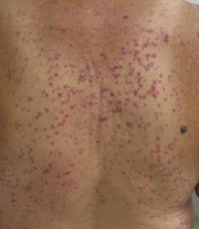
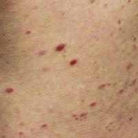
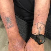
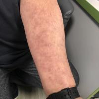
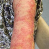
Lichenoid (figures 20-23)
-
Drug‐induced LP reactions represent approximately 10% of all LP cases
-
Chronology - usually months (even years) after the first exposure to the medication
-
Clinical features
-
Can be clinically indistinguishable from normal LP although is frequently more severe, Wickham's striae are often absent, and nail and mucosal change less common
-
In some cases the features are non‐specific, sometimes lesions may be scaly
-
Mucosal lichenoid reactions can also be caused by drugs
-
Photodistributed lichenoid lesions may occur with a number of drugs, including thiazide diuretics
-
Most commonly associated medications - gold, antimalarials, thiazides, ACEI, NSAIDs, betablockers, quinine, penicillamine, lithium, sulfa drugs (including sulfonylurea hypoglycaemic agents, dapsone, sulfasalazine), and tramadol. Hepatitis B, influenza and varicella-zoster are the most common immunisations associated with a lichenoid eruption
-
Skin histology is essential for the diagnosis of lichenoid drug eruptions
-
The diagnosis is proven by disease resolution on withdrawal of the offending drug. Disease resolution may take weeks to months
Lupus erythematosus (figure 24)
- 10% of cases of LE-like reactions are drug-induced
-
Chronology - usually months (even years) after the first exposure to the medication
-
Autoantibodies - anti-Ro / La may be positive, and anti-histone antibodies can be found in 95% of cases of drug-induced lupus compared with 50% of cases of idiopathic lupus
-
SCLE (subacute cutaneous lupus erythematosus)
-
Is more likely to be drug-induced when the rash is widespread ie also involves the trunk and distal extremities
-
Several medications have been implicated including PPI, calcium-channel blockers, thiazide diuretics, terbinafine, beta-blockers, ACE inhibitors, tamoxifen, leflunomide, acebutolol, some of the biologics, carbamazepine, cinnarizine, ticlopidine, naproxen, and gold
-
With some drugs there can be a delay of several months to years before the rash presents. The condition usually resolves after discontinuation of the drug
-
SLE-like (systemic lupus erythematous) syndromes
-
Many medications may rarely precipitate SLE in predisposed individuals. Symptoms generally take several months to develop, and drug-induced SLE does not usually involve the skin. The most frequent drugs implicated are hydralazine, carbamazepine, lithium, phenytoin, the sulphonamides, and minocycline
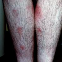
Erythema nodosum (figure 25)
-
Drugs are an uncommon cause of erythema nodosum
-
The most commonly associated drugs are the OCP and the sulfa drugs
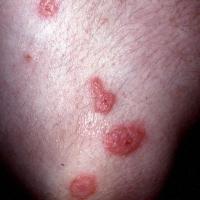
Erythema multiforme (figure 26)
-
Many cases of erythema multiforme are not related to medication. Those that are secondary to medication tend to cause more varied and less well-defined target lesions than when due to herpes simplex (antibiotics can be an exception)
-
The most commonly associated medications include - the sulfa drugs, penicillins, tetracyclines, aromatic anticonvulsants (eg phenytoin and carbamazepine), thiabendazole, aspirin and other NSAID, furosemide and thiazide diuretics, phenothiazines, quinine, progestogens and omeprazole
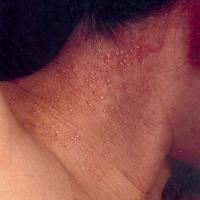
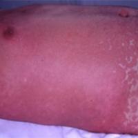
Pustular (figures 27-28)
-
Chronology - symptoms occur within a few days of commencing the medication
-
Pustules can be localised but are more often generalised and known as acute generalized exanthematous pustulosis (AGEP)
-
Clinical features of AGEP - a generalised erythematous eruption with superficial desquamation. Small sterile pustules mainly found in the axillary and inguinal areas. Can develop blisters. Associated with fever
-
Most commonly associated drugs - often an antibiotic eg aminopenicillins and quinolones. Also terbinafine, diltiazem, hydroxychloroquine, isoniazid and the antibiotic sulphonamides
-
Good outcome - settles within two weeks of stopping the drug
Purpura / cutaneous vasculitis (figures 29-30)
-
Chronology - symptoms tend to occur at around three weeks after the medication was commenced
-
Clinical findings - palpable purpura (thrombocytopenia is non-palpable)
-
Many drugs are implicated, the most commonly associated ones are the penicillins, minocycline, quinolones, 'sulfa' drugs, NSAID and other analgesics, thiazides and other diuretics, anticonvulsants, allopurinol, vaccines, cocaine and other illicit drugs
-
It is important to rule out other causes of purpura before concluding the rash is secondary to a drug
-
For more information refer to the chapter Vasculitis
Ulcers / infarction (figure 31)
-
Ulcers - two of the most commonly associated medications are hydroxyurea and nicorandil, the latter mainly affects the eyes, mouth, genitalia, perineal and perianal skin
-
Infarction - the main cause is warfarin
Drug-induced eczema
-
Rare. The vast majority of cases of eczema are not drug-induced
-
A systemic allergic contact dermatitis (SACD) caused by drug hypersensitivity
-
Many drugs implicated - α‐Blockers, captopril, calcium‐channel blockers, NSIADs, opiates, paracetamol, many antibiotics, sulphonamides, antihistamines, aciclovir, valaciclovir, clonidine, aminophylline, heparin, hydroxycarbamide, iodinated radiocontrast media, oestradiol, phenothiazines, rivastigmine
-
The latency from drug initiation to onset of eczema typically occurs at 7–14 days
-
On withdrawal of the offending drug, resolution of the clinical symptoms generally occurs in 1–3 weeks. Re‐exposure to the culprit would be expected to reproduce the same clinical picture
-
If there is any doubt about the diagnosis patch tests can be performed, which are commonly positive
Symmetrical drug related intertriginous and flexural exanthema (SDRIFE) (figure 32)
-
A symmetrical erythematous rash affecting the buttocks/natal cleft, peri-anal region or groins, usually along with one or more other flexural sites
-
The latency from drug intake to the onset of SDRIFE ranges from hours to a few days
-
Among the medications causing SDRIFE, beta-lactam antibiotics, especially amoxicillin, are most common, others include cephalosporins, clindamycin, erythromycin, valaciclovir, NSAIDS, codeine, hydrochlorothiazide, terbinafine, cimetidine, nystatin, fluconazole, risperidone, rivastigmine, heparin and pseudoephedrine
-
The reaction improves quickly on withdrawal of the culprit drug
-
Patch tests are positive in up to 50% with SDRIFE
Clinical Images
Please refer to notes on image rights at bottom of the page with regards to individual image
ownership.
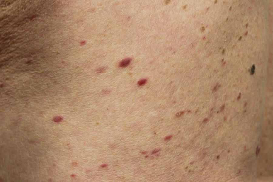
1
2
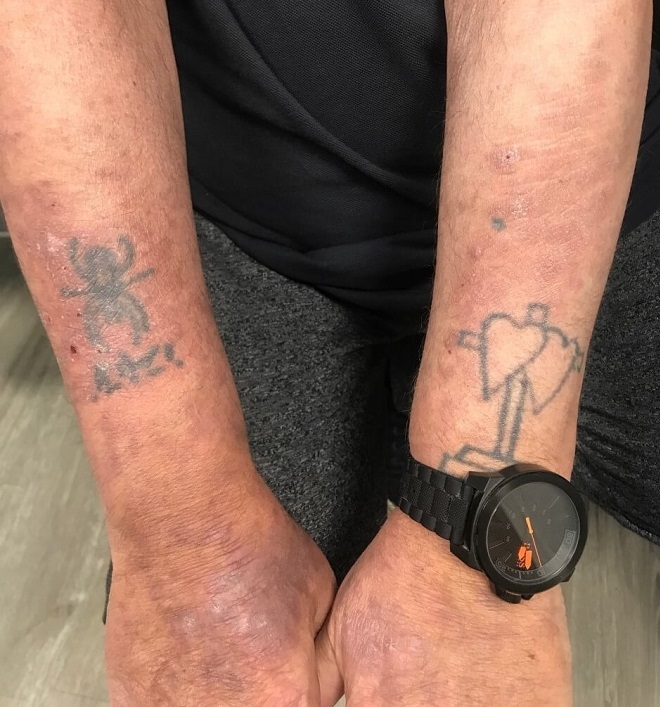
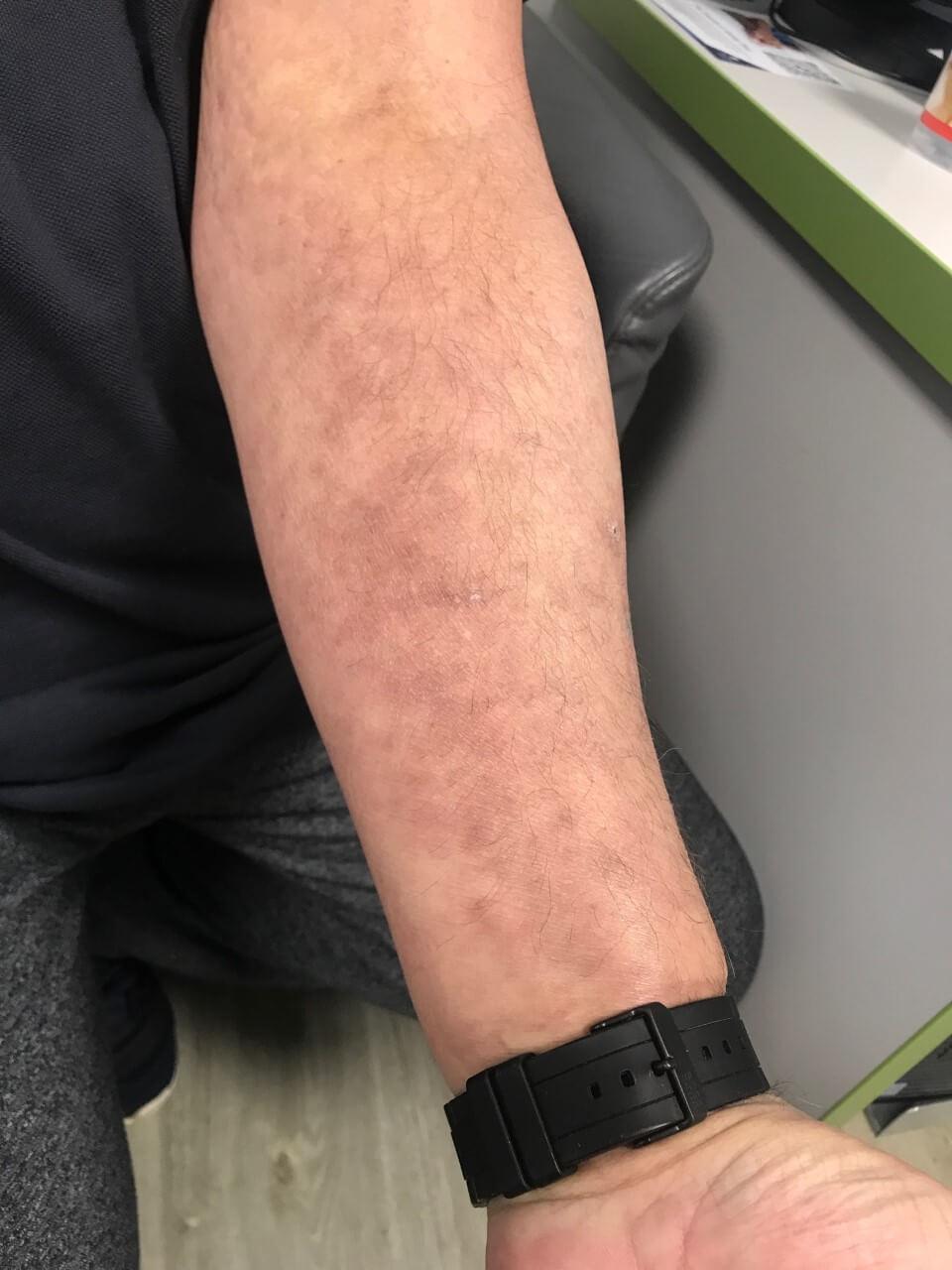
3
4
5
6
7
8
9
10
11
12
13
14
15
16
17
18
19
20
21
22
23
24
25
26
27
28
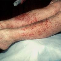
29
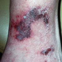
30
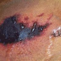
31
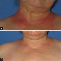
32
Investigations
-
Occasionally a biopsy is needed. Histology can show a number of features, partly dependant on whether the rash is in an acute or chronic stage. The most common histopathologic pattern of ACDR is a vacuolar interface dermatitis. Drug eruptions, like many other inflammatory diseases, are often associated with an infiltrate of eosinophils and/or neutrophils
Management
-
Stop the offending drug if possible
-
Supportive therapy eg emollients and topical steroids if needed
-
Cross-reactivity
-
10% of patients allergic to penicillin have cephalosporin allergy
-
Patients allergic to one sulphonamide antibiotic should avoid other antibiotics in the same group. However, the link between the antibiotic and non-antibiotic group is less strong and cross-reactivity is unlikely to arise, but some still advocate avoiding dapsone or sulphasalazine in patients allergic to sulphonamide antibiotics
Disclaimer - the author PCDS cannot accept responsibility for any misleading or incorrect statements, and the management of individual patients remains the direct responsibility of the individual doctor. We do however hope that visitors to this site can contact us regarding comments that are considered misleading or incorrect so that we can continue to improve the site.
Image Rights - The PCDS would like to thank Dermatoweb, DermQuest (Galderma), and others who have contributed images. All named individuals and organisations maintain copyright for the relevant images.