Annular erythema
LAST UPDATED: Nov 21, 2021
Introduction
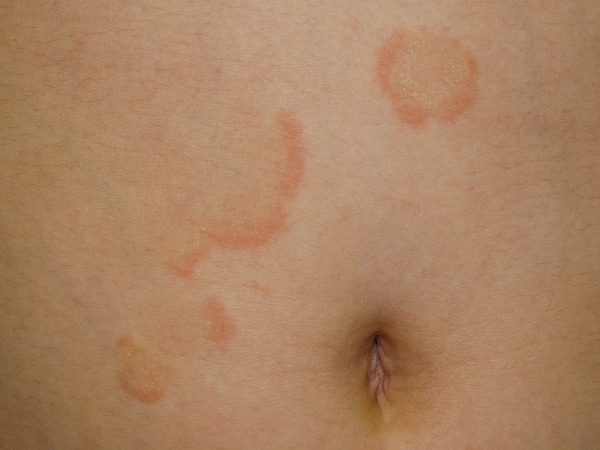
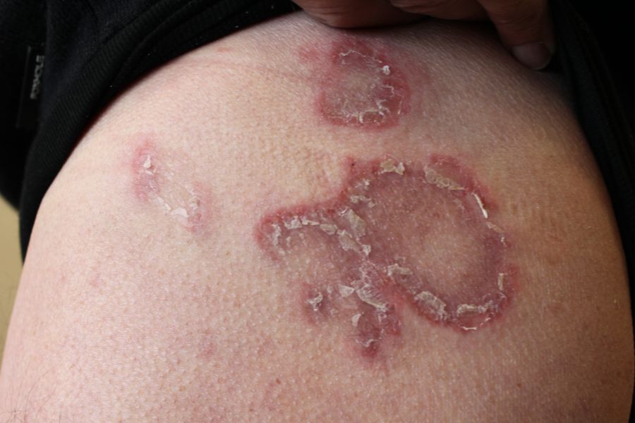
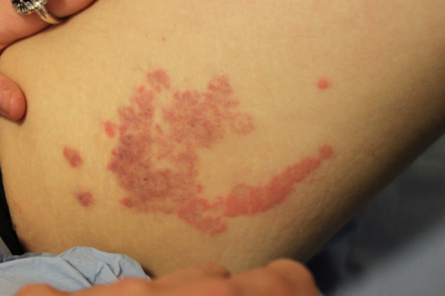
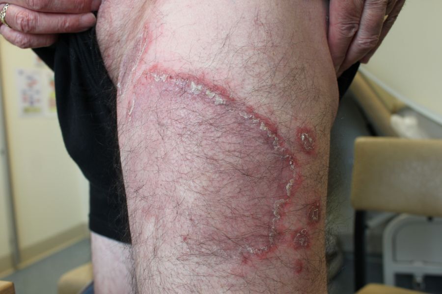
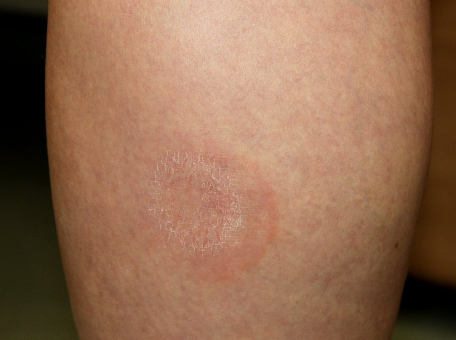
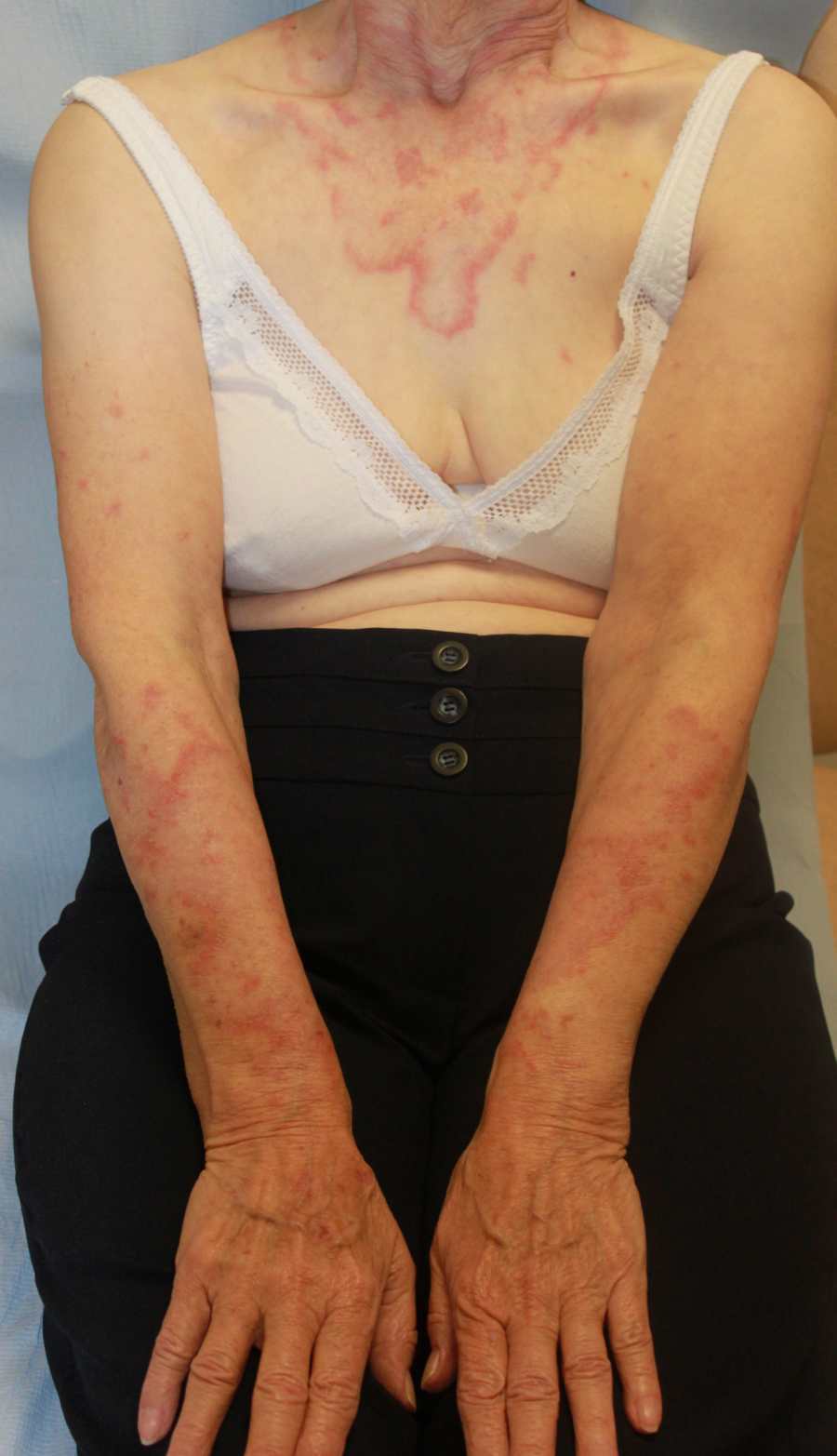
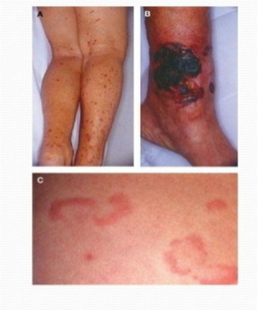
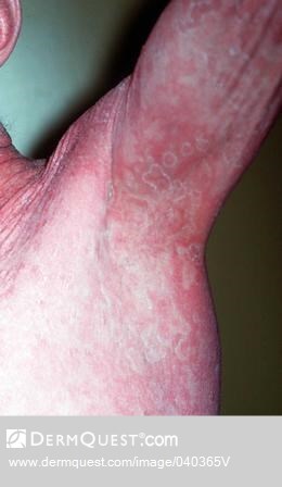
Annular erythema is not a diagnostic term but rather a description of a spreading erythematous eruption with an annular (ring-shaped) appearance. If the expansion of the rash is not uniform it may become arciform (arc-like) or polycyclic (two or more connected rings).
While there are many dermatoses that have an annular appearance, the term annular erythema is sometimes reserved for a group of uncommon skin conditions that include erythema (chronicum) migrans, erythema annulare centrifugum (EAC), ENA-positive annular erythemas, and erythema gyratum repens. This chapter discusses these diagnoses and is set out as follows:
Clinical findings
Erythema (chronicum) migrans - the cutaneous features of Lyme disease
-
Presents with a spreading erythema. It is usually round or oval, but can be triangular or linear. Often, a central punctum is evident at the bite site
-
The rash typically expands over days, although it may not be observed until it is already full size. As the rash expands clearing behind the leading edge may occur, so giving an annular appearance. In the United States, however, erythema migrans is more likely to have a uniform colour
-
Given the potential systemic complications of Lyme disease this condition requires particular attention. Please refer to the related chapter Lyme disease (and erythema migrans)
Erythema annulare centrifugum (EAC)
-
Introduction
-
EAC is a skin condition characterised by expanding, erythematous annular lesions usually lasting for several weeks, and often of unknown aetiology
-
Aetiology
-
Although infection, drugs and underlying malignancy, particularly haematological, have all been associated, in the large majority of cases no cause is found
-
History
-
EAC presents most commonly in young and middle-aged individuals, although any age can be affected including infancy
-
Itch is variable but seldom intense
-
Clinical
-
Distribution
-
Solitary or multiple lesions
-
The buttocks, thighs and upper arms are the most commonly affected sites. The face is rarely affected
-
Morphology
-
A pink-red papule slowly enlarges, as the central area flattens and fades the eruption becomes annular (ring-shaped)
-
The edge may be flat or slightly palpable
-
Lesions often have slight scaling behind the advancing edge, but can be smooth
-
Extension is usually 2-3 mm per day but can be quicker. If the rash does not grow uniformly then the lesion can become arciform or polycyclic
-
Occasionally lesions are more deep-seated and have no scale. This is sometimes referred to as a deep gyrate erythema, which is probably a variant of EAC
-
Lesions usually last a few weeks, but occasionally days or several months
-
EAC is often chronic with periodic fluctuations
-
Investigations
-
A thorough examination and FBC are required to exclude haematological malignancy
-
Skin scrapings may be required for mycology to exclude tinea
-
The diagnosis of EAC is made clinically, however, a biopsy may be necessary to confirm the diagnosis and to exclude an alternative diagnosis such as cutaneous T-cell lymphoma, the lesions of which tend to have a broader leading edge than in EAC. Histology shows a perivascular lymphocytic infiltration
-
Treatment
-
Lesions of EAC usually resolve spontaneously, although the more potent topical steroids may speed up resolution
-
For recalcitrant cases, some patients benefit from hydroxychloroquine, which has anti-inflammatory properties in the skin
ENA (extractable nuclear antigens) positive annular erythemas
-
Annular erythematous eruptions may occur in the following anti-Ro/SSA positive patients:
-
Lupus erythematosus, especially subacute cutaneous lupus erythematosus, and also neonatal lupus erythematosus
-
Sjögren's syndrome
-
Otherwise well patients
-
Cases of anti-La/SSB positive annular erythemas have also been reported
-
Lesions are often photosensitive, and may scale
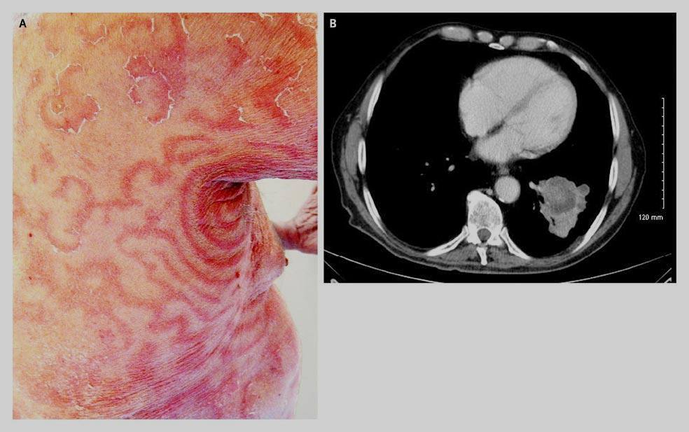
Erythema gyratum repens
-
80% of cases are reported as associated with internal malignancy, most commonly the lung. 20% of cases are found to have no cause
-
Morphology
-
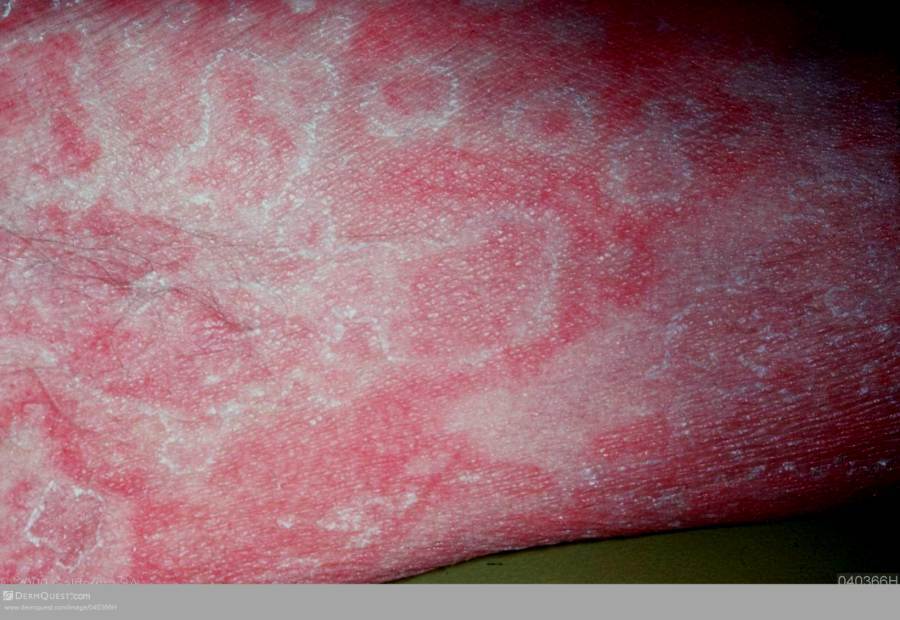
Regular waves of erythema spread over the body
-
Rings, swirls or waves appear within existing lesions to form a sequential pattern that produces a series of concentric, figurate bands resembling the grain of wood
-
Scaling, usually at the trailing edge is common
-
Management is directed at trying to find the underlying cause
Clinical Images
Please refer to notes on image rights at bottom of the page with regards to individual image
ownership.
Management
-
Refer to the section on clinical findings
Disclaimer - the author PCDS cannot accept responsibility for any misleading or incorrect statements, and the management of individual patients remains the direct responsibility of the individual doctor. We do however hope that visitors to this site can contact us regarding comments that are considered misleading or incorrect so that we can continue to improve the site.
Image Rights - The PCDS would like to thank Dermatoweb, DermQuest (Galderma), and others who have contributed images. All named individuals and organisations maintain copyright for the relevant images.