Basal cell carcinoma - an overview
LAST UPDATED: Dec 29, 2025
Introduction
Basal cell carcinoma (BCC) is the commonest from of skin cancer accounting for approximately 70% of all skin cancers. BCC are composed of cells similar to those in the basal area of the epidermis and its appendages. The estimated lifetime risk for BCC in the white population is 33-39% for men and 23-28% for women. Reliable statistics on the incidence of non-melanoma skin cancer (NMSC) are difficult to obtain, but it is estimated that around 100,000 people are diagnosed with NMSC each year in the UK.
While BCC very rarely metastasize, they can cause significant morbidity by local tissue invasion and destruction, particularly on the head and neck.
This chapter provides an overview on BCC and is set out as below. More detailed information can be found in the related chapters.
Aetiology
- Both environmental and genetic factors are important
- Although cumulative lifetime UV exposure (sunlight and artificial light) is a major risk factor for the development of BCC, it does not accurately predict the frequency of BCC development at a particular site on its own
- Risk increases with age
- Although BCC is observed in people of all races and skin types, it is most often found in fair-skinned individuals (skin types I and II). Those with skin type I are very fair and have red or blonde hair and freckles; these individuals always burn and never tan. Those with skin type II are fair and burn easily while tanning minimally. Whites of Celtic ancestry have the highest risk for BCC
- Basal cell carcinoma is much less common in other races; recent studies suggest it is the most common skin cancer in Skin of Colour. The estimated incidence of BCC per 100,000 population has been reported as:
- 0.32–6.4 in Chinese
- 0.7–1.4 in Asian Indians
- 1–2 in African American
- Men are affected twice as often as women. The higher incidence in men is probably due to increased recreational and occupational exposure to the sun
- Other risk factors include:
- Trauma appears to play a part in the development of some BCC and there are reports of tumours arising in sites of traumatic injury or burn
- Smoking
- Immunosuppression
- Rare genetic disorders can be associated with BCC. The most well-known is the Basal Cell Naevus syndrome (syn. Gorlin syndrome), an autosomal dominant condition, features of which can include:
- The development of BCC during adolescence or early adulthood (any patient developing 2 or more BCC under the age of 30 should be referred). The number of BCC that develop during a lifetime varies from zero to hundreds, with a mean of 160
- Most people develop benign jaw tumours called odontogenic keratocysts. These tumours usually first appear during adolescence; new tumours form until about age 30. If untreated, these tumours may cause painful facial swelling and tooth displacement
- Other features include small depressions (pits) in the skin of the palms of the hands and soles of the feet; an unusually large head size (macrocephaly) with a prominent forehead; and skeletal abnormalities involving the spine, ribs, or skull. These signs and symptoms are typically apparent from birth or become evident in early childhood
- Individuals have a higher risk than the general population of developing other tumours such as a medulloblastoma or fibroma
History
- BCC tend to be slow growing, often increasing in size by 2-4 mm a year; occasionally faster
- Most lesions are reported by the patient as never fully healing and periodically bleed or produce a scab
Clinical findings
Distribution
- Although the majority occur on the head and neck, any part of the body can be affected - BCC can be multiple so a thorough skin examination is needed
- The incidence of superficial BCC, especially on the trunk is increasing significantly, especially in younger patients
- BCC are uncommon on the forearms and hands, and are very rare on non-hair bearing parts of the body
Morphology - general
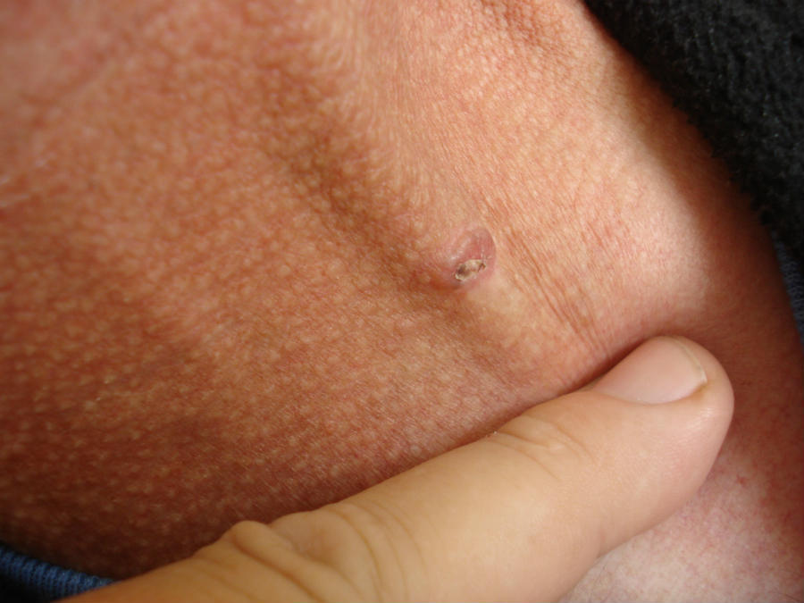
- Lesions often produce a bloody crust as opposed to the scale found in keratinising tumours such as actinic keratoses and SCC
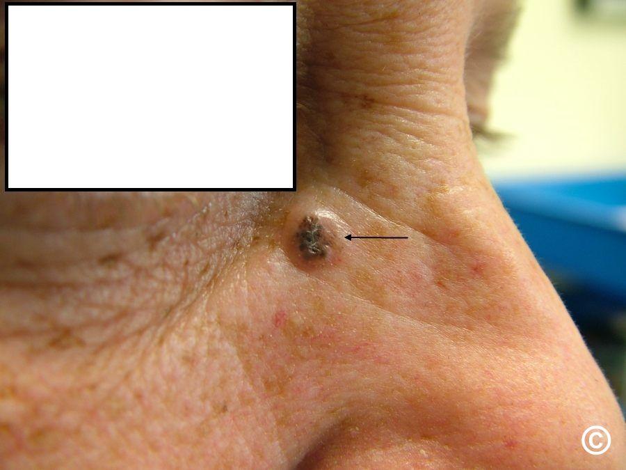
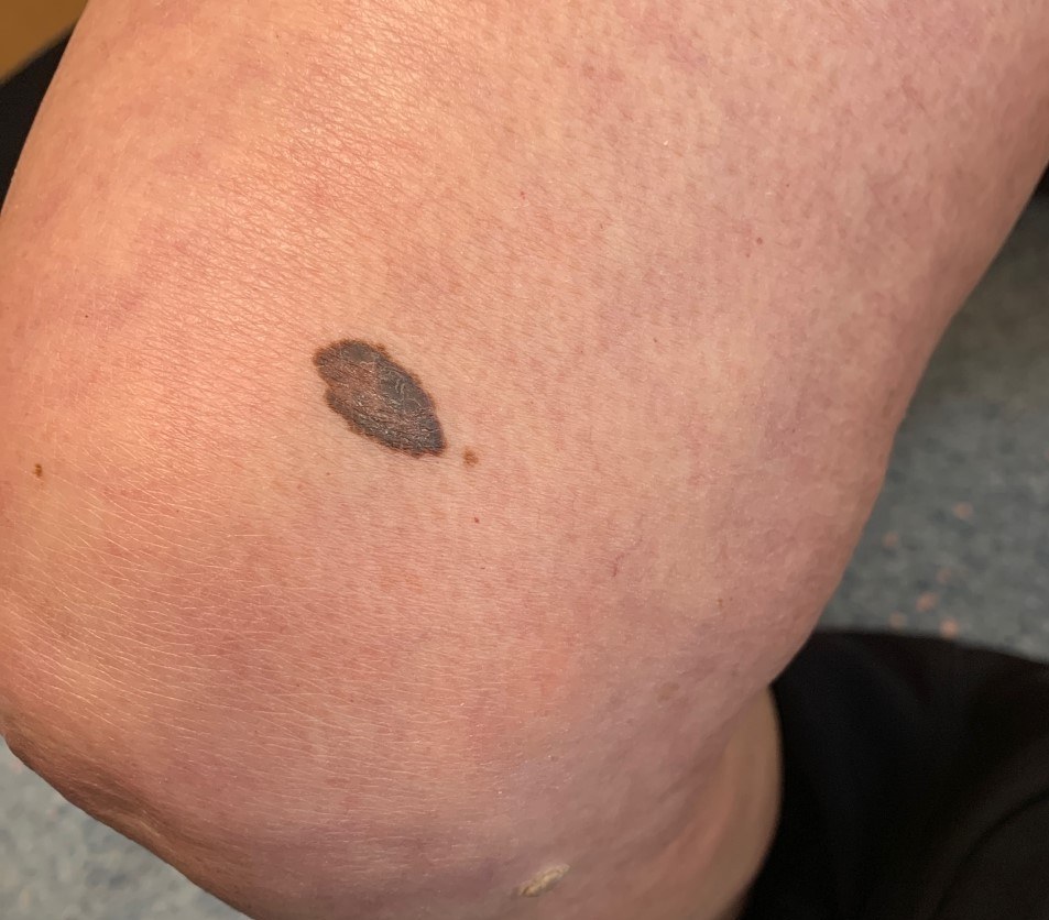
- May contain pigment
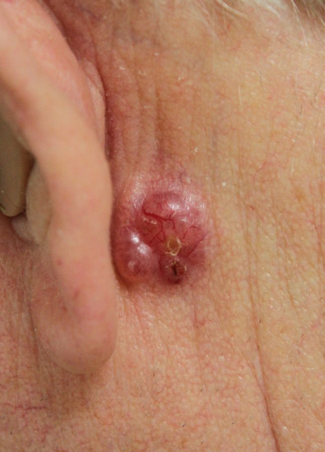
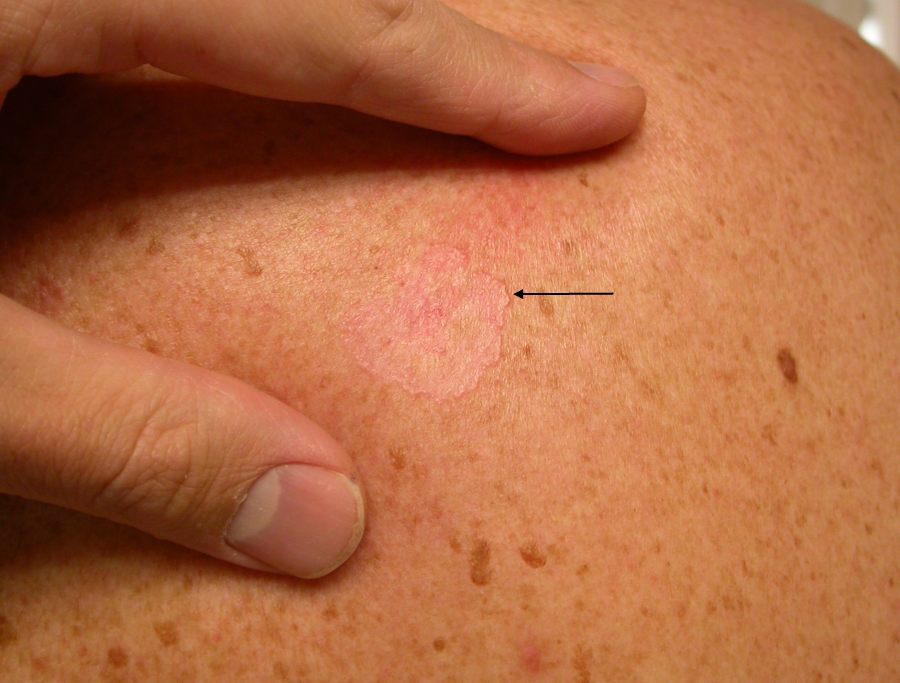
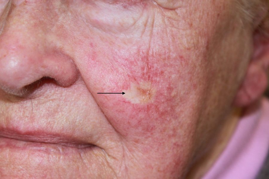
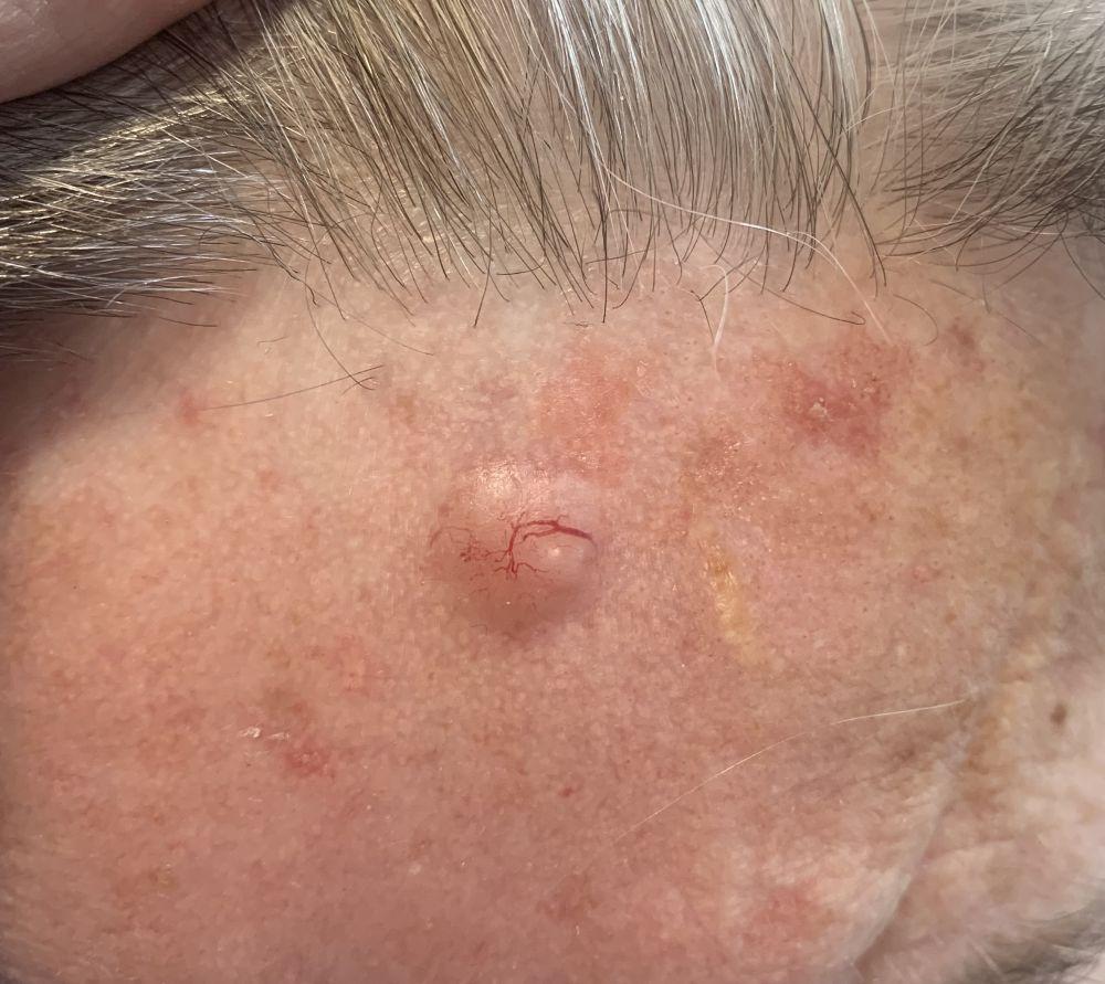
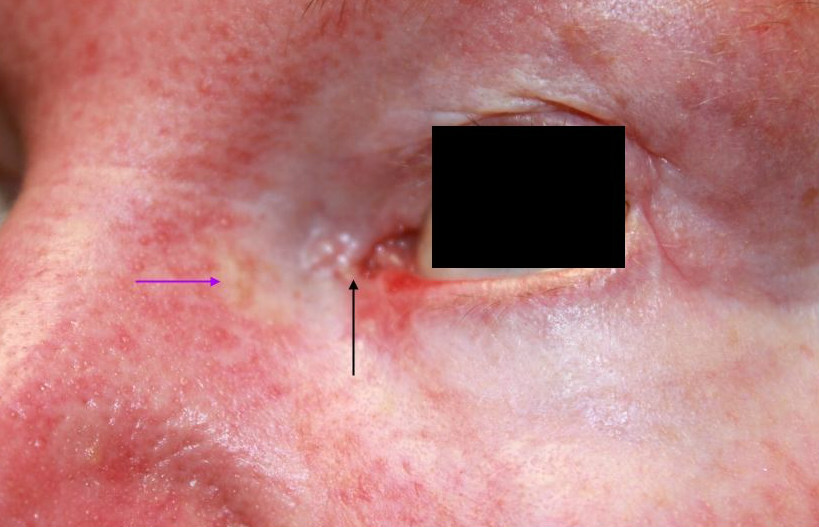
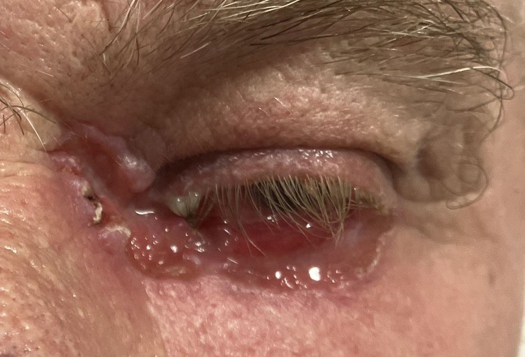
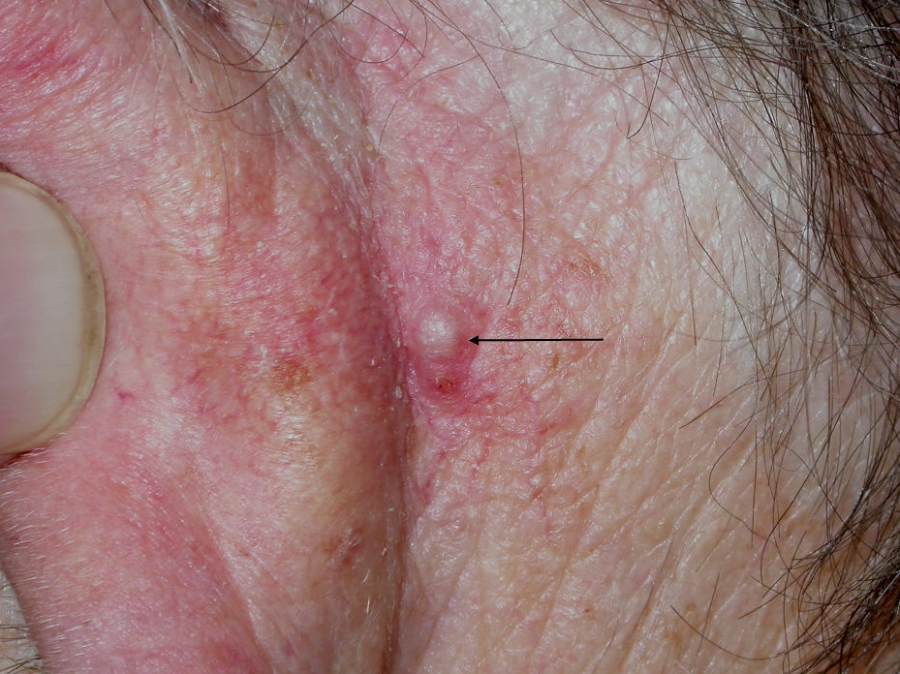
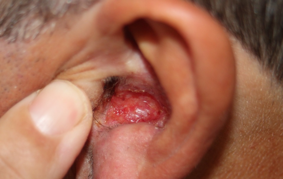
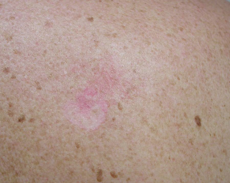
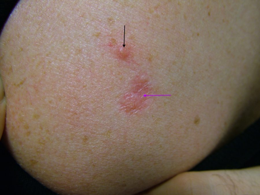
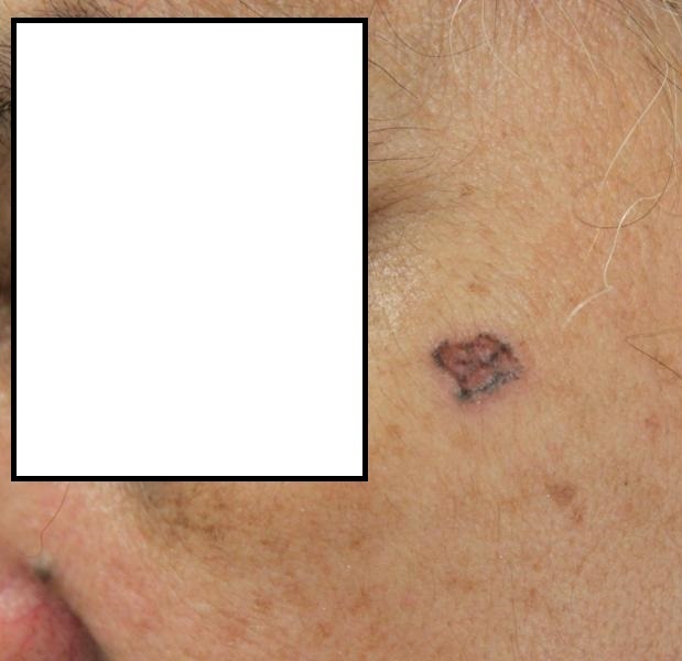
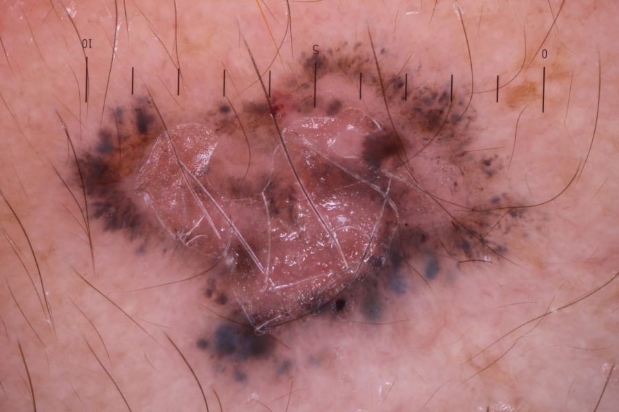
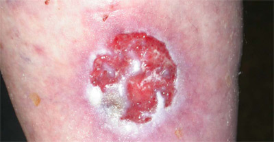
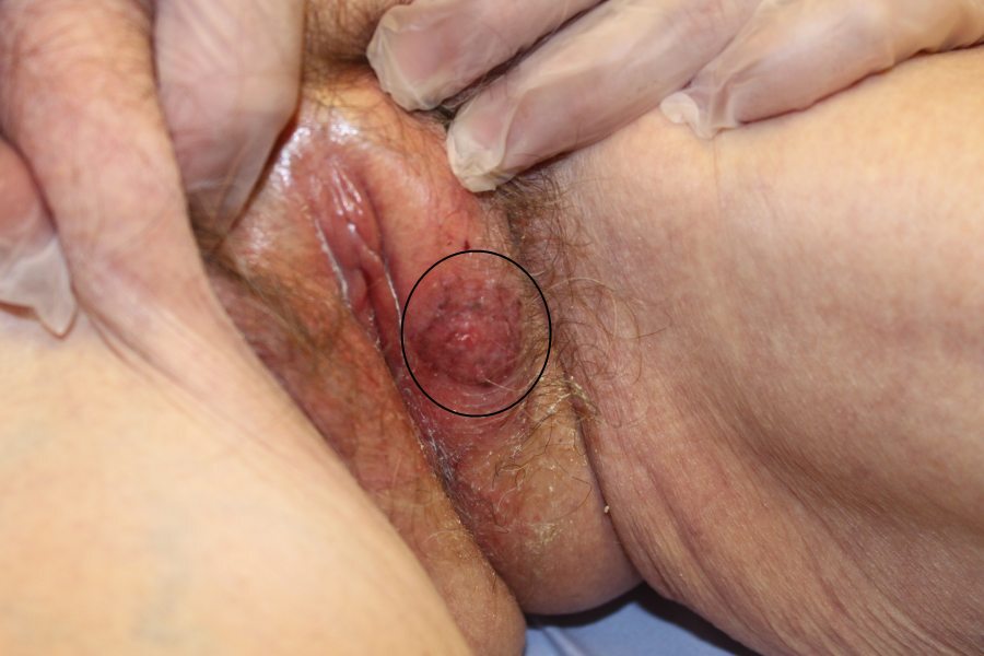
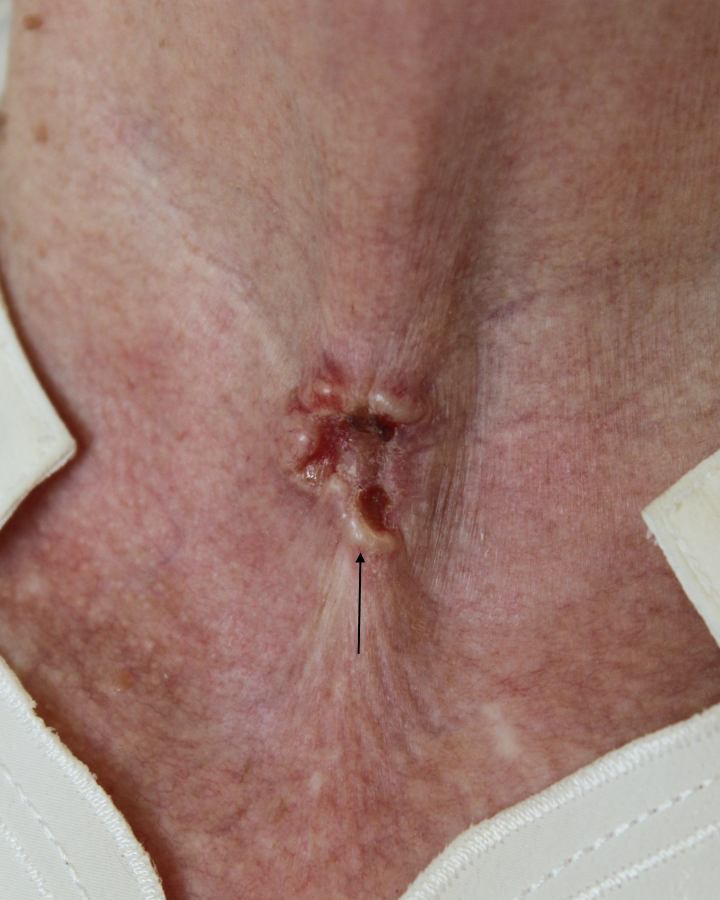
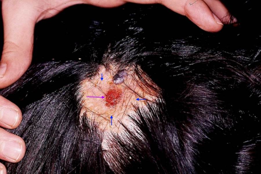
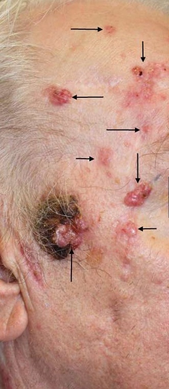
The exact appearance (naked-eye and dermoscopic) depends on the type - refer to the related chapters (top right of page).
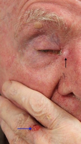
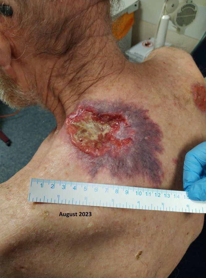
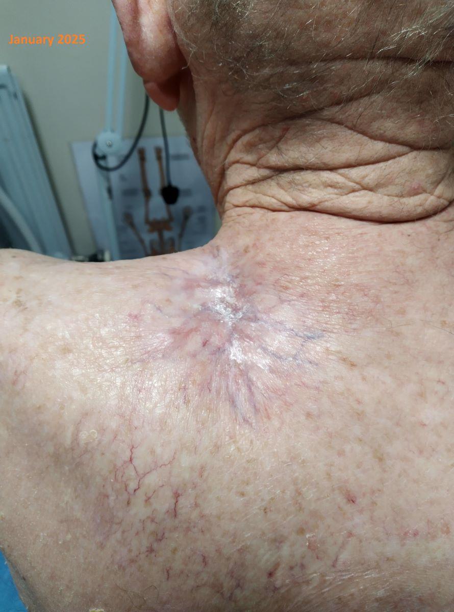
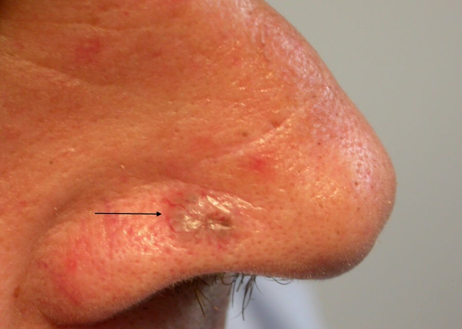
Clinical Images
Please refer to notes on image rights at bottom of the page with regards to individual image
ownership.
1
2
3
4
5
6
7
8
9
10
11
12
13
14
15
16
17
18
19
20
21
22
23
24
Investigations
- Lesions reported histologically as having a squamous component (basosquamous) should be managed as a squamous cell carcinoma
Management
General
Surgical treatment
- Is first-line for the majority of BCC, except for superficial BCC (refer to the related chapter). Just occasionally a patient may not be fit for surgery, but if on a high-risk site the case will still need to be discussed with a local consultant who is part of the skin cancer MDT, as other options such as skin radiotherapy or medical treatment may be recommended
- In standard skin surgery lesions should be excised with a 4 mm margin of normal surrounding skin. Some high-risk lesions are managed in Secondary Care with Mohs surgery (Mohs micrographic surgery), a very time consuming, yet accurate and tissue sparing technique (not available in all localities)
Who can manage BCC (England and Wales)?
- Diagnostic uncertainty - the patient should be referred to either a nationally accredited GPwER in Dermatology or a Secondary Care department skilled in dermoscopy
- Diagnostic certainty in low-risk BCC beneath the clavicle - if the lesion is not on the exclusion list (see below) the patient can be managed by nationally accredited Group 2 and 3 GPwERs, or, GPs meeting the requirements to perform skin surgery within the framework of the Direct Enhanced Services and Local Enhanced Services under General Medical Services or Personal Medical Services
- Diagnostic certainty in low-risk head and neck lesions - if the lesion is not in the exclusion list (see below) the patient can be managed by some nationally accredited Group 2 and 3 GPwERs (check locally), but not GPs
- High-risk BCC should be managed in Secondary Care, and only by individuals with appropriate surgical skills (plastic surgeons, some dermatologists, and in some regions the departments of Oral and Maxillofacial Surgery and Oculoplastics)
High-risk factors warranting referral to Secondary Care (exclusion list for Primary Care)
The patient
- Aged 24 years or under
- Immunosuppressed (especially renal and other transplants) or have Gorlin syndrome
All head and neck lesions that are any of:
- Poorly-defined
- > 1 cm diameter
- On/bordering the nose and lips (including nasofacial sulci and nasolabial folds), ears, or around the eyes (periorbital)
- Over important underlying anatomical structures (for example, major vessels or nerves)
- Morphoeic, infiltrative or basosquamous in type
All lesions beneath the clavicle that are any of:
- At a site where primary closure is unlikely to be achievable
- Morphoeic, infiltrative or basosquamous in type
GPs are also discouraged to excise BCC at sites of high cosmetic importance.
For more information on the management of BCC refer to the British Association of Dermatologists Guidelines.
Disclaimer - the author PCDS cannot accept responsibility for any misleading or incorrect statements, and the management of individual patients remains the direct responsibility of the individual doctor. We do however hope that visitors to this site can contact us regarding comments that are considered misleading or incorrect so that we can continue to improve the site.
Image Rights - The PCDS would like to thank Dermatoweb, DermQuest (Galderma), and others who have contributed images. All named individuals and organisations maintain copyright for the relevant images.