Bullous pemphigoid
LAST UPDATED: Aug 10, 2024
Introduction
Bullous pemphigoid is an uncommon blistering condition of the elderly, which often starts with itch and urticated and erythematous lesions. Later, large tense blisters develop on both erythematous and on normal skin and there may be mucosal involvement with blisters and erosions. The blisters are subepidermal. Bullous pemphigoid is the most commonly seen autoimmune blistering disease in the West with a reported incidence in the UK of 4.3 per 100,000 persons per year.
This chapter is set out as follows:
Aetiology
-
Bullous pemphigoid is an immunobullous condition
-
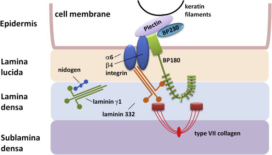
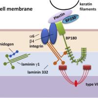
The immunobullous conditions are characterised by pathogenic autoantibodies directed at target antigens whose function is either cell-to-cell adhesion within the epidermis or adhesion of stratified squamous epithelium to the dermis or mesencyme. The target antigens involved are components of desmosomes or the functional unit of the basement membrane zone known as the adhesion complex
-
In bullous pemphigoid the autoantibodies, chiefly IgG, are directed to the basement membrane zone, particularly the bullous pemphigoid antigens BP180 and BP230
-
Drugs - although there are no well-defined causal agents, several drugs have been implicated including furosemide, spironolactone, sulphasalazine, penicillins, beta-blockers, penicillamine, antipsychotics, enoxoparin, DPP-4 inhibitors (Gliptins), and checkpoint inhibitors. Such patients tend to present at a younger average age. Prompt cessation of the drug results in rapid improvement
-
Bullous pemphigoid can occur in association with lichen planus
History
-
Onset is usually after the age of 60 years, with a mean of 80 years. It is extremely rare in children and young adults
-
Males and females are equally affected
-
Itch is a common feature, and may precede the rash by several weeks or months
-
Even in cases of extensive blistering, patients otherwise appear well
Clinical findings
-
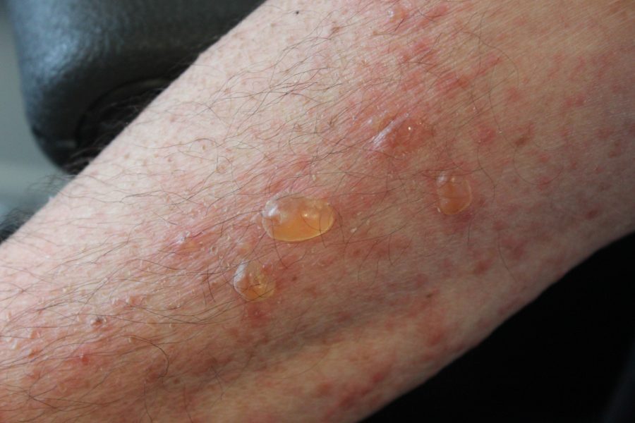
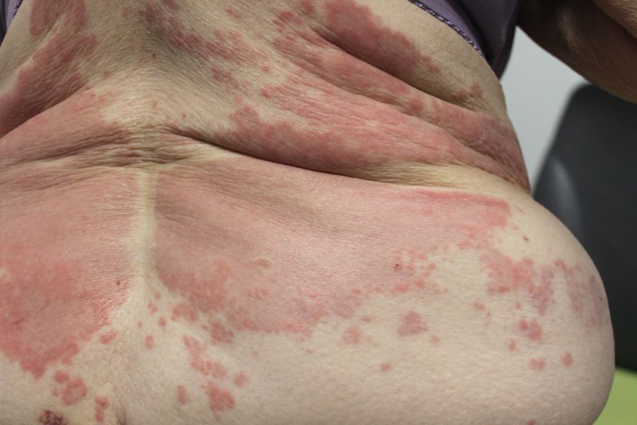
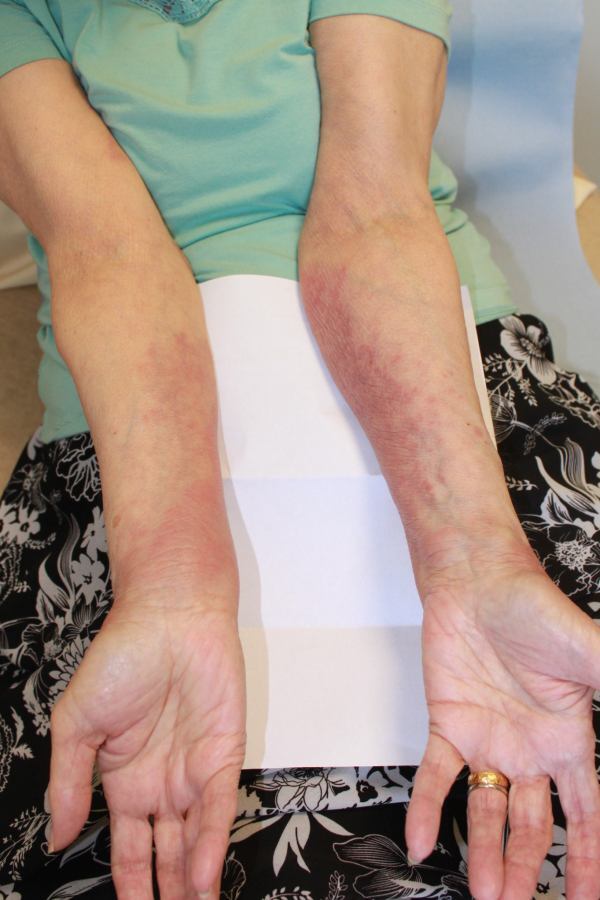
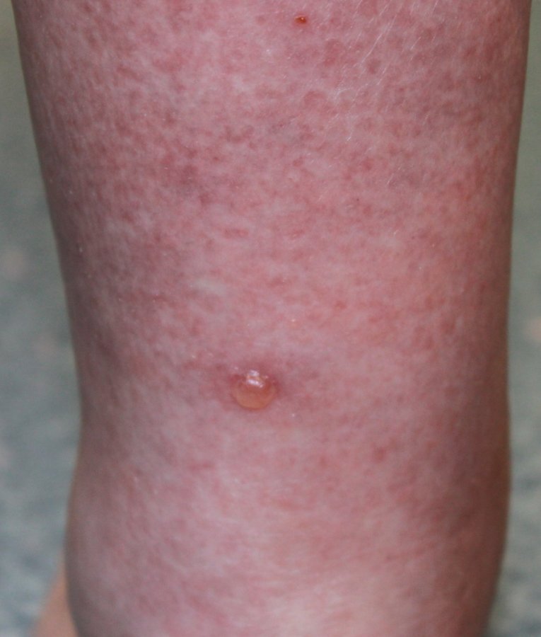
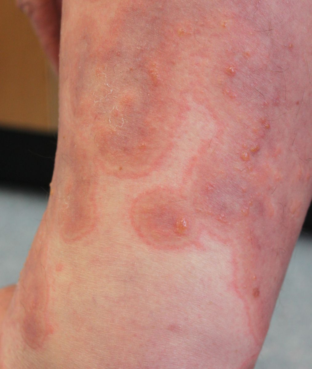
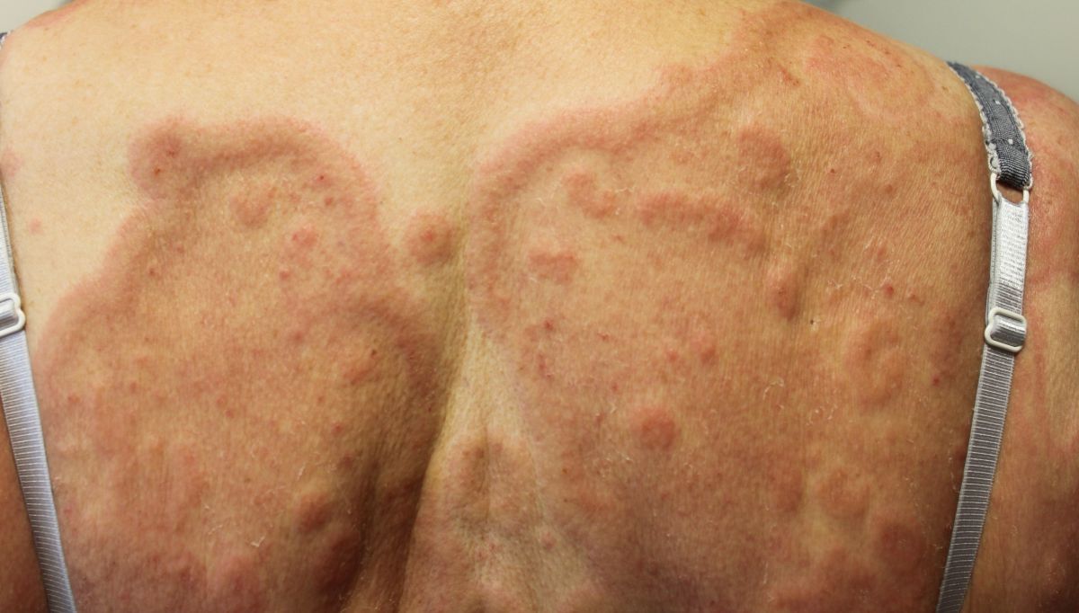
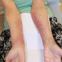
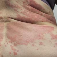
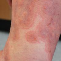
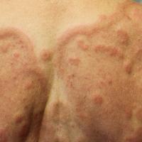
Bullous pemphigoid commonly starts with itching and fixed urticarial-like lesions or occasionally an eczematous rash, which predominantly affects the limbs
-
The itch may precede the rash for several months
-
An urticarial prodrome usually lasts 1-3 weeks before the blisters arise, whereas an eczematous prodrome may precede the blisters by several months
-
Distribution of blisters
-
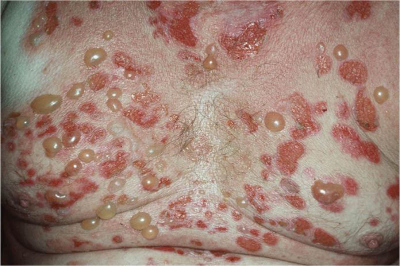
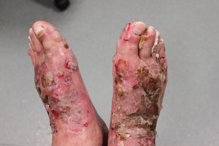
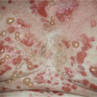
Generalised bullous pemphigoid is the most common form, blisters can form anywhere and are especially widespread on the trunk, proximal limbs and flexures. Once blisters arise they can become widespread within a matter of days
-
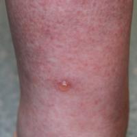
Localised bullous pemphigoid is less common, and is usually limited to the lower extremities. Occasionally this may progress to generalised disease
-
In infants lesions are often acral and in older children involvement of the genital region occurs in almost half
-
Morphology of blisters
-
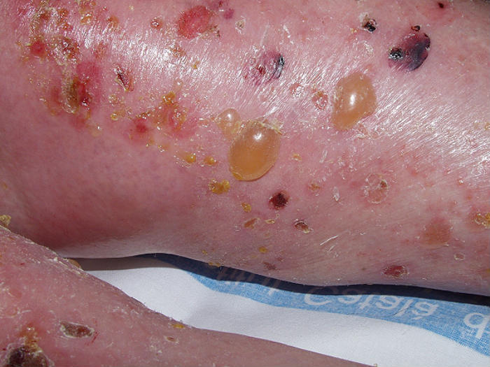
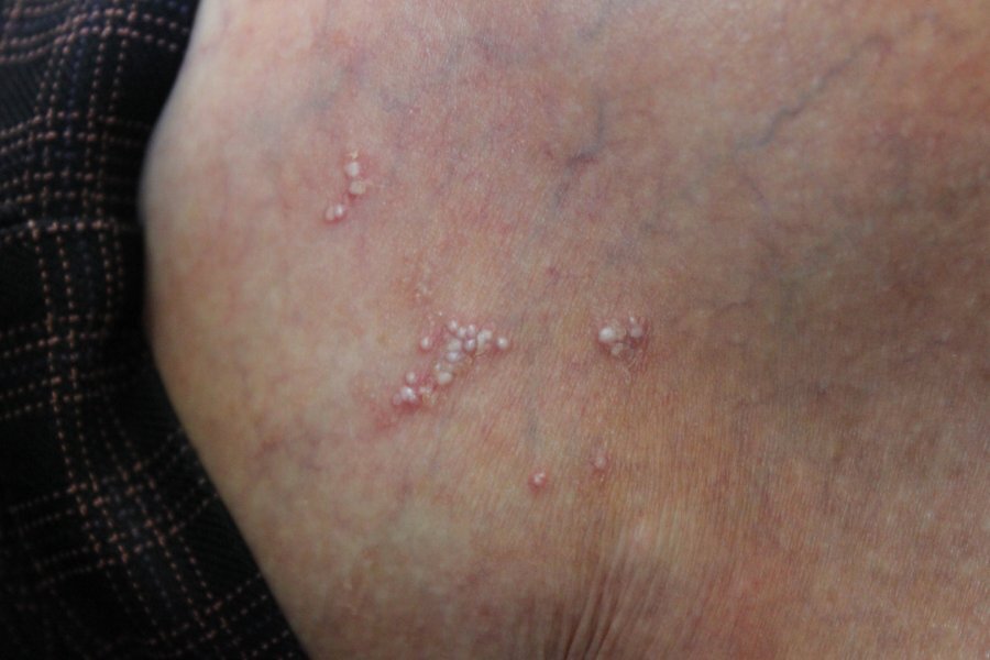
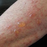
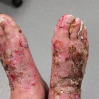
Crops of large, tense, fluid-filled blisters, which may arise from normal-looking or erythematous skin
-
They are generally clear but can be cloudy or blood-stained
-
Blisters may remain intact for several days
-
Lesions heal quickly and without scarring
-
Post-inflammatory hyperpigmentation and / or milia may follow, but these usually resolve within a few months
-
Other body sites
-
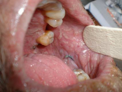
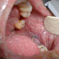
Mucosal involvement with small blisters is uncommon, and tends not to be clinically significant when it does occur. The blisters mainly affect the palate of the mouth
Clinical Images
Please refer to notes on image rights at bottom of the page with regards to individual image
ownership.
1
2
3
4
5

6
7
8

9
10
11
12
13
14
15
Investigations
Bloods tests - indirect immunofluorescence
-
A request for skin antibodies covers both pemphigoid (skin basement membrane) and pemphigus (skin desmosome) antibodies. In bullous pemphigoid, circulating pemphigoid antibodies are present in approximately three-quarters of patients
Skin biopsies - histology and direct immunofluorescence
-
Two skin biopsies are required
-
An intact blister should be excised and sent for histology
-
A second biopsy, of peri-lesional skin (within two cm of the blister), is also required for direct immunofluorescence (DIF). The sample must be put on top of a piece of plain gauze, which has been soaked in a small amount of normal saline, and placed into a dry pot, the specimen must be examined the same day. If the sample cannot be examined the same day it must be placed in a suitable transport media eg Michel's solution, in order to preserve the sample. The result of DIF can sometimes be false negative, and if needed a further biopsy sample can be taken from unaffected skin of the buttocks or thighs
-
Findings in bullous pemphigoid (reference: Rook's Textbook of Dermatology)
-
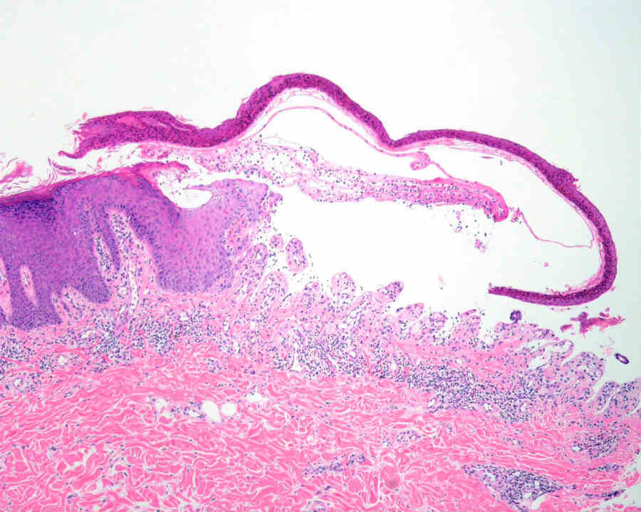
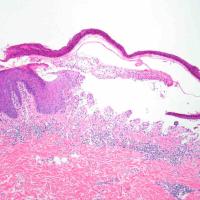
Histology - the blister is subepidermal and contains fibrin and large numbers of inflammatory cells including eosinophils, which may be very dense in areas and form small abscesses in the superficial dermis. The epidermis should be intact
-
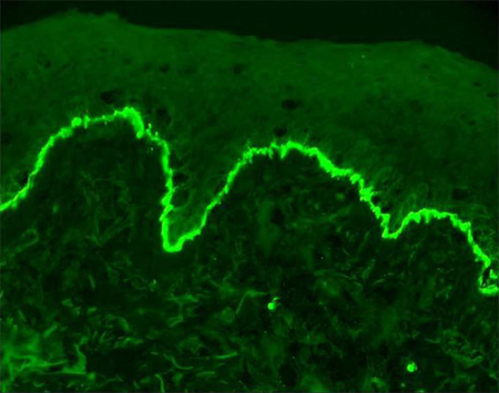
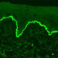
DIF shows linear deposition of IgG (most often IgG4 subtype) and C3 to BP180 and BP230 along the basement membrane. Deposition of IgA and IgM may also be seen
-
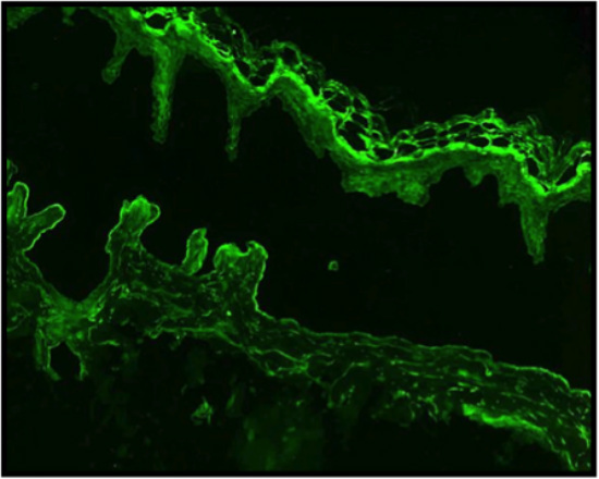
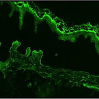
The use of salt-split skin immunofluorescence studies can be useful in differentiating between bullous pemphigoid and epidermolysis bullosa acquisita - the skin biopsy sample is placed in 1 mol/L salt prior to performing the immunofluorescence studies, which causes cleavage through the lamina lucida. Examination reveals IgG on the blister roof (epidermal side of split skin) in patients with bullous pemphigoid, while, in epidermolysis bullosa acquisita the IgG localises to the blister floor (dermal side of split skin)
Management
Step 1: general measures
-
The vast majority of patients need urgent referral to Secondary Care, and those with widespread blistering may require admission. A few patients, eg those with localised disease, may be suitable for management in Primary Care
-
Provide a patient information leaflet on pemphigoid
-
Supportive measures include wound care and reducing the risk of secondary infection by using antiseptic regimes, eg Dermol 500 lotion ® as a wash and / or topical emollient
Step 2: super-potent topical steroids
-
Dermovate ® cream (clobetasol) is a very effective treatment for localised disease. It can also be tried BD in more extensive disease, as the side effects are very much less then when compared to systemic steroids
Step 3: other treatments for localised bullous pemphigoid / other mild-moderate cases
-
Doxycycline 100 mg BD
- There is also some evidence for nicotinamide
Step 4: systemic steroids for moderate-severe bullous pemphigoid
-
Systemic steroids are the mainstay of treatment. The initial dose is as follows:
-
20 mg / day in localised-mild disease
-
40 mg / day in moderate disease
-
50-70 mg / day in severe disease
-
The dose of steroid can usually be reduced over a matter of weeks to 15-20 mg / day, and then more slowly after that. It is felt prudent to reduce the daily prednisolone dose by 1 mg / month once the dose is below 10 mg / day, if blisters start to break through the dose can be increased, and then after a period of stabilisation gradually reduced again. The additional use of Dermovate ® cream may enable a lower dose of prednisolone
-
Adverse effects
-
In addition to providing gastric protection with a PPI, treatment to help prevent osteoporosis should be initiated at an early stage
-
Monitor BP and glucose levels
Step 5: other treatments for moderate-severe bullous pemphigoid
-
Immunosuppressive drugs such as methotrexate and mycophenolate mofetil are generally only considered if the steroid dose cannot be reduced to an acceptable level
-
The role of biologic therapies remains unclear
Prognosis
-
Bullous pemphigoid is a serious condition, with an average duration of 3-6 years. It is occasionally fatal. Factors associated with a worse prognosis include generalised disease, low albumin, and high dose steroids
-
In contrast, localised bullous pemphigoid has a very good prognosis
Other resources
Disclaimer - the author PCDS cannot accept responsibility for any misleading or incorrect statements, and the management of individual patients remains the direct responsibility of the individual doctor. We do however hope that visitors to this site can contact us regarding comments that are considered misleading or incorrect so that we can continue to improve the site.
Image Rights - The PCDS would like to thank Dermatoweb, DermQuest (Galderma), and others who have contributed images. All named individuals and organisations maintain copyright for the relevant images.