Calcification of the skin and subcutaneous tissues (syn. calcinosis cutis)

LAST UPDATED: Jun 08, 2021

Introduction

Calcification occurs in the skin and subcutaneous tissues either as a consequence of the alteration of systemic calcium homeostasis or as a local side effect of inflammation, infection, trauma or neoplastic disease. In its most benign form, calcification is noticed as an incidental finding on histopathological analysis. The more severe forms, seen in scleroderma, dermatomyositis and calciphylaxis, lead to significant morbidity and mortality.

This chapter, which is set out as below, provides a brief overview of the various conditions that cause calcification.


Clinical findings

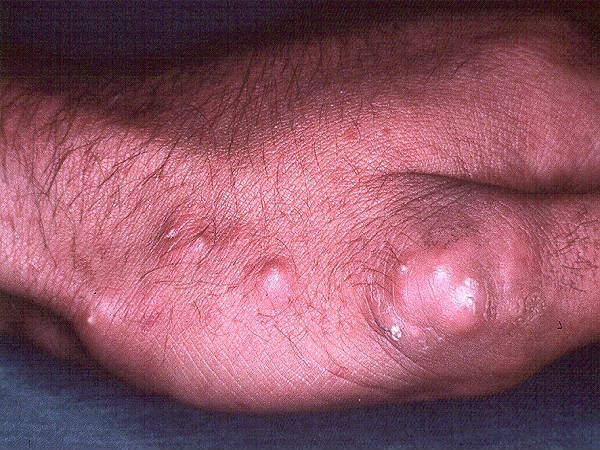
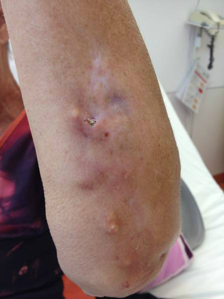
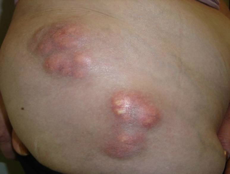
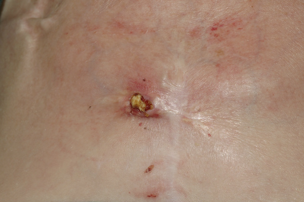
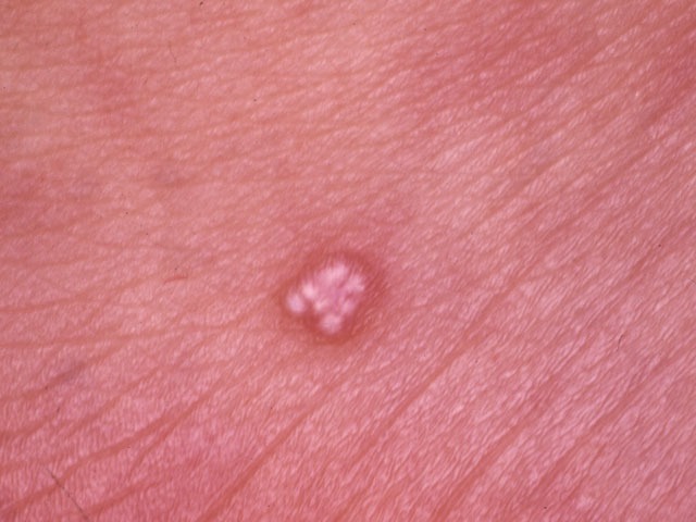
The signs and symptoms of calcinosis cutis vary according to the underlying cause. In many cases the lesions gradually develop and are often symptomless. The lesions usually appear as firm, white-yellow papules, plaques or nodules. A solitary lesion may develop, although multiple lesions are more common. Lesions may become tender and ulcerate, discharging chalk-like creamy material. Fingertip lesions may be painful, while lesions at other sites may restrict joint mobility and limit movement due to stiffening of the skin. In severe cases tissue necrosis may occur.

The different processes involved in calcification of the skin and subcutaneous tissues are as follows: 

  • Calcinosis may develop at a site of tissue damage, a process known as dystrophic calcification. This process is probably a consequence of cell death, with the release of alkaline phosphatase, intracellular calcium and an alteration of local pH resulting in the precipitation of calcium
     
  • Calcification and ossification of tumours - the best recognised tumour is the pilomatricoma, which calcifies in 75% of cases and is readily identified as a mobile, hard, subcutaneous papule or nodule
     
  • Idiopathic (ie unknown):
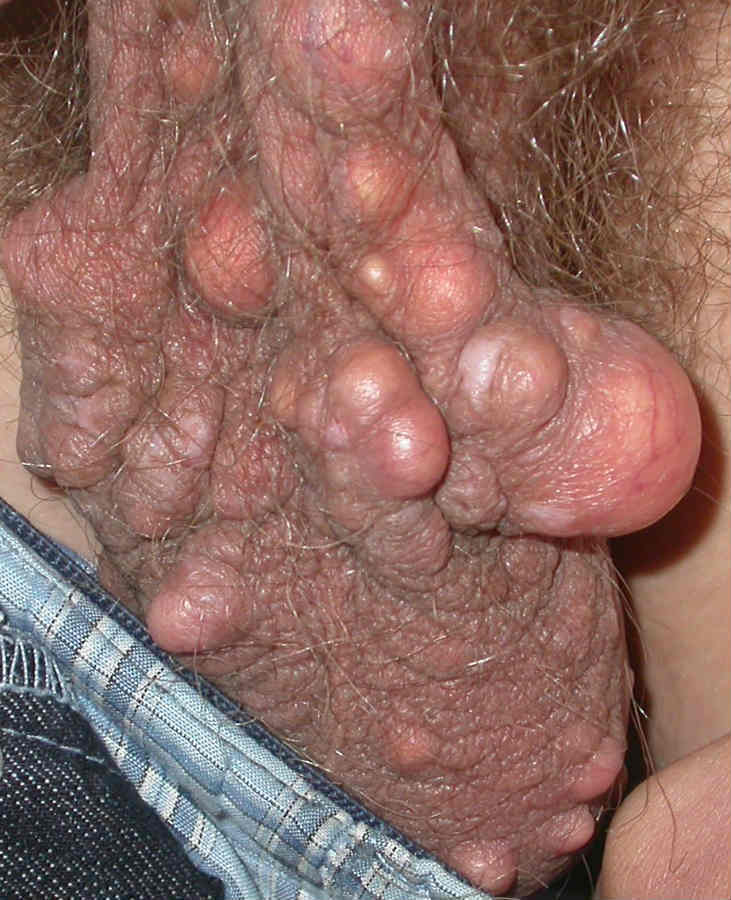
    • Scrotal calcinosis is the best recognised of these conditions presenting in men aged 20-40 years, as multiple, white-yellow, firm, subcutaneous nodules, sitting within the scrotal skin
    • Other examples include subepidermal calcified nodules, tumoral calcinosis (large subcutaneous masses around the major joints, most usually seen in adolescents) and miliary calcinosis cutis 
       
  • Inflammatory, particularly autoimmune, processes in the skin may result in calcification. Dermatomyositis and scleroderma (especially the CREST syndrome - calcinosis, Raynaud's phenomenon, oesophageal dysfunction, sclerodactyly and telangiectasia) are the best recognised and most troublesome. Calcinosis has been described in several variants of lupus erythematosus, particularly lupus panniculitis and, in contrast, is often asymptomatic
     
  • Metastatic calcification results from an underlying disorder of systemic calcium homeostasis, and is seen in hyperparathyroidism, hypervitaminosis D, milk alkali syndrome, sarcoidosis and bone metastases. It presents with predominantly periarticular calcification in addition to signs and symptoms of hypercalcaemia (eg abdominal pain, renal failure, cardiac arrhythmias and depression)
     
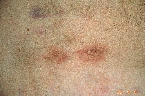
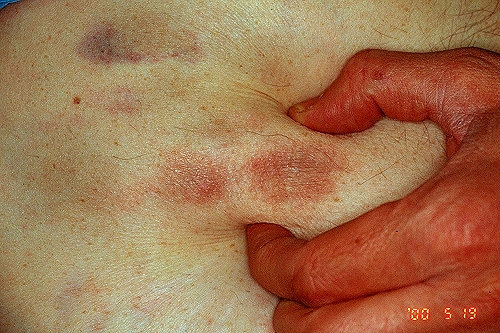
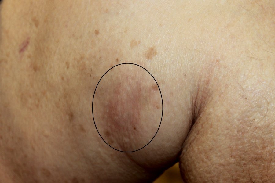
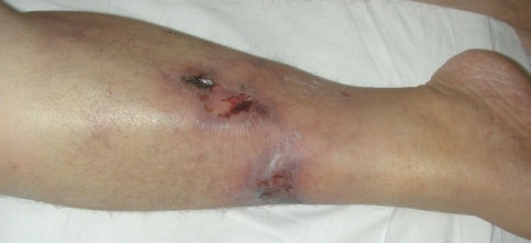
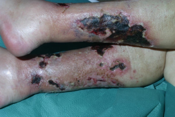
  • Calciphylaxis remains a highly morbid and poorly understood condition of vascular calcification and tissue necrosis, most frequently of the skin, but also affecting other organs such as the heart and gastrointestinal tract. While most frequently described in patients with end-stage renal disease (1-4% of this population), calciphylaxis is seen in other settings, such as multiple myeloma, polyneuropathy, organomegaly, endocrinopathy, cirrhosis, and rheumatoid arthritis. Clinically, nodules or plaques tend to progress quickly to livedo-like areas and then ulcerate. The thighs, abdomen and buttocks are the most commonly affected sites. Ulceration is associated with severe pain. In patients with renal failure, lesions on the trunk are associated with a much higher mortality rate than lesions on the legs

Clinical Images

Please refer to notes on image rights at bottom of the page with regards to individual image ownership.
Investigations

  • As well as serological assessment for connective tissue disease, full biochemical assessment is required to exclude any abnormality of systemic calcium homeostasis - this should include serum calcium, phosphate, alkaline phosphatase, vitamin D and parathyroid hormone (PTH) estimation

Management

  • The management depends on the underlying cause 

Disclaimer - the author PCDS cannot accept responsibility for any misleading or incorrect statements, and the management of individual patients remains the direct responsibility of the individual doctor. We do however hope that visitors to this site can contact us regarding comments that are considered misleading or incorrect so that we can continue to improve the site.

Image Rights - The PCDS would like to thank Dermatoweb, DermQuest (Galderma), and others who have contributed images. All named individuals and organisations maintain copyright for the relevant images.

Quick Links

The following pharmaceutical companies have had no involvement in the content of this website or in our conference programmes


Almirall
Galderma
Glenmark
Johnson & Johnson
La Roche-Posay
LEO Pharma
Pierre Fabre
Schuco