Erythema multiforme
LAST UPDATED: Nov 16, 2021
Introduction
Erythema multiforme (EM) is a hypersensitivity reaction usually triggered by infections, most commonly herpes simplex virus. Clinically, EM is characterised by macular, papular or urticated lesions, as well as the classical 'target lesions' distributed preferentially on the distal extremities. Mucosal surfaces may be involved.
EM is self-limiting, usually resolving without complications, and is now regarded as distinct from Stevens-Johnson syndrome and toxic epidermal necrolysis.
This chapter is set out as follows:
Aetiology
-
EM appears to be caused by an immunological reaction to numerous triggers, although in up to 50% of cases no provoking factor is identified
-
Herpes simplex virus (HSV type 1 is more commonly associated than type 2) is the most common cause, and usually precedes the skin eruption by 3-14 days, although the infection can be subclinical
-
Mycoplasma pneumonia is the next most common trigger
-
There are many other identifiable triggers including:
-
Viral infections eg the parapoxvirus (orf, milkers' nodules), varicella (chickenpox, shingles), Hepatitis B
-
Bacterial infections eg Rickettsia
-
Fungal infections eg tinea
-
Drugs - probably account for less than 10% of EM cases. While many drugs have been implicated, eg antibiotics, NSAID and sulphonamides, too often the evidence is inadequate to be certain if the drug was the cause
History
-
Although EM can affect any age group, including neonates, it arises most commonly in young adults
-
Prodromal symptoms are usually absent or mild
-
EM is not usually itchy
Clinical findings
Typical EM (sometimes referred to as EM minor)
Distribution
-
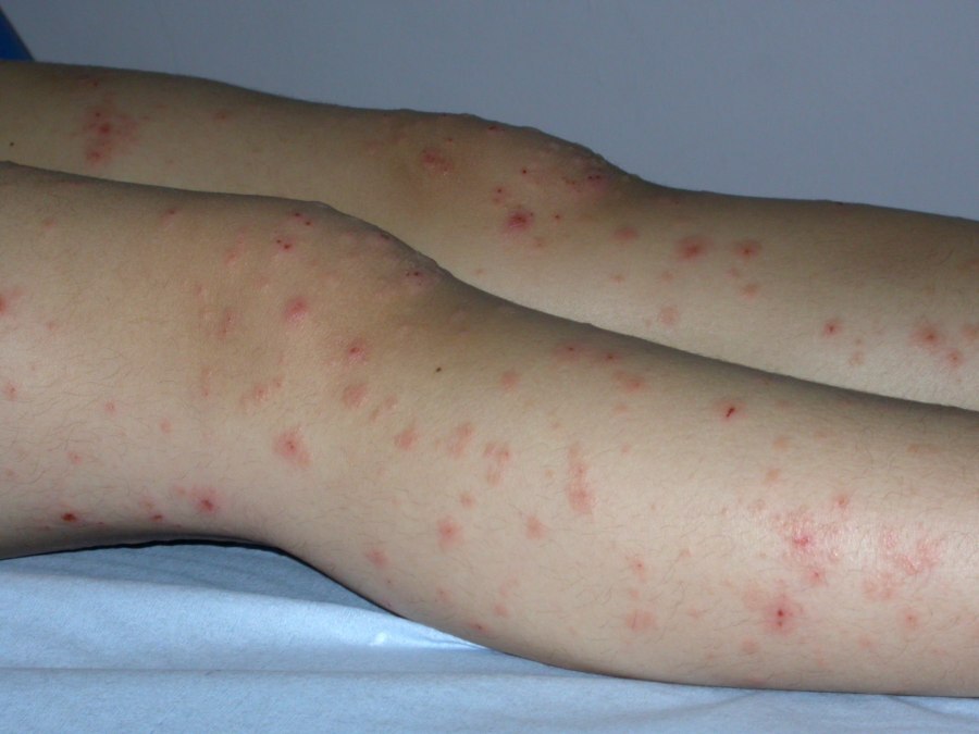
Lesions of EM arise quite abruptly, and tend to do so in successive crops over a few days. There may be few or very many lesions
-
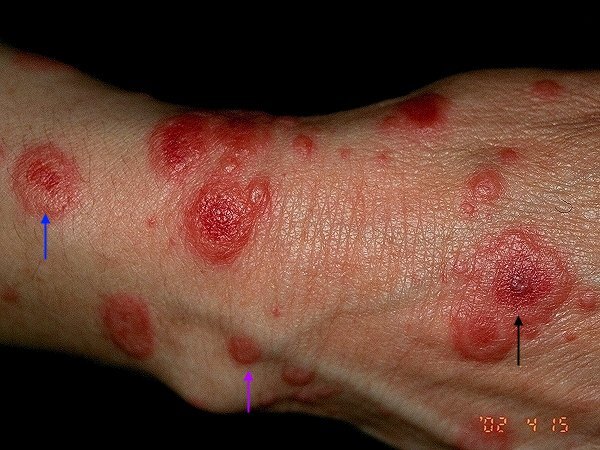
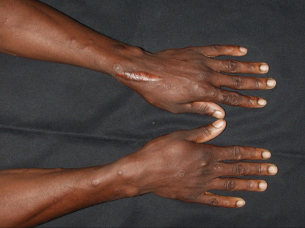
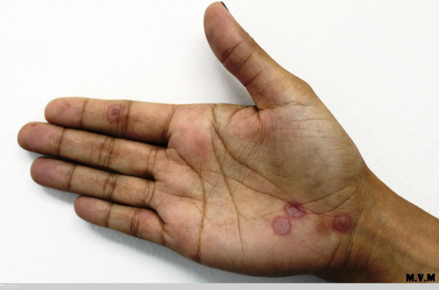
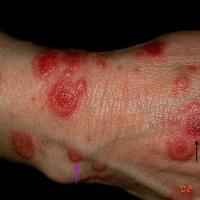
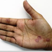
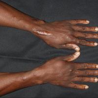
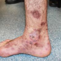
EM tends to arise on the distal extremities, especially the hands, with the upper limbs more commonly affected than the lower limbs. The palms, trunk, and mucosal surfaces may also be affected. The face is less commonly affected
Morphology
-
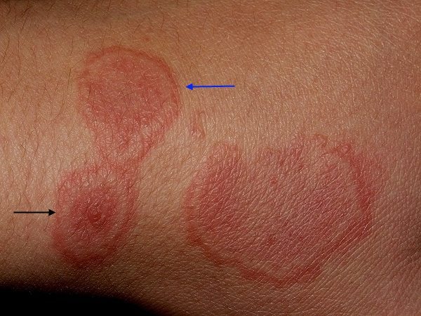
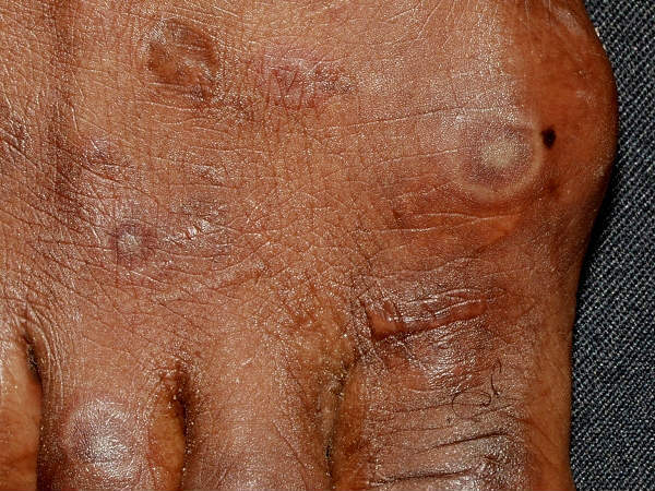
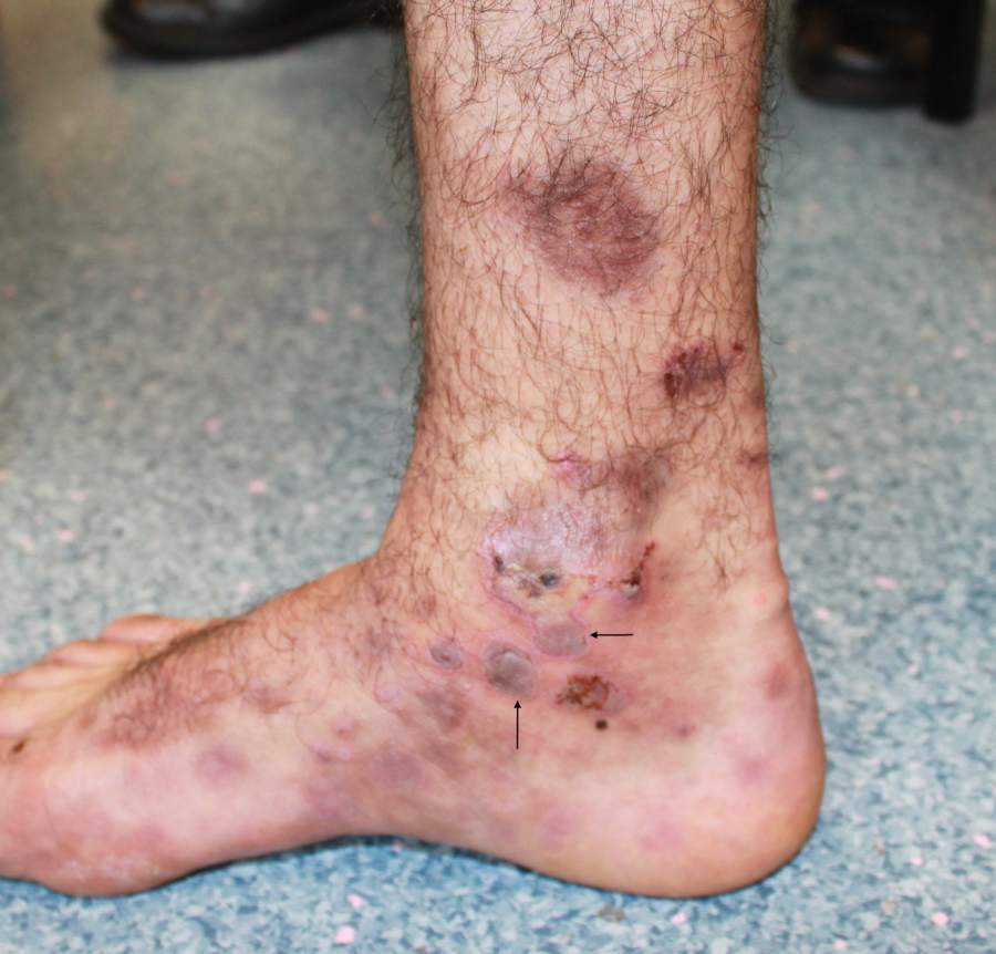
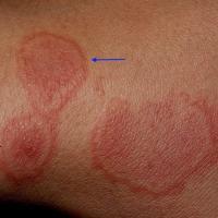
Lesions are initially flat and gradually become raised. They are red-pink, circular and have a well-demarcated edge, measuring 1-3 cm in diameter
-
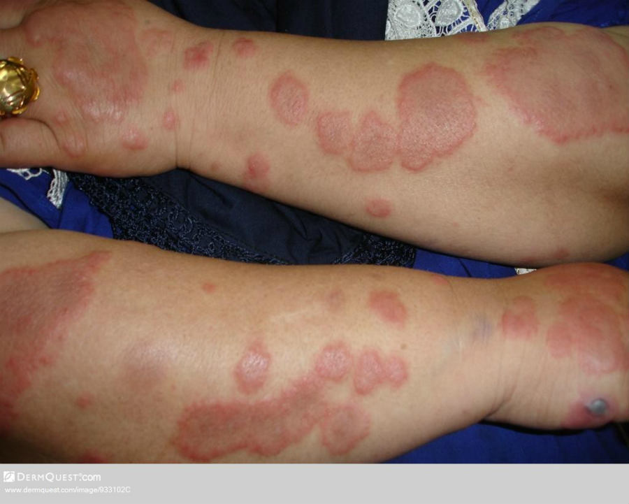
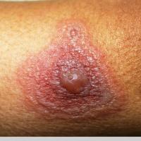
The typical lesion of EM is described as targetoid, or iris, with a central dark red zone and lighter outer zones (lesions can have two or three zones). Lesions may coalesce to develop polycyclic patterns
-
The Koebner phenomenon, with lesions developing at sites of trauma, is a relatively common feature
-
Lesions of EM tend to fade within two weeks
Mucosal lesions in typical cases of EM
-
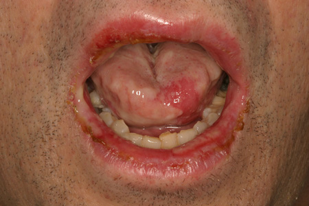
Mucous membrane involvement is mild or absent. If present it is usually limited to one site, which in order of frequency is:
-
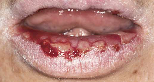
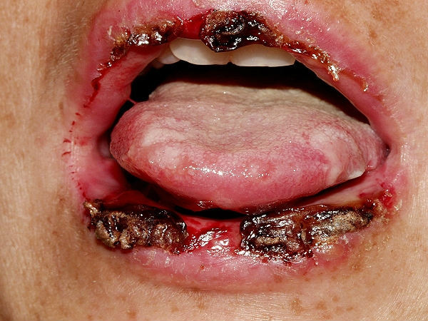
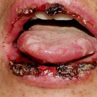
The oropharynx (lips, palate, and gingiva), often with sparing of the gingiva. Lots of small lesions arise, which then coalesce
-
Conjunctivae - eye involvement is usually mild
-
Genitalia - can be severe
-
Anus
-
Tracheobronchial tree
-
Oesophagus
-
Bowel
Vesiculobullous EM
-
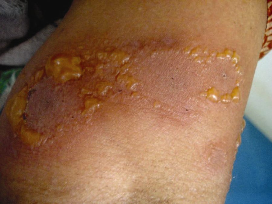
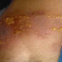
Lesions are fewer in number and predominantly involve acral sites and mucosal surfaces
-
They have a central bullae and may have peripheral vesicles
EM major
-
This is a more serious condition with extensive target lesions, mucous membrane involvement and greater systemic upset
-
More severe erosions-ulcers of at least two mucosal surfaces are seen, with characteristic haemorrhagic crusting of the lips
-
Lesions heal without scaring
Clinical Images
Please refer to notes on image rights at bottom of the page with regards to individual image
ownership.
1
2
3
4
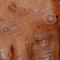
5
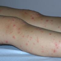
6
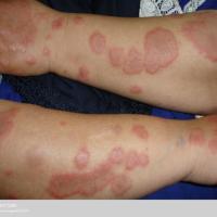
7
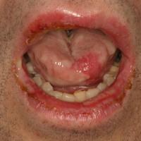
8
9
10
11
12
Investigations
-
For the majority, EM is a clinical diagnosis
-
A biopsy is sometimes required to exclude other conditions
Management
Step 1: general measures
Step 2: eye involvement
-
Usually requires the early help of an ophthalmologist
Step 3: moderate-severe EM
-
Recurrent cases of moderate-severe mucosal lesions - provide a mouth wash, and for those already on prophylactic aciclovir (see step 4), consider prednisolone starting at 40 mg OD and reducing down over 10-14 days
-
In more severe cases, eg EM major, hospital admission is usually required for intensive nursing care. The role of systemic steroids remains controversial
Step 4: prevention
-
Recurrent EM secondary to herpes simplex should be treated with prophylactic oral aciclovir. The standard adult dose is 400 mg BD, in children the dose is 10 mg/kg/day in divided doses. Even in patients where herpes simplex appears absent, if attacks are multiple consider prophylactic aciclovir as some cases of herpes simplex are believed to be subclinical. Patients may require prophylactic treatment for 1-2 years or longer
-
If EM is felt to be secondary to a drug, then it should be withdrawn
Disclaimer - the author PCDS cannot accept responsibility for any misleading or incorrect statements, and the management of individual patients remains the direct responsibility of the individual doctor. We do however hope that visitors to this site can contact us regarding comments that are considered misleading or incorrect so that we can continue to improve the site.
Image Rights - The PCDS would like to thank Dermatoweb, DermQuest (Galderma), and others who have contributed images. All named individuals and organisations maintain copyright for the relevant images.