Leg ulcers (and disorders of venous insufficiency)
LAST UPDATED: Oct 27, 2024
Acknowledgements: This chapter was developed by Dr Brian Malcolm (a GP principal, trainer and Associate Specialist in Dermatology, in Barnstaple, North Devon), and further enhanced by Geraldine Bellerby (dermatology nurse specialist at the Middlesbrough Primary Care Skin Service).
Introduction
Leg ulcers are of huge socio-economic importance. Approximately over 500,000 people live with a form of venous ulceration in the UK, this has doubled in the last 10 years. A leg ulcer is not a diagnosis; it is a manifestation of an underlying disease process and so the concept should be of the patient with the leg ulcer.
This chapter discusses leg ulcers, along with disorders of venous insufficiency such as lipodermatosclerosis, and is set out as follows:
Aetiology
A working definition of a leg ulcer would be an area of epidermal discontinuity lasting in excess of two weeks:
-
70-80% of leg ulcers are venous
-
10-20% are of mixed venous / arterial
-
10% are arterial
-
A small number are related to other causes
Venous ulcers
-
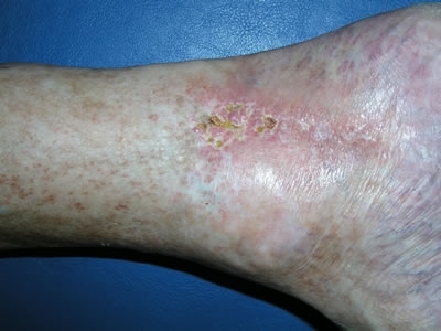
The pathogenesis of venous ulceration is not completely understood but is generally agreed to be the end result of a combination of chronic venous hypertension resulting from venous incompetence and calf muscle insufficiency evidenced by the tissue changes of induration, haemosiderin deposition, fibrosis and reticulate scarring (atrophie blanche) collectively known as lipodermatosclerosis
Arterial ulcers
-
Arterial ulceration is due to a reduced arterial blood supply to the lower limb. The most common cause is atherosclerotic disease of the medium and large sized arteries. Other causes include diabetes, thromboangiitis, vasculitis, pyoderma gangrenosum, thalassaemia, and sickle cell disease, some of which may predispose to the formation of atheroma. Further damage to the arterial system occurs with concurrent hypertension through damage of the intimal layer of the artery. The reduction in arterial blood supply results in tissue hypoxia and tissue damage. Thrombotic and atheroembolic episodes may contribute to tissue damage and ulcer formation. Arterial ulcers cannot be treated by lower leg compression
Ulcers of mixed aetiology
-
Ulceration of mixed aetiology is not uncommon: patients may have a combination of venous and arterial diseases, resulting in ulcers of mixed aetiologies, which will limit the degree of compression (if any) that can be used to treat the ulcers
Neuropathic ulcers and diabetic patients
-
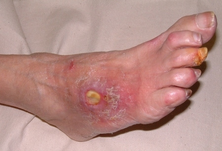
Nerve damage, most commonly due to diabetes, leads to altered or complete loss of sensation in the foot and/or leg. Pressure from shoes, and bandages, or injury to the foot, may subsequently go unnoticed and eventually leads to the development of a neuropathic ulcer. In diabetic patients the picture is often further complicated by damage to the small blood vessels feeding the skin (microangiopathy), giving an additional arterial component to the ulcer
Other causes of leg ulcers
-
There are many other causes of legs ulcers including those secondary to vasculitis, and rheumatoid disease, the latter group have a multifactorial aetiology and are particularly difficult to manage
History
-
A full medical history needs to be obtained with particular respect given to co-morbid pathologies, and social factors such as smoking, nutrition, and dehydration
-
The history must include medications, as drugs such as beta-blocker, steroids, non-steroidal anti-inflammatories (NSAID) and immunosuppressive agents can all adversely influence ulcer healing. Nicorandil can both cause ulcers and delay healing
-
A history of allergy eg to neomycin or lanolin is also important when the choice of appropriate dressings and topical agents is being considered
Clinical findings
Venous disease
The spectrum of skin changes with venous insufficiency includes the following:
-
Varicose veins - assess the whole leg up to the groin, whilst the patient is standing
-
Varicose eczema - caused by irritation from blood by-products that have leaked into the skin
-
Venous oedema - tends to be pitting and to go down when the patient elevates their legs at night, may be unilateral. Should not be confused with cardiac or renal oedema which are also pitting, but will always affect both legs
-
Ankle flare - tiny varicose veins on the inner aspect of the ankle
-
Atrophy blanche - venous congestion causes swollen congested capillaries, sometimes visible as tiny red “dots” under the skin. Where the capillaries cannot sustain this pressure they atrophy, leaving white “lacy” areas of avascular tissue
-
Haemosiderin staining - a red / brown discolouration caused by leakage of haemoglobin from the engorged capillaries into the skin
- Hyperkeratosis - thickening of skin
-
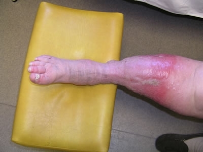
Lipodermatosclerosis - typically arises in patients with venous insufficiency. Obesity is a common factor. It can present in two ways:
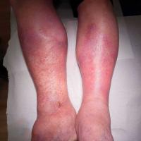
- Sclerosing panniculitis - this is the acute form characterised by painful inflammation of the lower legs, above the ankles, resembling cellulitis. It can affect one or both legs
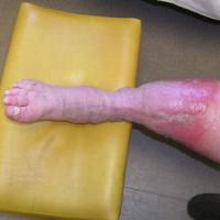
- Chronic lipodermatosclerosis - may follow an acute episode or develop gradually. Features of chronic lipodermatosclerosis include pain, hardening of the skin, colour change - red / brown and atrophie blanche. Over time a hard layer of fibrosis tissue develops below the skin’s surface caused by leakage of fibrin from the engorged capillaries, which can be localised around the ulcer, but often collects around the ankle preventing it from swelling and instead causing oedema to collect in the calf giving rise to the ''inverted champagne bottle” or “bowling pin” appearance. Lipodermatosclerosis tends to be painful and can prevent patients from tolerating compression therapy. Management may require the use of potent or superpotent topical steroids to reduce the inflammation before the gradual introduction of compression therapy along with adequate analgesia
- Venous ulcers
- Slow onset
- Pain in legs and ulcers - worse when first put leg out of bed in the morning
- Throbbing, aching heavy feeling in legs - improves with elevation and rest
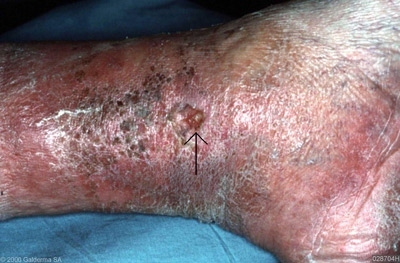
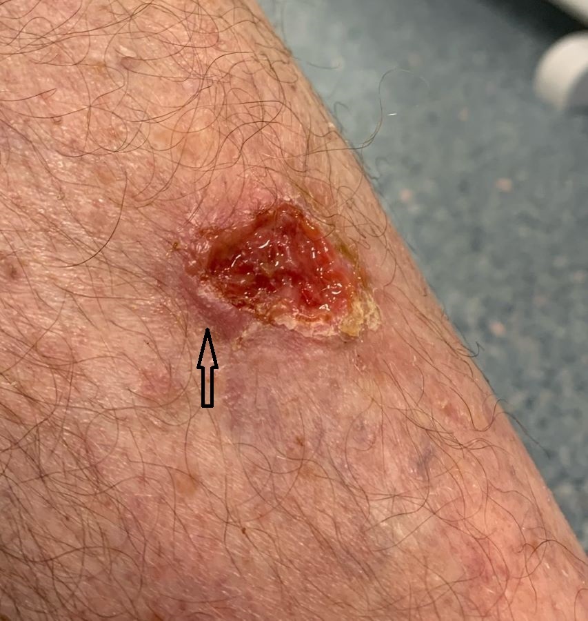
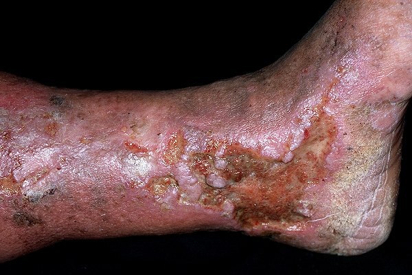
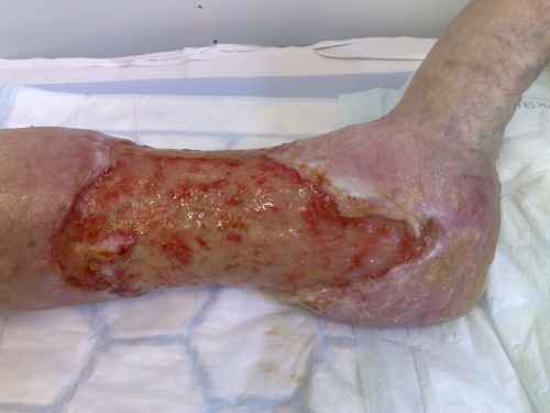
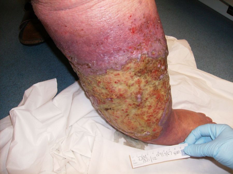
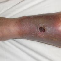
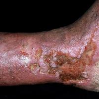
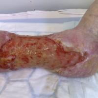
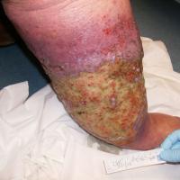
- Typically affect the medial aspect of gaiter region (ankle to mid-calf)
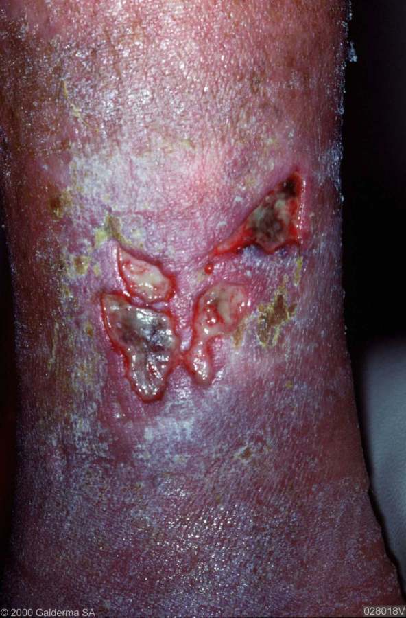
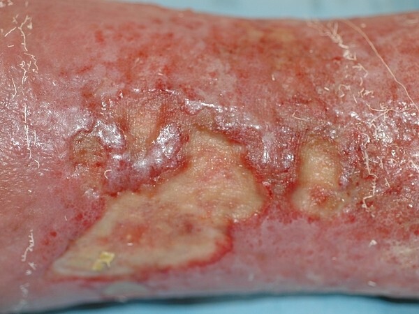
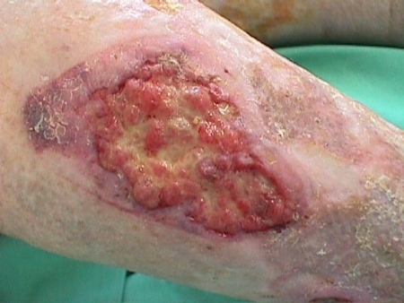
- Often large, shallow, and irregular in shape with ill-defined edges
- Usually have slough at the base with granulation tissue
- Moderate-heavy exudate
Arterial disease
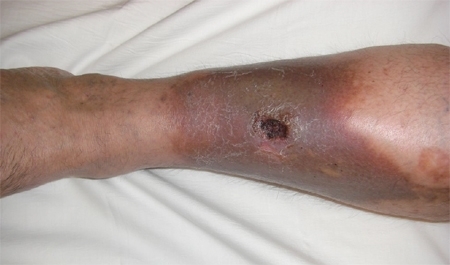
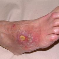
- Usually on the foot or the lateral aspect of the leg, but may occur anywhere on the lower limb
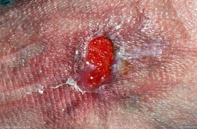
- Punched out appearance with well-defined edges
- Often small in diameter; deeper than venous ulcers and very painful (unless co-existent neuropathy)
- Unhealthy appearance of wound bed
- Presence of necrotic tissue or fixed slough
- Low exudate, unless ulcers infected
- There may also be signs of arterial compromise such as pallor, loss of hair, nail dystrophy, coldness and diminished capillary refill. Pulses may be impalpable
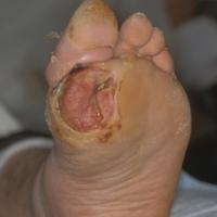
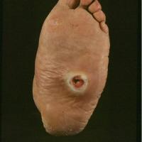
Neuropathic ulcers
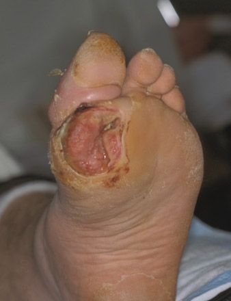
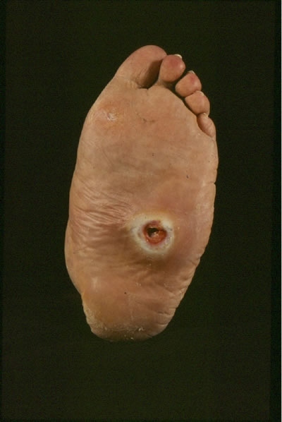
- Develop over pressure areas eg metatarsal heads, sole of foot, balls of toes
- The shape is often irregular, and corresponds to the shape of pressure point that has become exposed
- The edge and base is often clean and may be deep with exposure of bone and tendon
- Neuropathic ulcers may also have features of arterial ulcers if there is co-existent vascular insufficiency
Other causes of leg ulcers
-
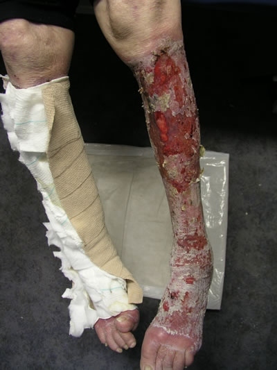
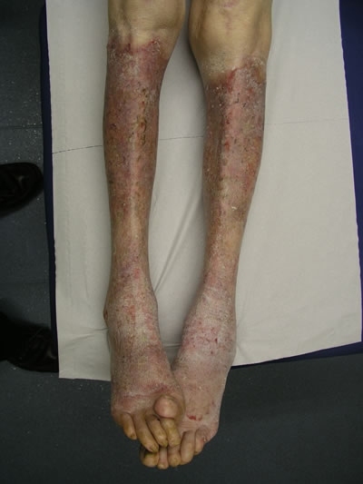
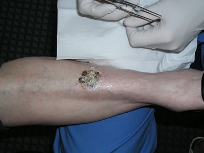
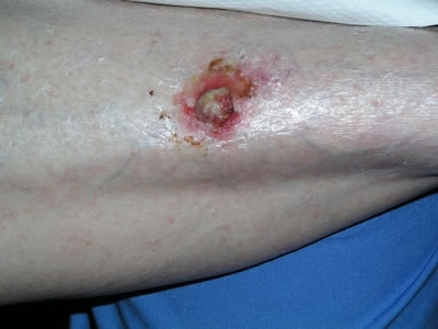
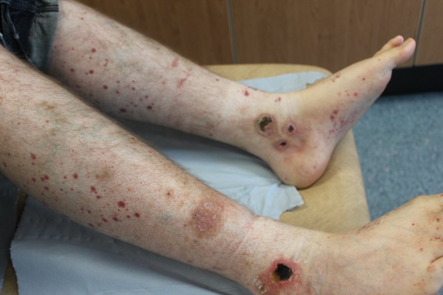
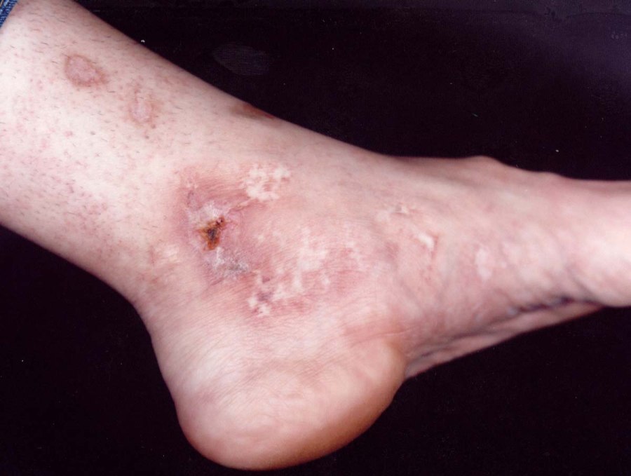
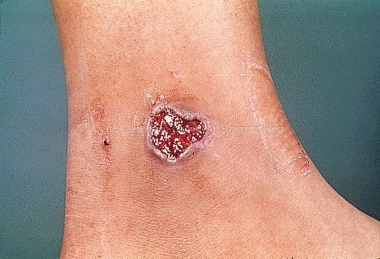
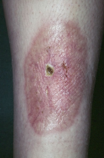
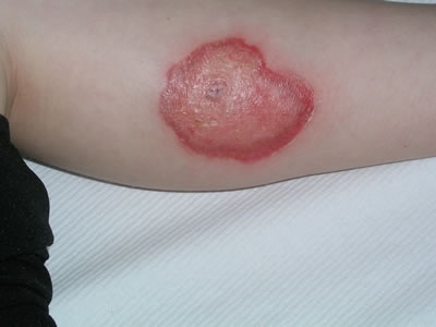
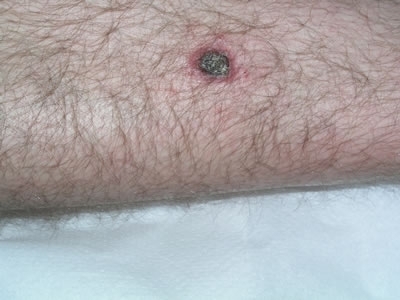
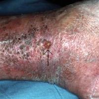
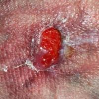
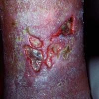
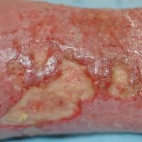
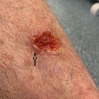
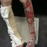
There are many other causes of leg ulcers, please refer to the images at the end of this chapter for more information
Clinical Images
Please refer to notes on image rights at bottom of the page with regards to individual image
ownership.
1
2
3
4
5
6
7
8
9
10
11
12
13
14
15
Management
Treatment
Step 1: general measures
Patients with all forms of ulceration often have very significant co-morbidities such as cardiac failure, hypertension, peripheral vascular disease, diabetes mellitus and connective tissue disease that require a holistic approach if there is to be successful ulcer healing. The cornerstones of wound management are:
-
Define and treat underlying causes
-
If the history or clinical features are atypical exclude other causes of leg ulcers such as skin cancer (refer to the images at the bottom of this page)
-
Check the patient has an up to date FBC, U+E, LFT, TFT, glucose and lipid profile
-
Strict control of factors affecting healing, for example oedema and infection
-
Appropriate dressings
-
Compression therapy where appropriate
-
Long-term maintenance of successful healing
- For venous leg ulcers provide a patient information leaflet
Step 2: dressings
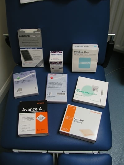
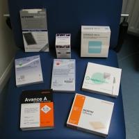
Do not heal wounds. All dressings, however, have the common goal of achieving an optimal environment for wound healing, which is moist but not macerated, with an optimal ph and temperature, permeable to air, and free of infection, toxins, allergens and irritants. It is important that doctors should have a basic understanding of the different categories of dressings in order to have an informed conversation with their nursing colleagues. The dressing types include:
-
Films eg Opsite ® - these are polyurethane membranes, waterproof, transparent, flexible and permeable to gas and water vapour. They are non-absorptive and thus not suitable for heavily exuding wounds
-
Foams eg Lyofoam ® - heat-treated polyurethane, very absorbent and insulating. Suitable for a wide range of granulating wounds both flat and cavity
-
Hydrogels eg Intrasite ® - organic polymers with an increased water content, hydrating and absorbent and capable of absorbing large amounts of exudates
-
Alginates eg Kaltostat ® - derived from seaweed, very absorbent, hydrating and haemostatic. These can be used in moderate or heavily exuding wounds
-
Hydrocolloids eg Granuflex ® and Comfeel ® - polymers in fine suspension, absorbent and promote autolysis. Available in pastes, granules and wafers
-
Inert eg any dressings and gauze - secondary dressings with primarily protective role. These provide an optimal environment for moist wound healing
-
Proteolytic eg Varidase ® - their major indication is for use in the early healing of sloughy or infected wounds
-
Antimicrobials eg honey, silver and PHMB dressings / antiseptics eg iodine based dressings, for critically colonised or infected wounds
For more information please find attached a wound-dressing guide.
Step 3: Doppler assessment
-
The Doppler ultrasound detects the flow of blood in the blood vessels This must ONLY be performed by a skilled clinician that has received training and been deemed as competent. They must perform Doppler assessment regularly and maintain their skills. The use of the handheld Doppler provides an objective assessment of peripheral perfusion. To date, the gold standard for arterial screening has centred on the use of the hand-held continuous wave Doppler ultrasound and the measurement of ankle systolic blood pressure from which the ankle brachial pressure index (ABPI) can be derived
-
Management of ABPI readings:
- Normal ratio is equal to or greater than 1.0 but not greater than 1.3
- >1.0-1.3 apply full compression
- 0.8-1.0 mild peripheral vascular disease apply reduced compression therapy with caution
- 0.5-0.8 significant arterial disease do not compress refer to specialist nurse
- < 0.5 severe arterial disease do not compress refer urgently to vascular surgeon
- > 1.3 refer to specialist nurse
- Addtional notes on ABPI:
- Diabetic patients - all patients with foot ulcers must be referred to a diabetes foot clinic. For leg ulcers extra care is needed as ABPI readings can be falsely high
- Rheumatoid patients - with ulcers can be very complicated. Dopplers may give false readings, there is often additional small vessel disease, and the long-term medications used for treating rheumatoid arthritis can delay ulcer healing. Subsequently such patients are best referred for management
Step 4: compression therapy
-
Introduction
-
Is the most important step in the management of venous leg ulcers, and sometimes those with mixed vascular disease
-
There is an array of multi-layer bandaging systems available that are wrapped from the toes / foot to below the knee
-
In the best hands 70% of venous ulcers can be healed in compression bandaging within three months
- Types of bandaging systems
- Full compression - refer to notes above on Doppler assessment
- Reduced compression - refer to notes above on Doppler assessment
-
Rules of compression therapy
-
All bandages must be applied from toe to knee (Finnie, 2002) with the foot flexed at 90 degrees
-
All ankle measurements must be taken and the correct bandage system applied according to the ankle size
-
The calf must also be measured and be at least 10 cm larger than the ankle, this can be obtained by using extra padding / wadding
-
All compression bandages must be applied over a layer of padding (Edwards, 2003)
-
The shape of the limb needs to be assessed and any bony prominences identified. Excessive pressure applied to bony prominences could lead to pressure necrosis, which may extend to the tendons and bone (Moffatt, 1992). Protection of these areas can be gained by applying a protective layer eg pad, foam or orthopaedic wool. This will redistribute the pressure away from these bony areas, and fill in the troughs behind the malleoli (Wright et al, 1988)
-
Compression on the foot when required reduces the risk of gravitational oedema to the toes
-
If the ankle circumference is less than 18 cm extra padding should be used, as compression must never be applied to an ankle with a circumference less than 18 cm. An ankle circumference of greater than 25 cm may require an extra bandage when using short stretch, or a different size when using 4-layer systems. The ankle must be 10 cm smaller than the calf
-
Repeat ankle and calf measurement a week later, as a reduction in oedema could reduce the ankle circumference and the bandage regime may need to be changed accordingly
-
Short stretch bandaging is inelastic and provides a low resting pressure. Therefore in the absence of rest pain it is suitable for mixed aetiology ulcers (Marston, Vowden. 2003)
-
Under-compression is ineffective, over-compression can cause pressure trauma
-
Following application, if the patient shows signs of pain, reduced colour or numbness on their extremities, the bandage must be removed immediately
-
Caution is needed when using compression in patients with cardiac failure or with respiratory conditions as there can be a risk of fluid overload if not closely monitored, specialist advise and a multidisciplinary approach is often needed
Step 5: systemic treatments
-
Evidence regarding the various merits of low-dose aspirin and drugs such as Trental ® (Oxypentifylline) remain contradictory and confused
Complications
Infection
-
True pathogens such as beta-haemolytic strep have to be treated energetically with appropriate antibiotics
Over-granulation
-
Can be treated both physically with traditional silver nitrate or with potent topical fluorinated steroids such as Dermovate ® cream
Malignant change
-
Squamous cell carcinoma is a rare complication of chronic wounds (also known as Marjolin’s ulcer)
-
More commonly some chronic ‘ulcers’ are misdiagnosed and are indeed primary malignancies. It is important to de-crust persistent lesions in order to identify the possibility of underlying neoplasia
-
Any suspicious lesion should be referred urgently as a 2 Week Rule to a dermatologist
Eczema / cellulitis
-
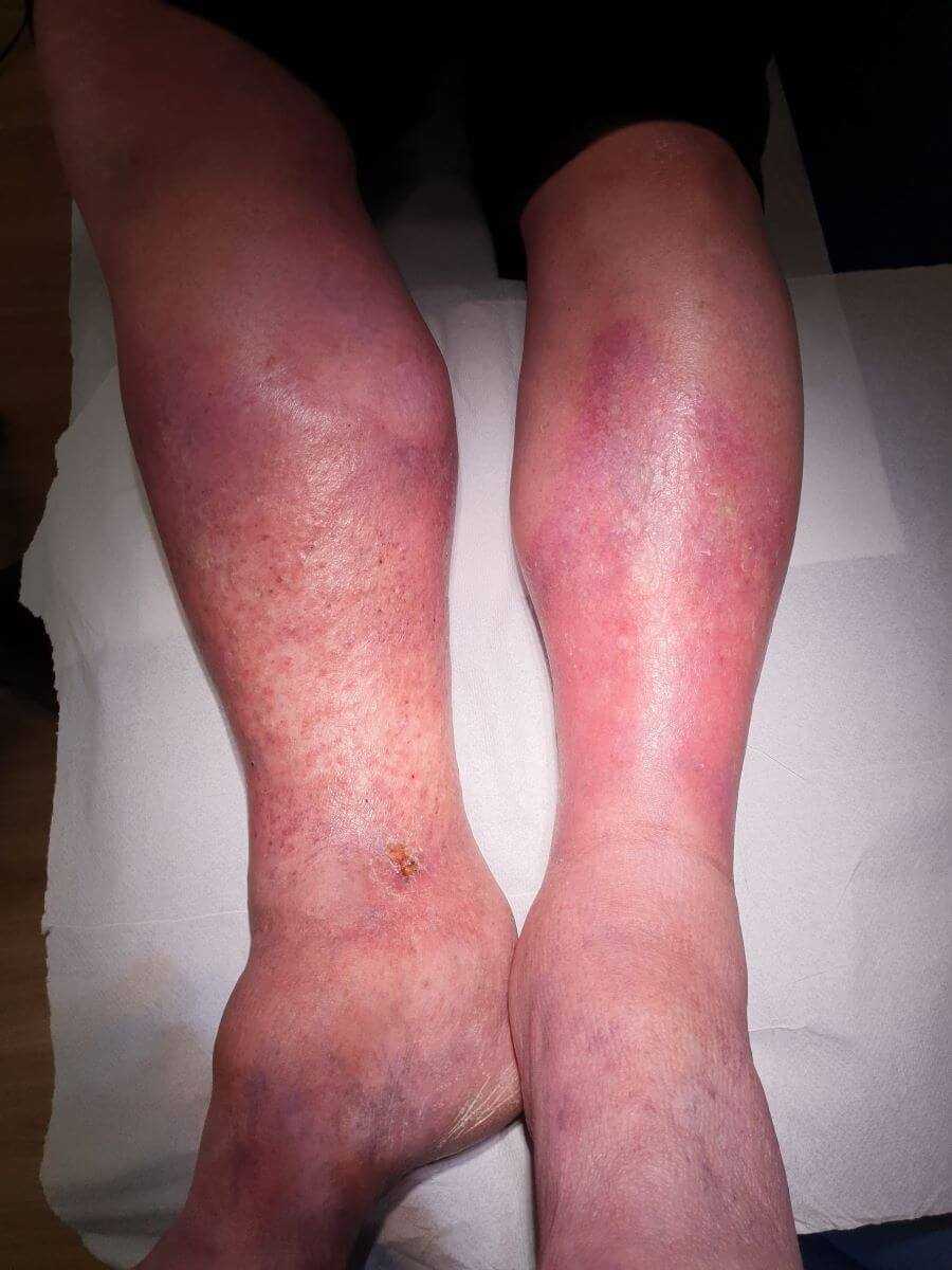
Patients with skin problems of the lower legs are frequently misdiagnosed as having ‘bilateral cellulitis’ and are treated inappropriately with several weeks of systemic antibiotics
-
Cellulitis is very unlikely to be bilateral and most of these patients instead have a chronic superinfected eczema
-
Such patients are systemically well and often have a symmetrical bilateral distribution of well-demarcated stable erythema on their legs. If there is doubt a CRP can be useful to exclude the diagnosis of cellulitis
-
Eczema can be improved with the use of an appropriate topical steroid eg Betnovate-C ® cream until symptoms improve. Patients must not be left on topical steroids indefinitely
-
If eczematous changes persist consider the possibility of a contact allergic dermatitis to one of the topical treatments / dressings - patch tests may be required
Who to refer
Referral must be considered in the following cases:
-
Venous ulcers failing to progress at three months or which have not healed by twelve months
-
All ulcers of an arterial or mixed aetiology - assessment needed for reconstructive surgical / radiological procedures
-
All diabetic and rheumatoid ulcers
-
Failure to adequately control underlying pathologies
-
Suspected malignant change (refer urgently as a 2 Week Rule to dermatology)
-
Suspected cases of contact allergic dermatitis
-
Post-healing - consideration should be given to the referral for further investigations of venous leg ulcers to see if surgical intervention would reduce the risk of re-ulceration. Such a referral would depend on local resources, and is particularly relevant for patients developing ulcers from a young age
Most patients needing referral should be directed to community leg ulcer (tissue viability) clinics or dermatology departments. However, patients in groups 2 or 7 should be referred to vascular surgeons, and diabetic ulcers to the diabetic clinic.
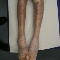
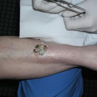
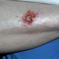
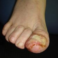
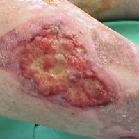
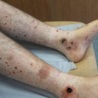
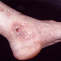
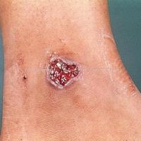
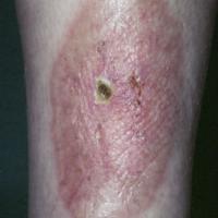
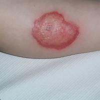
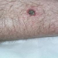
Remember that leg ulcers can result from diverse pathology, the images below are just a few examples.
Additional images
1
2
3
4
5
6
7
8
9
10
11
12
13
Disclaimer - the author PCDS cannot accept responsibility for any misleading or incorrect statements, and the management of individual patients remains the direct responsibility of the individual doctor. We do however hope that visitors to this site can contact us regarding comments that are considered misleading or incorrect so that we can continue to improve the site.
Image Rights - The PCDS would like to thank Dermatoweb, DermQuest (Galderma), and others who have contributed images. All named individuals and organisations maintain copyright for the relevant images.