Molluscum contagiosum

LAST UPDATED: Mar 23, 2026

Patient Information Leaflet
Link: Molluscum contagiosum

https://pcds.org.uk/patient-info-leaflets/molluscum-contagiosum
Introduction

Molluscum contagiosum is a common infection of the skin caused by a Poxvirus. It is largely, if not exclusively a human disease.

This chapter is set out as follows:


Aetiology

  • Infection with molluscum follows contact with infected persons or objects - the use of swimming pools appears to carry an increased risk
  • Sexual transmission of molluscum is also possible

History

  • Mainly presents in young children, although it rarely presents under the age of one year. There is also a later peak in young adults, some of which is attributable to sexual transmission

Clinical findings

Distribution

  • Any part of the body can be affected
  • Lesions are usually multiple but occasionally solitary lesions can be found
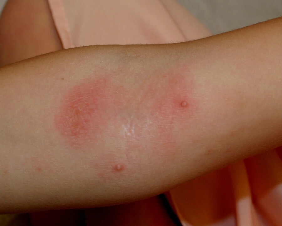
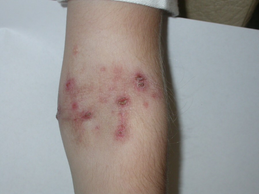
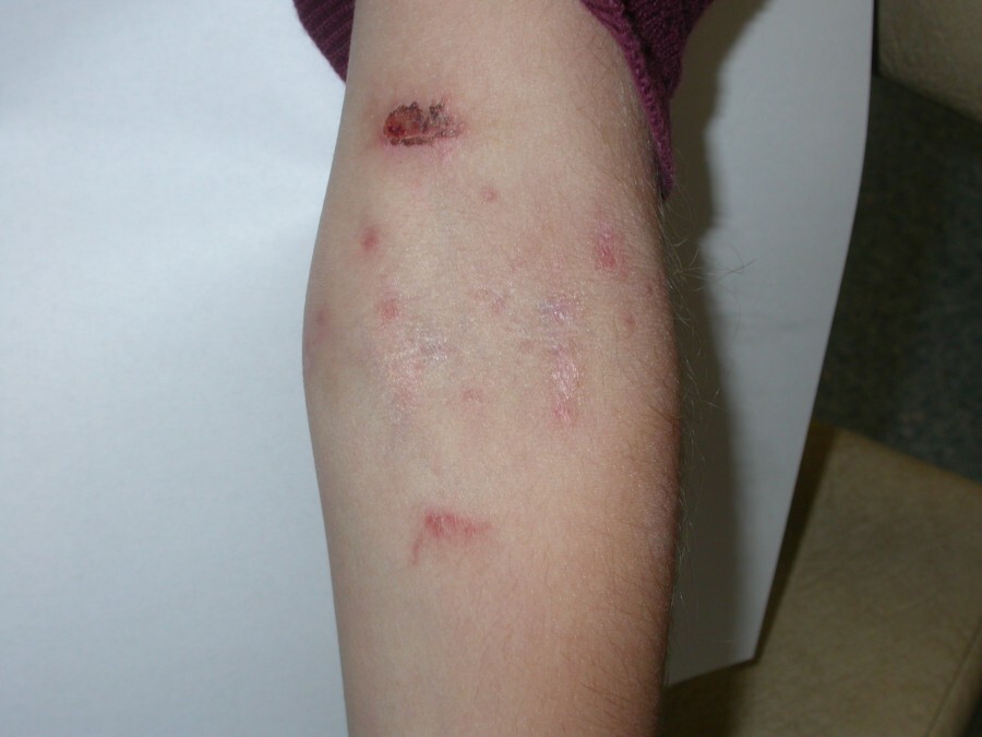
  • In temperate parts of the world molluscum are commonly found on flexural skin such as the neck, the axillae, the groin and behind the knees. Lesions tend to be more numerous and last longer in patients with atopic eczema. In addition some patients with an atopic tendency develop patchy eczema around areas of molluscum
  • In the tropics lesions are most common on the limbs
  • Infection of children though sexual abuse is presumably possible. However, to a greater extent than warts, molluscum is seen quite commonly on the genital and perineal skin, and abuse should not be regarded as the likely cause unless there are other suspicious features
  • Widespread lesions can be found in patients with HIV and other forms of immunosuppression, as well with sarcoidosis

Morphology

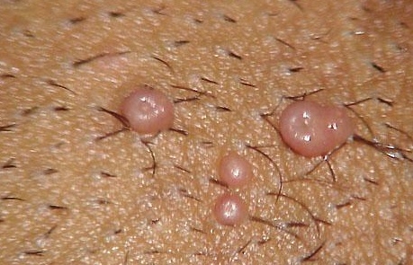
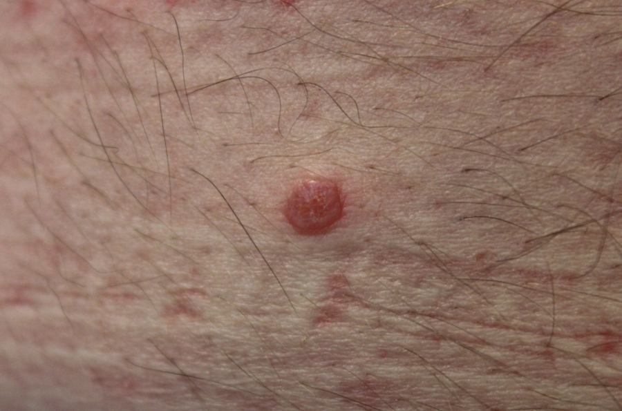
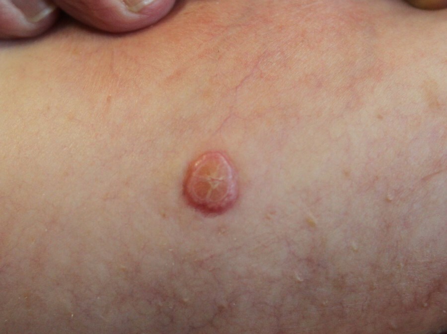
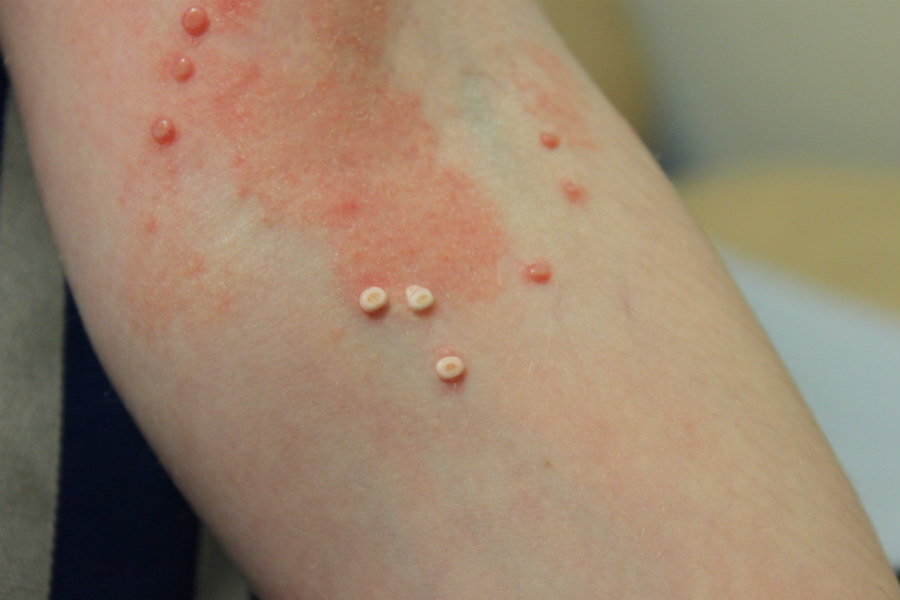
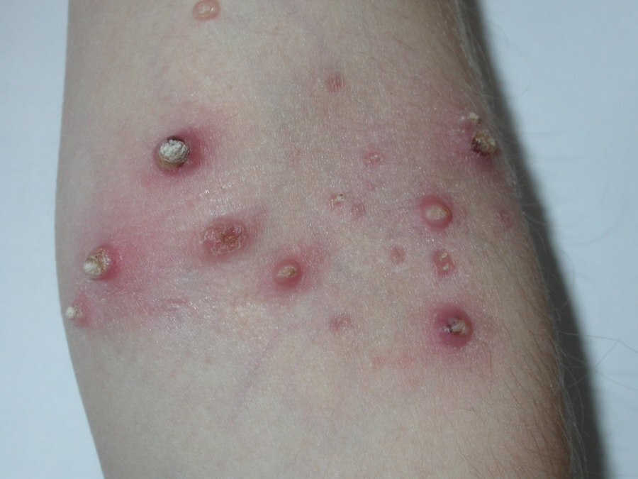
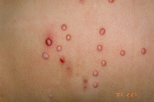
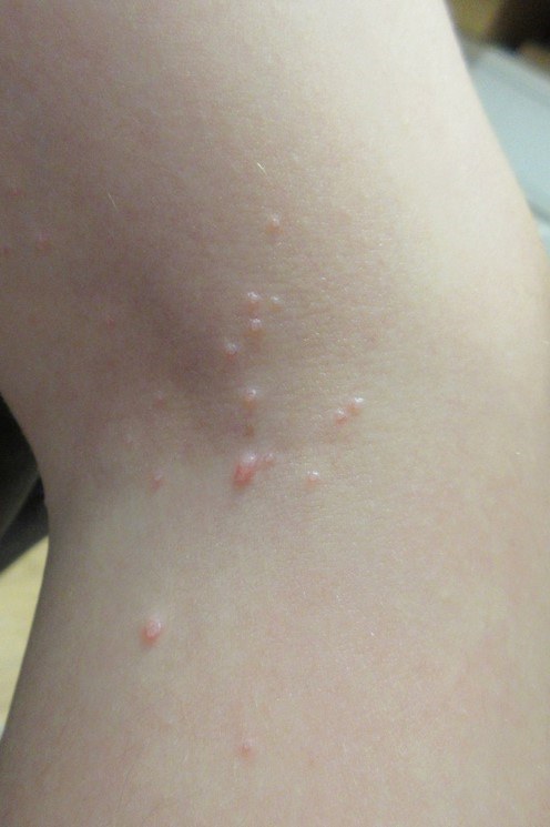
  • Clusters of small shiny papules with an umbilicated centre
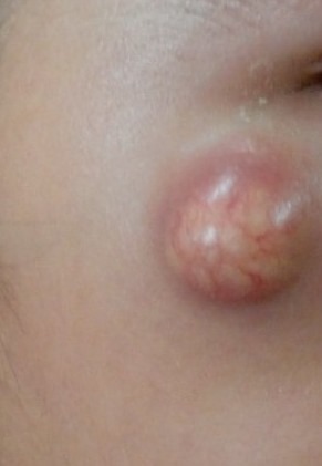
  • Most papules are 2-5 mm in size. Occasionally lesions can reach 1 cm or greater in diameter, this is particularly so with solitary lesions
  • As lesions resolve they become inflamed and crusty
  • Lesions may arise in sites of skin injury and grow in a row, this is known as the Koebner phenomena

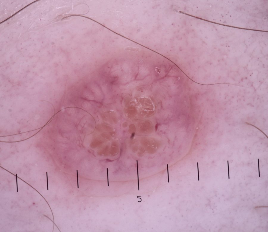
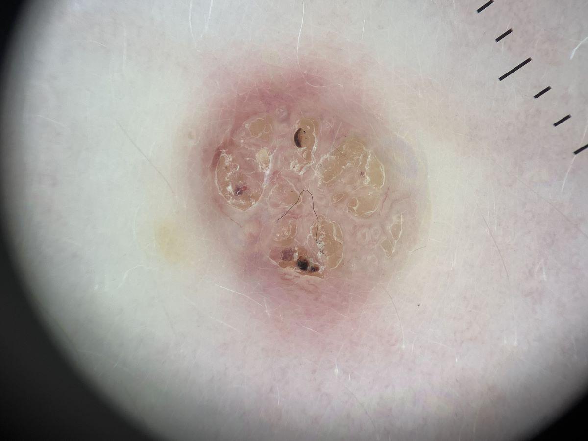
Dermoscopic appearance (hover over terminologies for description)

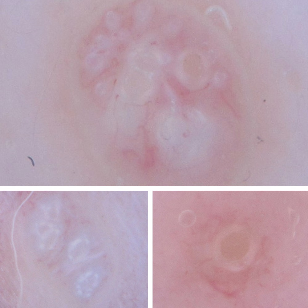
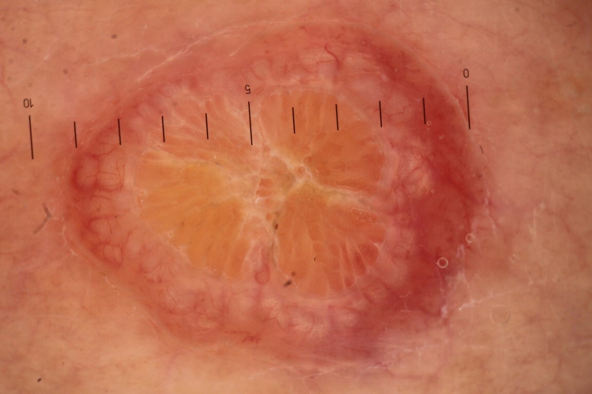
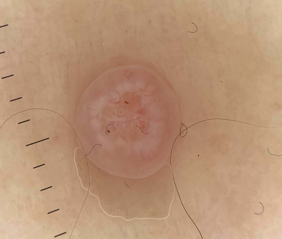
  • Central umbilication
  • White circles around a structureless area correspond to “molluscum bodies” on histology. Polarisation artefact may make the circles look “broken up''
  • Moderate-large sized lesions often have central white-yellow clods (looking like grapefruit segments)
  • The vascular patterns found in 188 lesions confirmed histologically were crown (72.34%), radial (54.25%) and punctiform patterns (20.21%). Half of the 188 lesions had a combination of vascular patterns (for more information on vessels refer to the chapter Vessel patterns in dermoscopy)

Clinical Images

Please refer to notes on image rights at bottom of the page with regards to individual image ownership.
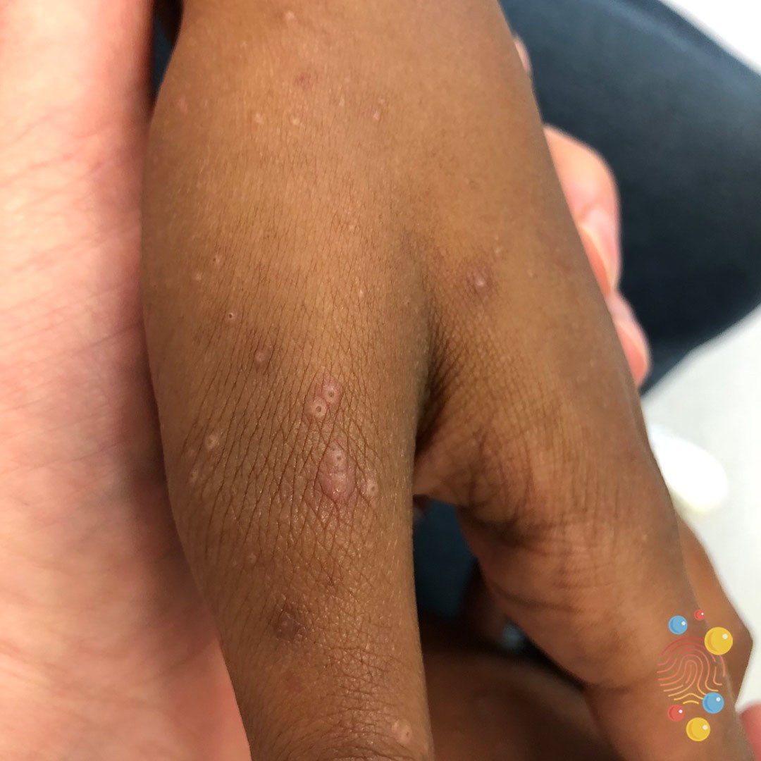
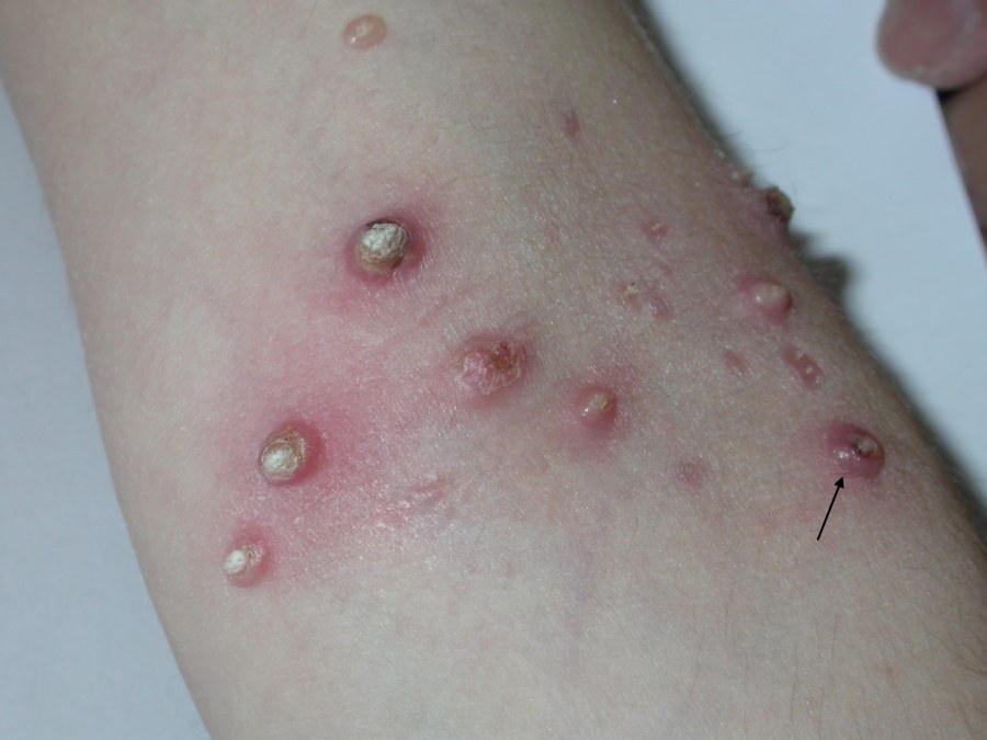
Investigations

  • Normally no investigations are needed
  • In very widespread, persistent, and atypical molluscum contagiosum consider immunosuppression as a cause. Patients with the acquired immunodeficiency syndrome (AIDS) and low CD4 T-lymphocyte counts have a particularly high distribution of lesions around the face and perioral skin

Management

  • Refer to the top right of the page for a patient information leaflet available through a QR code or printable PDF

  • Most cases are self-limiting with resolution of lesions occurring over 6-9 months, although some cases may last for up to four years. Where possible the parents of such children should be encouraged to let the condition run its natural course
  • Consider topical potassium hydroxide:
    • MolluTinc ® (10% potassium hydroxide) can be purchased over the counter
    • Molludab ® (5% potassium hydroxide), a topical treatment licensed for use from the age of two years. It is used BD until the lesions become inflamed, which often takes around five days, at which stage the treatment can be stopped. If there is no inflammation by day 14 the treatment should be stopped
  • Cryotherapy can be a very effective treatment for older children and adults, younger children rarely tolerate it. Often only a single freeze-thaw cycle of 5-10 seconds is needed
  • Curettage for histology may be needed for larger solitary lesions where there is diagnostic uncertainty
  • Underlying eczematous changes can be managed with standard eczema treatment

Disclaimer - the author PCDS cannot accept responsibility for any misleading or incorrect statements, and the management of individual patients remains the direct responsibility of the individual doctor. We do however hope that visitors to this site can contact us regarding comments that are considered misleading or incorrect so that we can continue to improve the site.

Image Rights - The PCDS would like to thank Dermatoweb, DermQuest (Galderma), and others who have contributed images. All named individuals and organisations maintain copyright for the relevant images.

Quick Links

The following pharmaceutical companies have had no involvement in the content of this website or in our conference programmes


Almirall
Galderma
Glenmark
Johnson & Johnson
La Roche-Posay
LEO Pharma
Pierre Fabre
Schuco