Necrobiosis lipoidica
LAST UPDATED: Jul 04, 2022
Introduction
Necrobiosis lipoidica is an uncommon skin condition characterised by sharply demarcated, atrophic yellowish patches or plaques on the shins. It was first described in patients with insulin-dependent diabetes but it can affect non-diabetics as well.
This chapter is set out as follows:
Aetiology
-
Vascular changes may play a role
-
Necrobiosis lipoidica is associated with diabetes, the reasons remain unclear
History
-
Necrobiosis lipoidica has a gradual onset
-
Although necrobiosis lipoidica can affect any age, it most commonly presents in young adults and early middle age. Patients with insulin-dependent diabetes tend to present at an earlier age when compared to non-insulin-dependent patients
-
Females are affected more than males in a 3:1 ratio
-
In many cases lesions are asymptomatic, although some patients experience pain
-
The main complaint is the unsightly appearance of the lesions
Clinical findings
Distribution
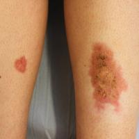
- Lesions typically affect both shins, although necrobiosis lipoidica is sometimes unilateral and other sites can be affected
Morphology
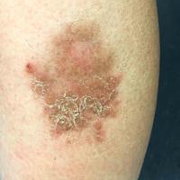
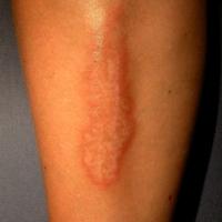
- Typically, one or more yellowish patches / plaques
-
Lesions grow with an erythematous edge, and a centre that becomes shiny and atrophic with prominent telangiectasia
-
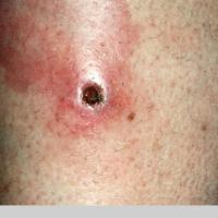
Ulceration can occur
-
Slow expansion over many years is usual, although lesions may go through periods of being more, and less, active
Clinical Images
Please refer to notes on image rights at bottom of the page with regards to individual image
ownership.
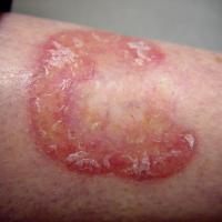
1
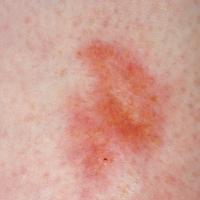
2
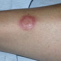
3
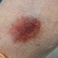
4
5
6
7
8
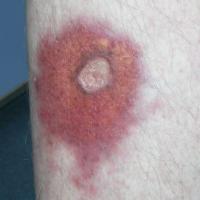
9
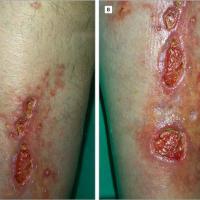
10
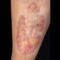
11
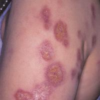
12
Investigations
-
It is important to investigate for diabetes mellitus
-
The diagnosis is usually clinical, although sometimes a biopsy is needed if the presentation is atypical as cutaneous sarcoid can occasionally mimic necrobiosis lipoidica
- Histology shows changes involving the full thickness of the dermis, often extending into the subcutaneous fat. Early lesions show a perivascular and interstitial mixed inflammatory cell infiltrate. A granulomatous inflammatory reaction is seen around areas of destroyed collagen. Areas of necrobiosis are usually more extensive and less well-defined than in granuloma annulare
Management
Step 1: general measures
-
Provide a patient information leaflet
-
Smoking cessation may help
-
Due to the increased risk of ulceration, advise the patient to avoid traumatising the skin
-
Not all cases require treatment
Step 2: topical steroids
-
A decision to treat necrobiosis lipoidica needs careful discussion with the patient - at best, individuals can expect a mild-moderate improvement, and one has to weigh up the possible risks of steroid atrophy
-
If treatment is given use a potent topical steroid, eg 0.1 % betamethasone cream (or ointment) once a day, and advise application only to the leading inflammatory edge and not the atrophic centre. The effectiveness of treatment can sometimes be enhanced by using occlusion eg Clingfilm
-
Photograph the lesion / lesions and review three months after starting the treatment to assess response
Step 3: steroid sparing therapy
-
If there are concerns with regards to steroid atrophy, consider a trial of 0.1% tacrolimus ointment BD
Step 4: ulcerated lesions
-
Consider treating more aggressively, for example:
- 0.1% betamethasone (or Dermovate ®) cream / ointment under occlusion with Clingfilm, Zipzocs ®, or Viscopaste ® bandages
- Again, photographs are useful to monitor progress
Step 5: Secondary Care
-
Patients with troublesome, recalcitrant lesions, especially if ulcerated, should be referred to Dermatology
- Phototherapy (especially PUVA) leads to improvements in some cases
- Other options include the careful administration of intralesional steroids in to the expanding edge, and short-term ciclosporin
-
Occasionally a chronic, ulcerated lesion can transform in to a squamous cell carcinoma. Refer suspect lesions urgently (two-week wait)
Disclaimer - the author PCDS cannot accept responsibility for any misleading or incorrect statements, and the management of individual patients remains the direct responsibility of the individual doctor. We do however hope that visitors to this site can contact us regarding comments that are considered misleading or incorrect so that we can continue to improve the site.
Image Rights - The PCDS would like to thank Dermatoweb, DermQuest (Galderma), and others who have contributed images. All named individuals and organisations maintain copyright for the relevant images.









