
LAST UPDATED: Jul 30, 2021
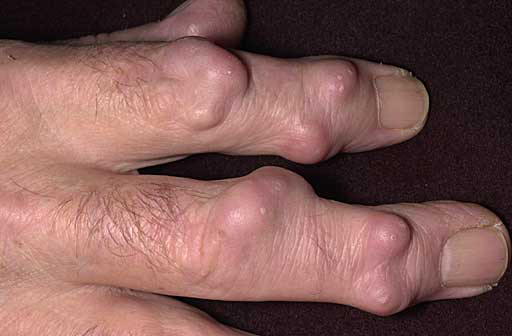
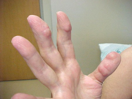
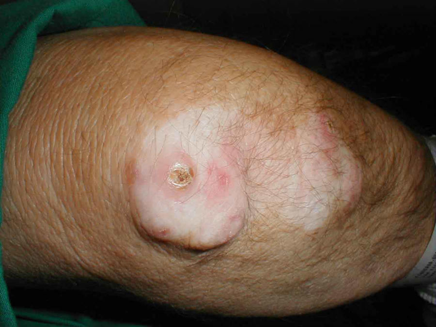
Rheumatoid nodules are the most common extra-articular manifestation of rheumatoid arthritis (RA), occuring in approximately 20–25% of patients with the condition. They are universally associated with a positive rheumatoid factor (RF).
Rheumatoid nodules are clinical predictors of more severe arthritis, joint erosions and rheumatoid vasculitis, and their presence often suggests a need for more aggressive treatment of the underlying RA. Rheumatoid nodules are not exclusive to RA, and histologically similar nodules can be a feature of subcutaneous granuloma annulare, necrobiosis lipoidica, systemic lupus erythematosus and foreign body granulomas.
This chapter is set out as follows:
Please refer to notes on image rights at bottom of the page with regards to individual image ownership.

There are very few treatment options for rheumatoid nodules. Large nodules can be excised, but they frequently recur within scar tissue, especially if subjected to repetitive trauma. Injecting corticosteroids directly into the lesion sometimes reduces its size. While this procedure is most effective for deep lesions in the olecranon bursa, nodules on the buttocks and feet tend to ulcerate and are likely to become infected. Once they are infected, surgical excision or drainage is required. Oral corticosteroids and hydroxychloroquine can also be used, but their effects on rheumatoid nodules vary and many patients with RA already receive these medications for the underlying condition. Rheumatoid nodules occasionally resolve without medical or surgical intervention
Disclaimer - the author PCDS cannot accept responsibility for any misleading or incorrect statements, and the management of individual patients remains the direct responsibility of the individual doctor. We do however hope that visitors to this site can contact us regarding comments that are considered misleading or incorrect so that we can continue to improve the site.
Image Rights - The PCDS would like to thank Dermatoweb, DermQuest (Galderma), and others who have contributed images. All named individuals and organisations maintain copyright for the relevant images.
It is important for the PCDS to build its own image bank, as such we welcome original images from our readers, especially in skin of colour:
Please start typing below the word of any medical term that you do not understand.
The following pharmaceutical companies have had no involvement in the content of this website or in our conference programmes







