Telangiectases

LAST UPDATED: Oct 25, 2024

Introduction

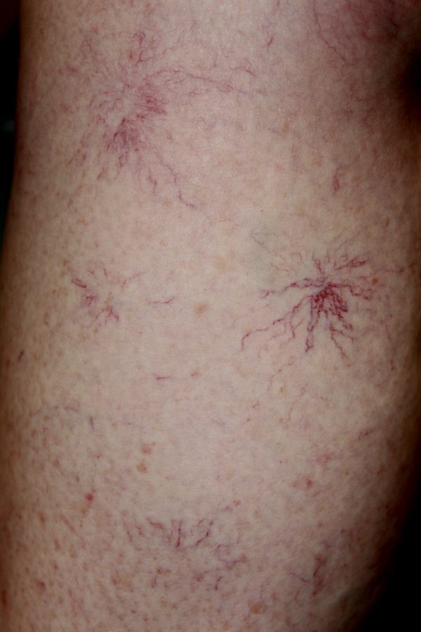
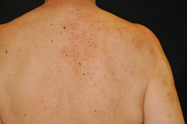
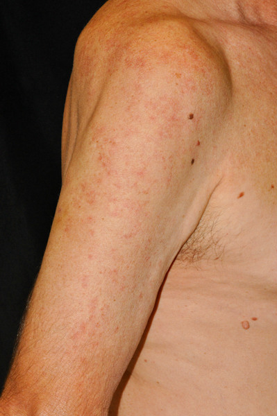
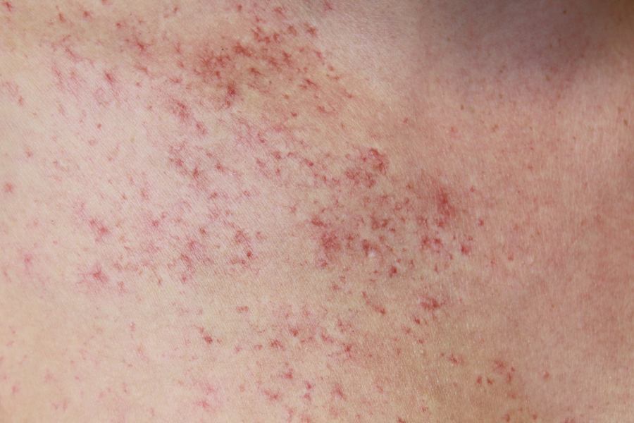
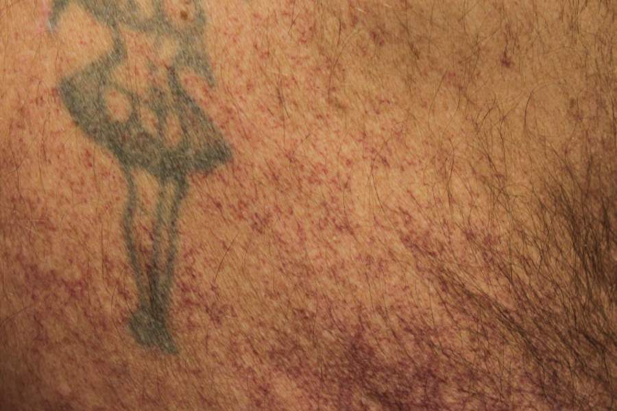
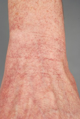
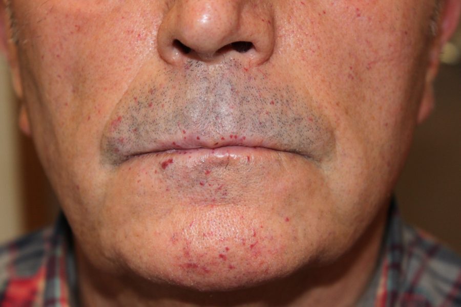
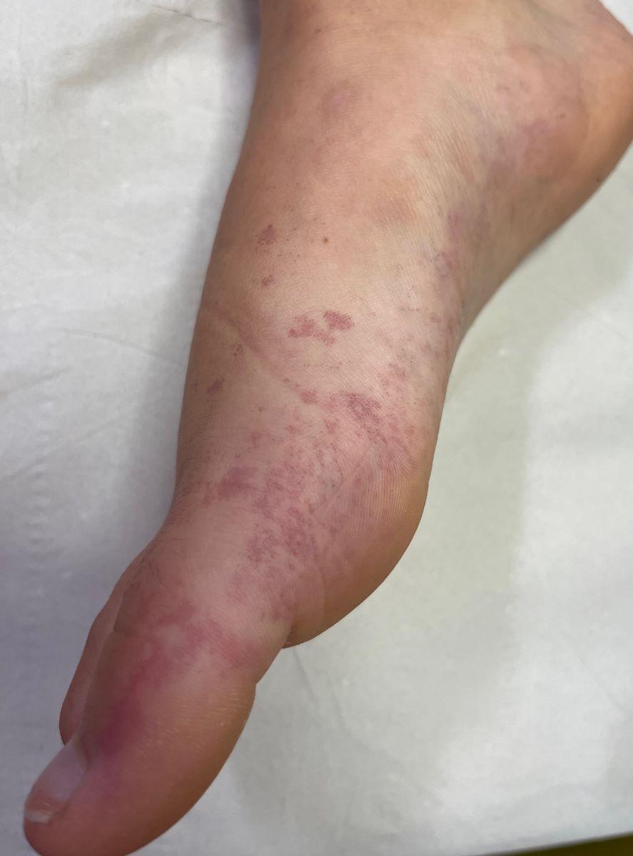
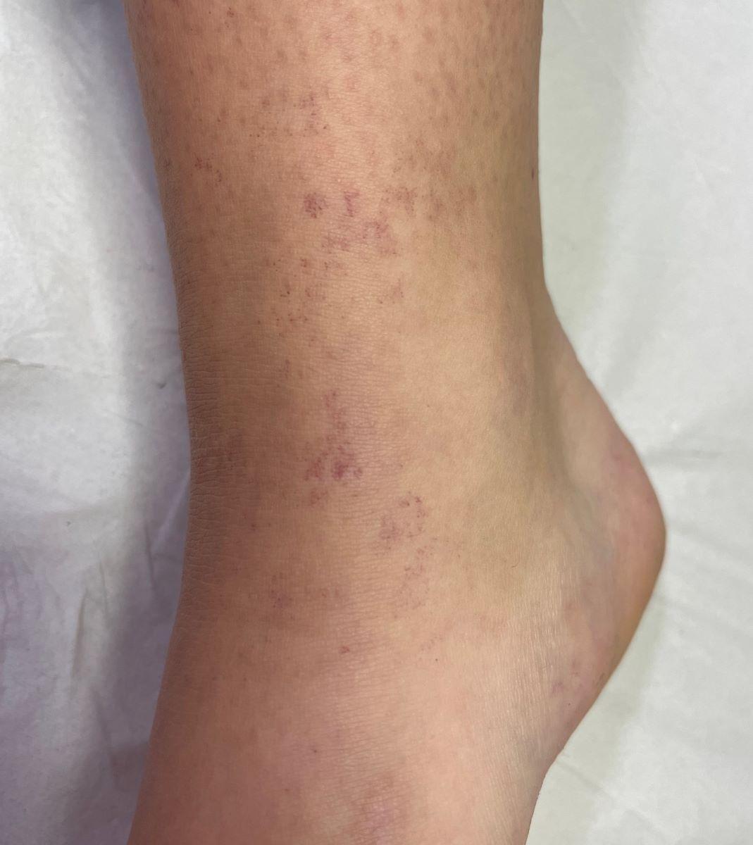
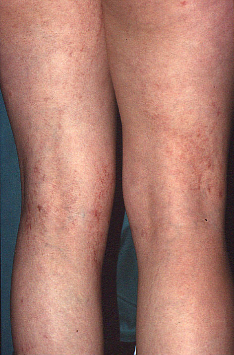
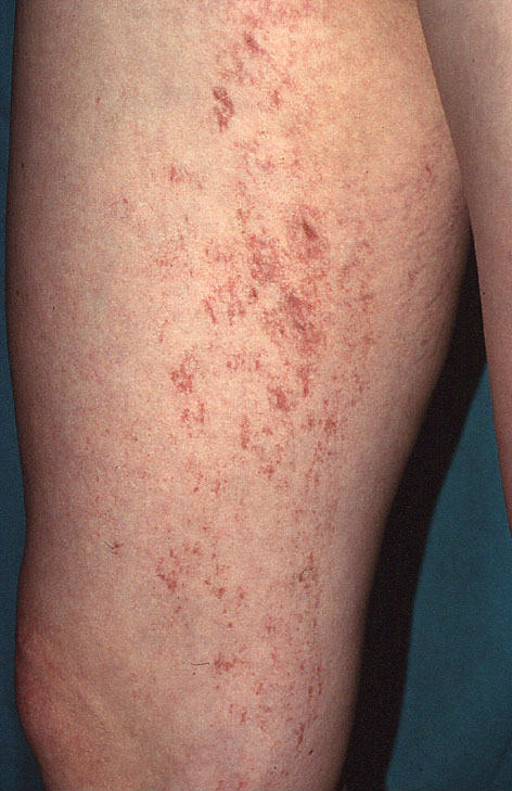
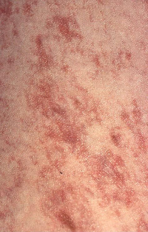
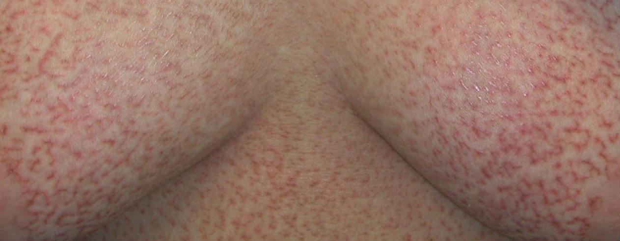
Telangiectases result from the chronic dilatation of pre-existing capillaries or venules. They appear on the skin and mucous membranes as small red, linear, stellate or punctate markings. Telangiectases can be primary or secondary in origin and while most are harmless a few types are associated with serious underlying conditions.

This chapter is set out as follows:


Aetiology

Primary telangiectases

While there are different types, this chapter will only discuss the following:

  • Spider telangiectases (syn. spider naevus)
  • Costal fringe 
  • Generalised essential telangiectasia
  • Hereditary haemorrhagic telangiectasia (syn. Rendu-Osler-Weber syndrome)
  • Angioma serpiginosum
  • Unilateral naevoid telangiectasia
  • Poikilodermatous mycoses fungoides (syn. poikiloderma atrophicans vasculare)

Secondary telangiectases 

  • There are many causes eg rosacea, prolonged UV exposure, steroid atrophy, radiodermatitis and connective tissue disorders

Clinical findings

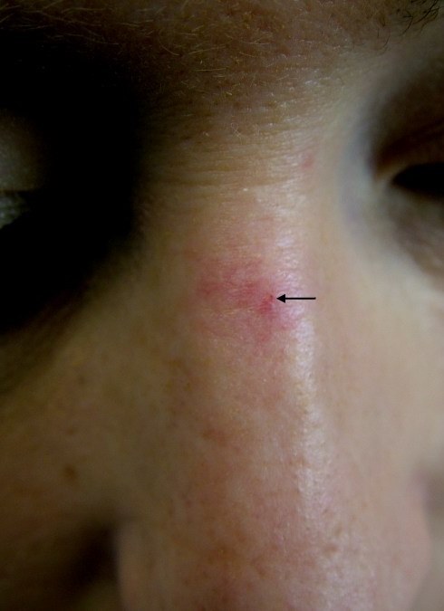
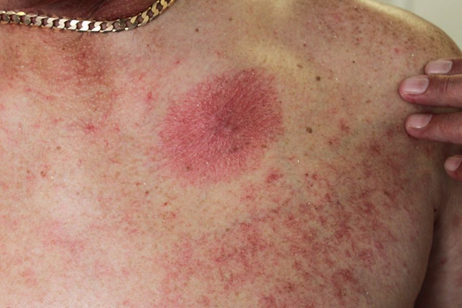
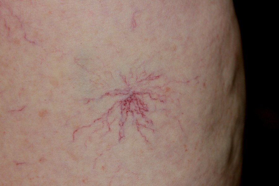
Spider telangiectases

  • History
    • Occur in up to 15% of normal people, and more frequently in children and in pregnancy
    • They can also be a feature of liver disease
    • The majority of lesions arising in pregnancy disappear spontaneously, in others lesions tend to persist
  • Distribution 
    • The upper half of the body, especially the face, neck, upper chest and hands
  • Morphology
    • ​An arcade of vessels radiates out from a central arteriole - compressing the central point will blanche the arcade

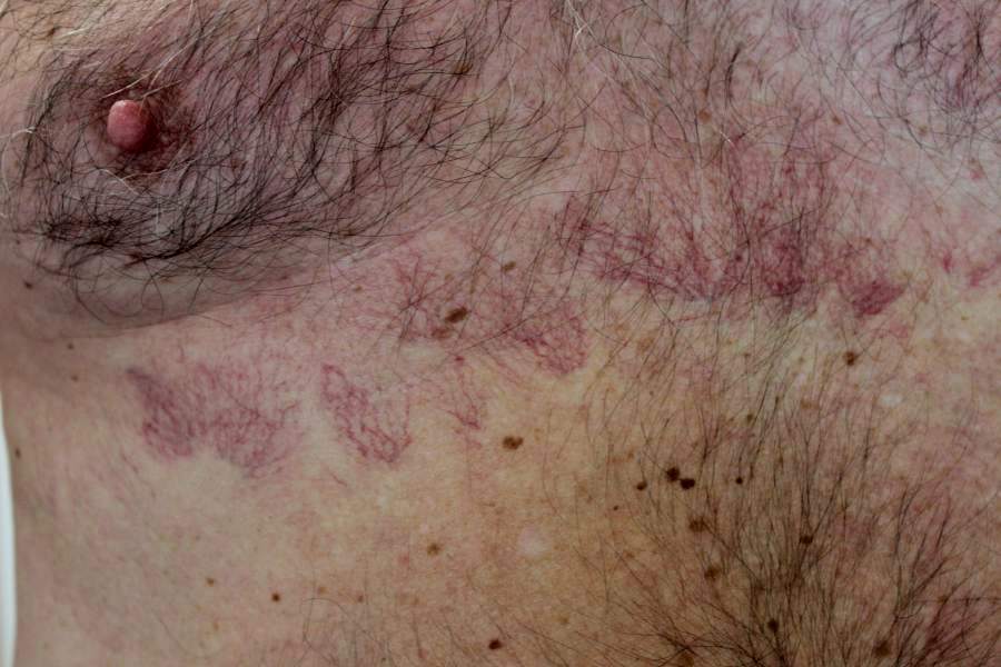
Costal fringe

  • History
    • It is a common and easily recognised entity, usually seen in elderly men
  • Distribution
    • Along the anterolateral costal margin
  • Morphology
    • A bandlike pattern of venous telangiectases

Unilateral naevoid telangiectasia

  • History
    • Uncommon
    • Mainly acquired, arising more commonly in women and unmasked during periods of relative oestrogen excess, such as puberty, pregnancy, and during oral contraceptive use. Also in chronic liver disease
    • Occasiaonally congenital, in which case it has male predominance
  • Distribution
    • It develops in a segmental pattern
    • The third and fourth cervical dermatomes are the most common sites, but the thoracic dermatomes and scattered distant sites may also be involved
  • Morphology
    • Patches of telangiectases may be small to large and few to numerous

Generalised essential telangiectasia

  • History
    • Starts in late childhood or early adulthood
    • More common in females
  • Distribution
    • Trunk or limbs
  • Morphology
    • Extensive sheets of telangiectases

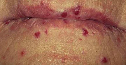
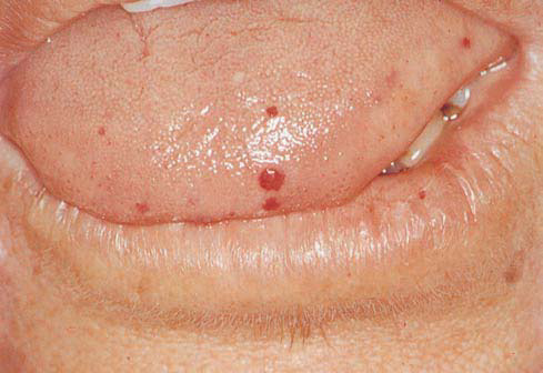
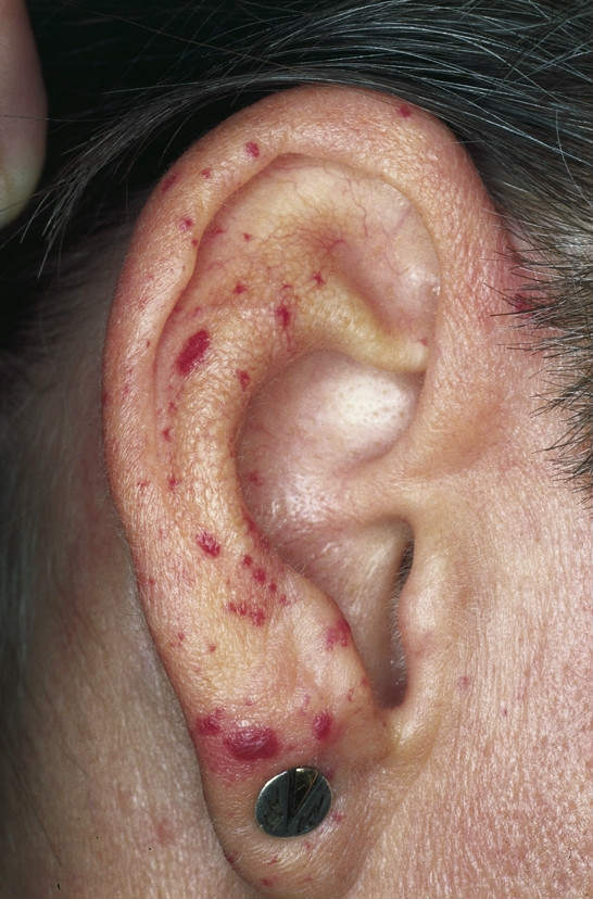
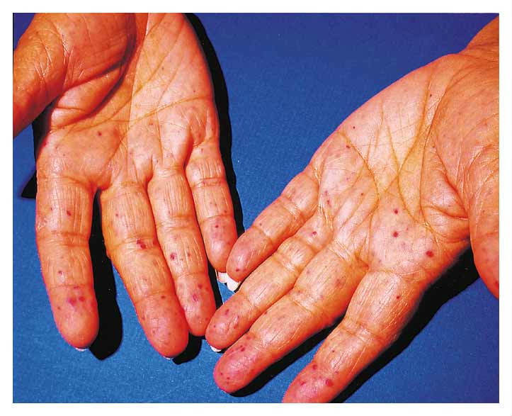
Hereditary haemorrhagic telangiectasia (syn. Rendu-Osler-Weber syndrome)

  • History
    • A rare and potentially serious autosomal dominant condition of blood vessels distinguished by cutaneous telangiectasia and resulting in widespread haemorrhage
    • Usually presents after puberty with nose bleeds, gastrointestinal, pulmonary or genitourinary haemorrhage 
    • The cutaneous lesions often arise in adult life 
  • Distribution
    • ​Oral lesions, the face, lips and ears. The nasal septum is also affected 
    • Chest, hands and feet
  • Morphology
    • Widespread, small telangiectases

Angioma serpiginosum

  • History
    • A rare unilateral disorder of the superficial blood vessels affecting a limb or buttock
    • Female predominance
    • Usually arises in childhood and then extends for a year or more before stopping
    • In adult life there may be further growth or complete / partial regression
  • Distribution
    • ​A serpiginous or ring-shaped pattern along a limb or buttocks. Occasionally it can be more extensive
  • Morphology
    • Multiple pinpoint, non-blanching red papules, which often coalesce in to sheets of affected skin

Poikilodermatous mycoses fungoides (syn. poikiloderma atrophicans vasculare)

  • Distribution
    • One or a limited number of patches may occur, especially over the breasts or buttocks. Can be more widespread
  • Morphology
    • Poikilodermatous changes ie the combination of telangiectasia, atrophy and hyper / hypopigmentation 

Clinical Images

Please refer to notes on image rights at bottom of the page with regards to individual image ownership.
Management

  • Most telangiectases are self-limiting 
  • For cosmetically sensitive areas of the body:
    • Gentle cautery can be used for small numbers of lesions
    • Laser therapy is also an effective treatment and can be used for larger numbers of lesions
  • Patients with potentially more serious conditions such as hereditary haemorrhagic telangiectasia or poikilodermatous mycoses fungoides need referring, the latter may need observation and multiple biopsies before the condition is diagnosed

Other resources


Disclaimer - the author PCDS cannot accept responsibility for any misleading or incorrect statements, and the management of individual patients remains the direct responsibility of the individual doctor. We do however hope that visitors to this site can contact us regarding comments that are considered misleading or incorrect so that we can continue to improve the site.

Image Rights - The PCDS would like to thank Dermatoweb, DermQuest (Galderma), and others who have contributed images. All named individuals and organisations maintain copyright for the relevant images.

Quick Links

The following pharmaceutical companies have had no involvement in the content of this website or in our conference programmes


Almirall
Galderma
Glenmark
Johnson & Johnson
La Roche-Posay
LEO Pharma
Pierre Fabre
Schuco