Sweet’s syndrome (syn. acute febrile neutrophilic dermatosis; Gomm-Button disease)
LAST UPDATED: Nov 19, 2021
Introduction
Sweet’s syndrome is a neutrophilic dermatosis, characterised by fever, peripheral neutrophil leucocytosis and the acute onset of painful, erythematous papules, nodules or plaques.
This chapter is set out as follows:
Aetiology
There are three main groups of Sweet's disease: classical, malignancy-associated, and drug-induced.
Classical
-
Typically affects women in the third to fifth decade
-
Associated with infection (eg streptococcal URTI, salmonella and yesinial GIT infection, and mycobacteria), IBD and pregnancy, although sometimes no cause is found
Malignancy-associated
-
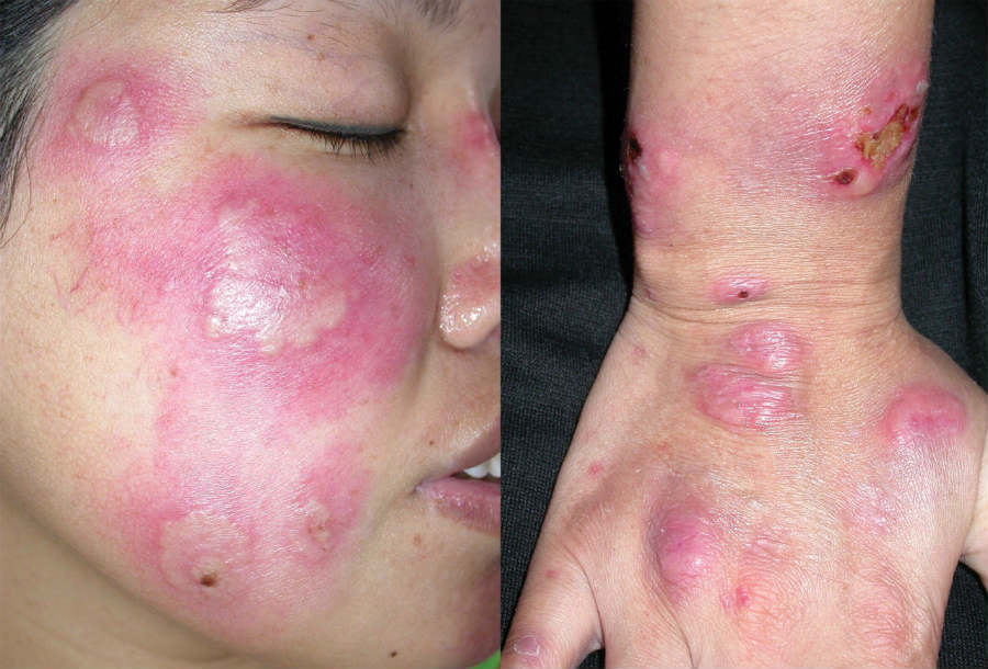
Comprises approximately one quarter of cases, and is more likely if the rash affects the face
-
Men and women are equally affected
-
Most tumours are haematological, especially AML, however a few are solid tumours, especially genitourinary, breast or gastrointestinal
-
Sweet's syndrome may be the initial manifestation of malignancy or may precede the diagnosis by months or years. Additional episodes of Sweet’s may also be an indication of cancer recurrence
Drug-induced
-
Mainly arise with granulocyte colony-stimulating factor
-
Other drugs include minocycline, trimethoprim, carbamazepine and oral contraceptives
Occasionally Sweet's can present in a subacute fashion that is sometimes referred to as Chronic relapsing and remitting Sweet's syndrome - refer below for further information.
Other conditions reported in association with Sweet’s include sarcoid, RA and connective tissue disorders, thyroid disease, other neutrophilic dermatoses eg Bechet’s disease, and erythema nodosum.
History
Sweet's syndrome presents with an acute onset and is characterised by some or all of the following:
-
Painful skin lesions
-
Moderate-high fever (can be absent in malignancy-related cases)
-
Malaise / fatigue
-
Arthralgia
-
Eye symptoms
-
Mouth ulcers
-
Headache
-
Occasionally internal organs may also be affected eg the nervous system (neuro-Sweet's)
Clinical findings
Sweet's syndrome
- Distribution
-
Lesions usually multiple
- Classically the arms, face and neck but may occur anywhere
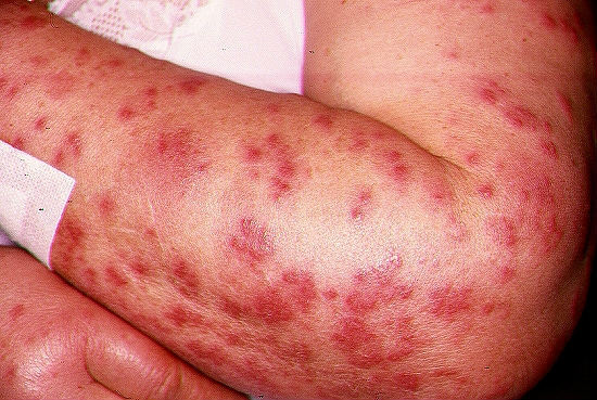
- Can become widespread, especially if secondary to malignancy
- Morphology
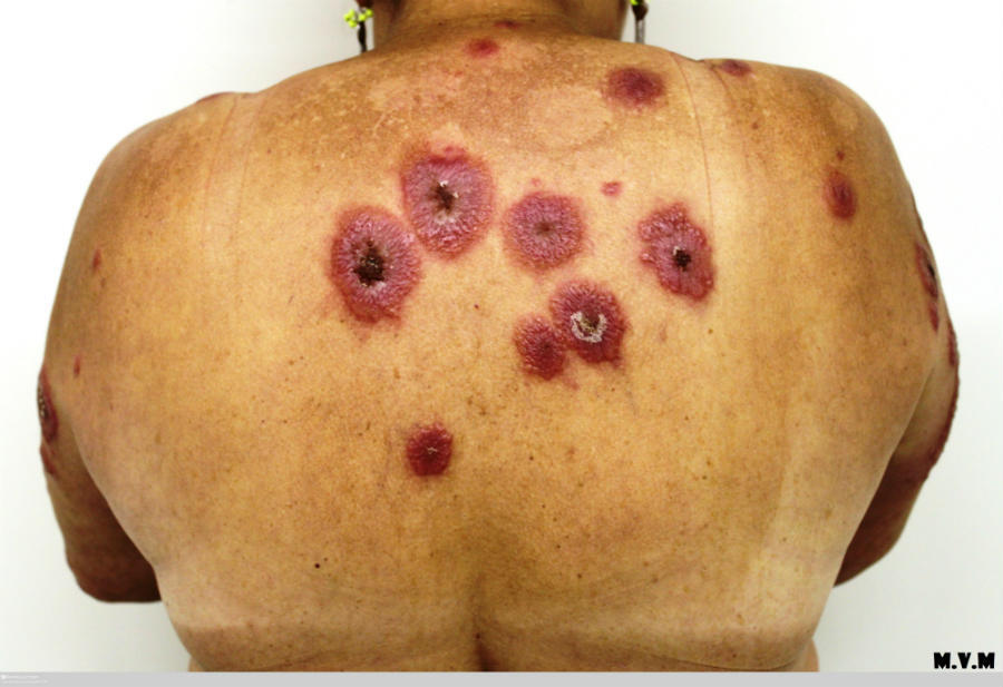
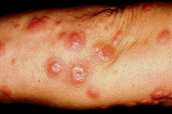
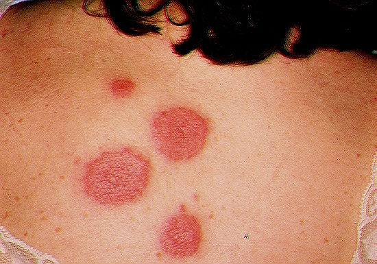
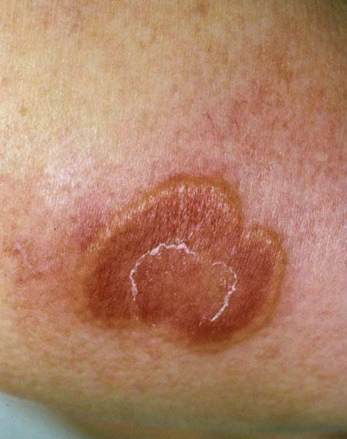
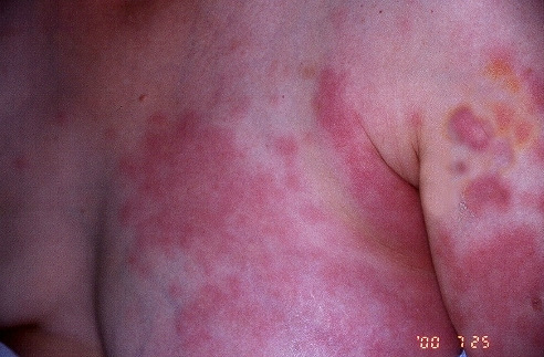
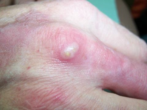
- The typical findings are those of red-purple, tender papules and nodules that coalesce to form plaques, which in some cases take on a targetoid appearance
- Lesions can become studded with pustules, and occasionally become bullous
- Healing normally occurs without scarring
- Other features
- Ocular manifestations eg conjunctivitis, episcleritis, uveitis and glaucoma
- Oral erosions / ulcers are mainly associated with underlying haematological malignancies
Chronic relapsing and remitting Sweet's syndrome
-
Fever / rigor is variable but polyarthralgia and tiredness are common. Some describe intermittent conjunctival congestion
-
Clinically urticated pink plaques and papules on trunk and proximal extremities predominate. The face and neck can be involved in some cases, especially the mastoid region. Lesions are variable from completely asymptomatic to intensely itchy, the painful tender oedematous “juicy” plaques seen in classical Sweet's is uncommon
Neutrophilic dermatosis of the (dorsal) hands
-
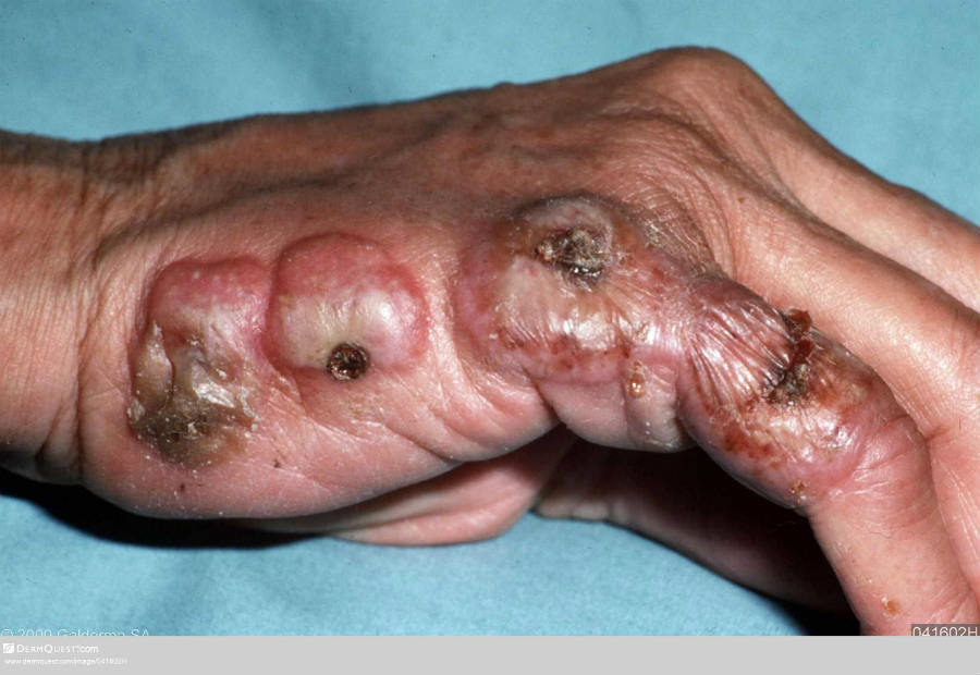
In this rare condition, patients develop pustular or ulcerative plaques and nodules on the dorsal hands. The lesions are more blue-grey than the red-purple colour seen in classical Sweet's
-
There remains debate as to whether or not this condition is a localised from of Sweet's syndrome
Clinical Images
Please refer to notes on image rights at bottom of the page with regards to individual image
ownership.
Investigations
-
Blood tests
- FBC - a neutrophil leukocytosis
- Inflammatory markers - can be raised during an acute episode
- Routine biochemistry screen, autoantibodies and rheumatoid factor, immunoglobulins and plasma protein electrophoresis
- In cases of associated early myelodysplasia the MCV can be high with variable cytopenia (leucopenia, thrombocytopenia, anaemia), however, the FBC can be completely normal for 2-3 years, sometimes even longer before it shows clues of early myelodysplastic syndrome
- Sweet's syndrome
- A biopsy shows a dense, predominantly neutrophilic, infiltrate located in the superficial dermis
- Recent studies suggest that leucocytoclastic vasculitis can be found in some cases, although some debate this is a consequence of intense inflammation, as opposed to being a primary vasculitis
- Chronic relapsing and remitting Sweet's syndrome
- The typical features described above are uncommon. Some initially start with a predominantly lymphocytic infiltrate that is difficult to diagnose histologically as Sweet's. As time passes some will evolve to typical histological features, others have a mononuclear cell infiltrate with atypical morphology resembling histiocytes (Histiocytoid), these cells can be highlighted on IHC CD68 and MPO stains
Management
-
Patients need urgent referral to dermatology for diagnosis and treatment
-
First-line treatment
-
A 4-6 week course of prednisolone is usually sufficient to settle symptoms down, but occasionally longer-term low dose treatment is needed
-
Other treatment options include:
-
Dapsone, colchicine 500 mcg tds, indomethacin 150 mg per day week one then 100 mg per day for two weeks, and ciclosporin
-
Long-term management
-
If no underlying cause was found at the onset, given the association with haematological disease it is advisable to re-check a FBC on an annual basis after the Sweet's resolves
-
Up to one-third of patients may develop recurrent episodes. This is more likely in patients who have underlying myelodysplasia or malignancy
Disclaimer - the author PCDS cannot accept responsibility for any misleading or incorrect statements, and the management of individual patients remains the direct responsibility of the individual doctor. We do however hope that visitors to this site can contact us regarding comments that are considered misleading or incorrect so that we can continue to improve the site.
Image Rights - The PCDS would like to thank Dermatoweb, DermQuest (Galderma), and others who have contributed images. All named individuals and organisations maintain copyright for the relevant images.