Tinea corporis (body), cruris (groin) and incognito (steroid exacerbated)
LAST UPDATED: Jun 02, 2023
Introduction
This chapter discusses tinea corporis, tinea cruris and tinea incognito, and is set out as follows:
Aetiology
-
Refer to the related chapter on Tinea - an overview
History
-
Tinea can affect any age
-
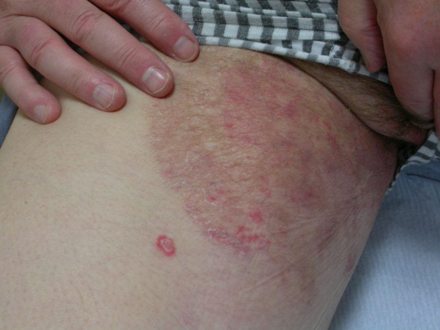
While both sexes are affected, tinea cruris affects more men than women
-
Itch may be present
Clinical findings
Typical features of tinea corporis / tinea cruris
Distribution
- Trunk or limbs - can be asymmetrical, although commonly bilateral
- The feet can act as the source of infection and so should also be examined and treated if they are involved
Morphology
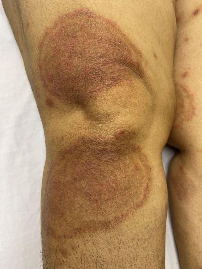
- Lesions can be single or multiple and over time can grow to become quite large
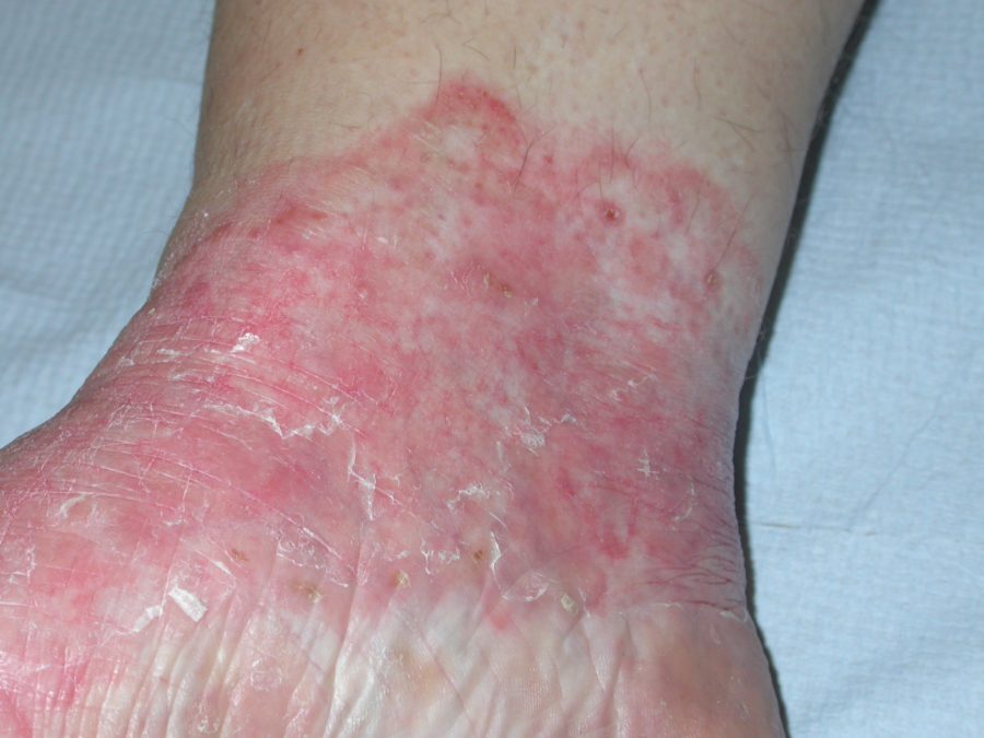
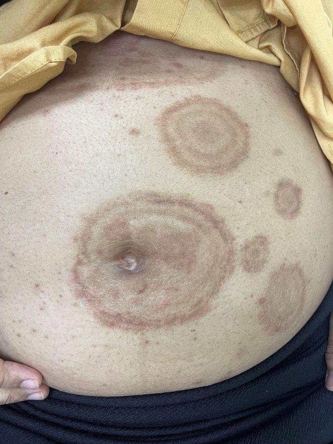
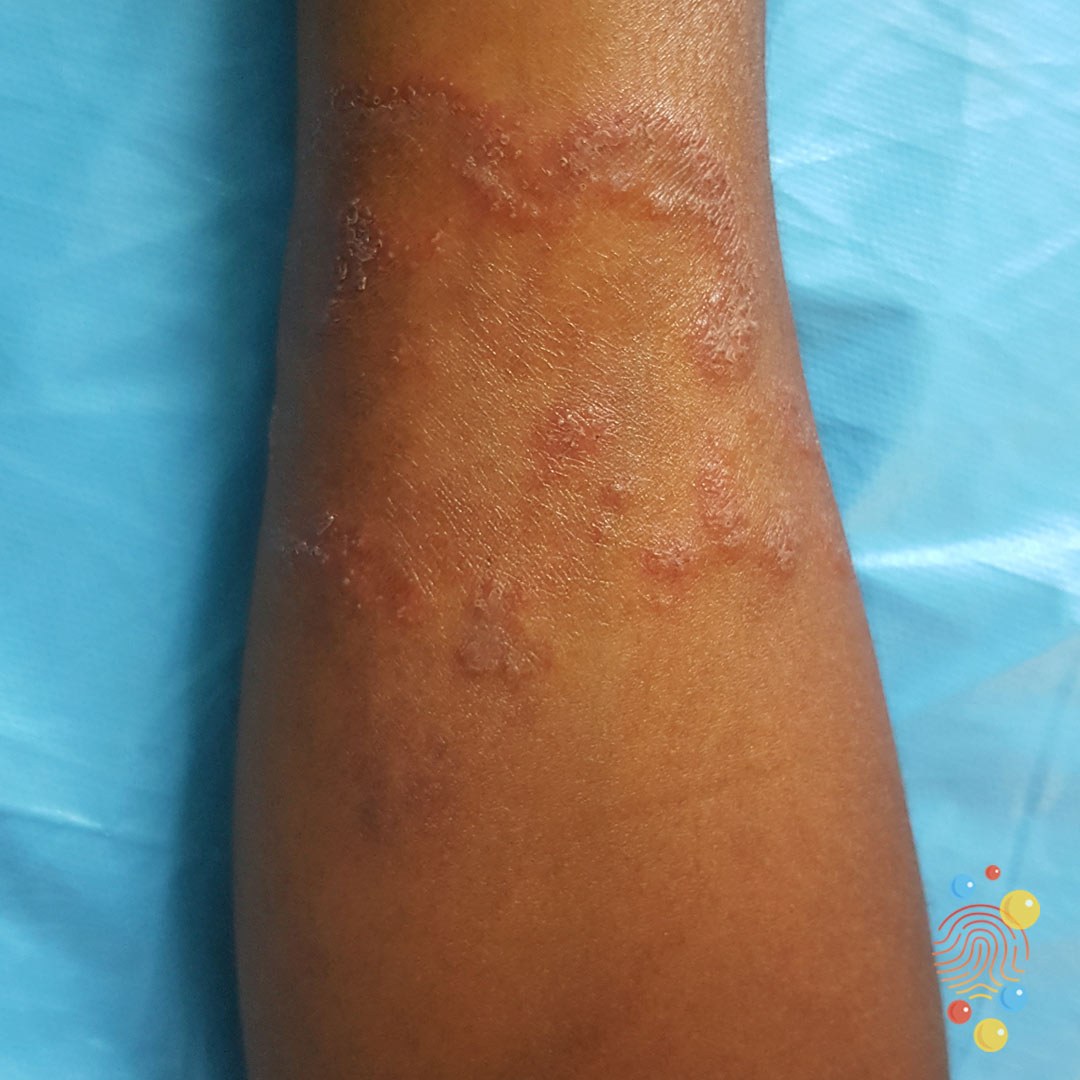
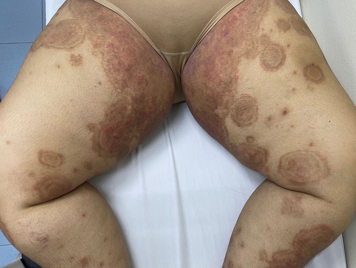
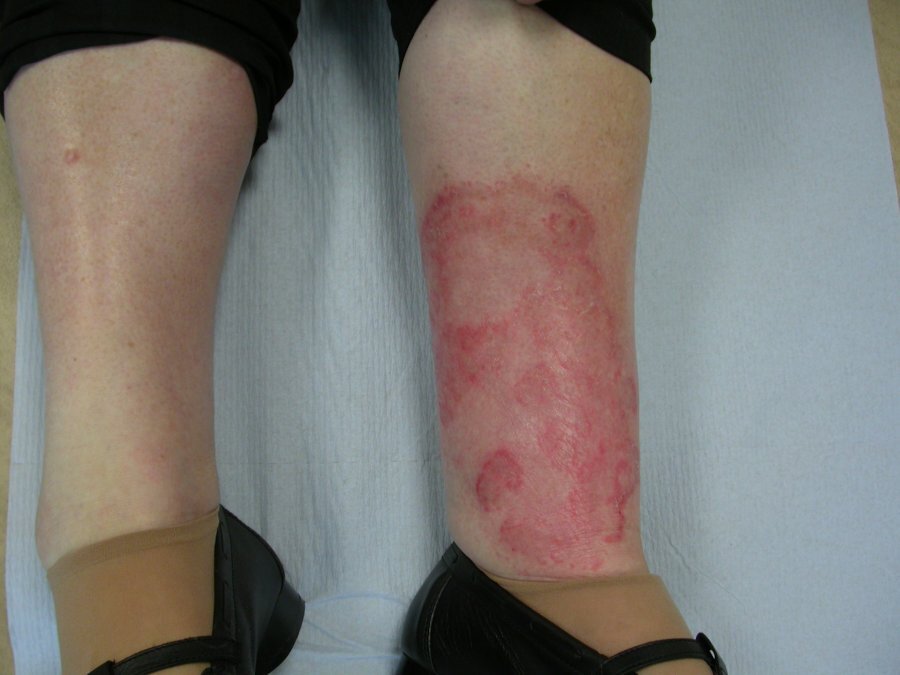
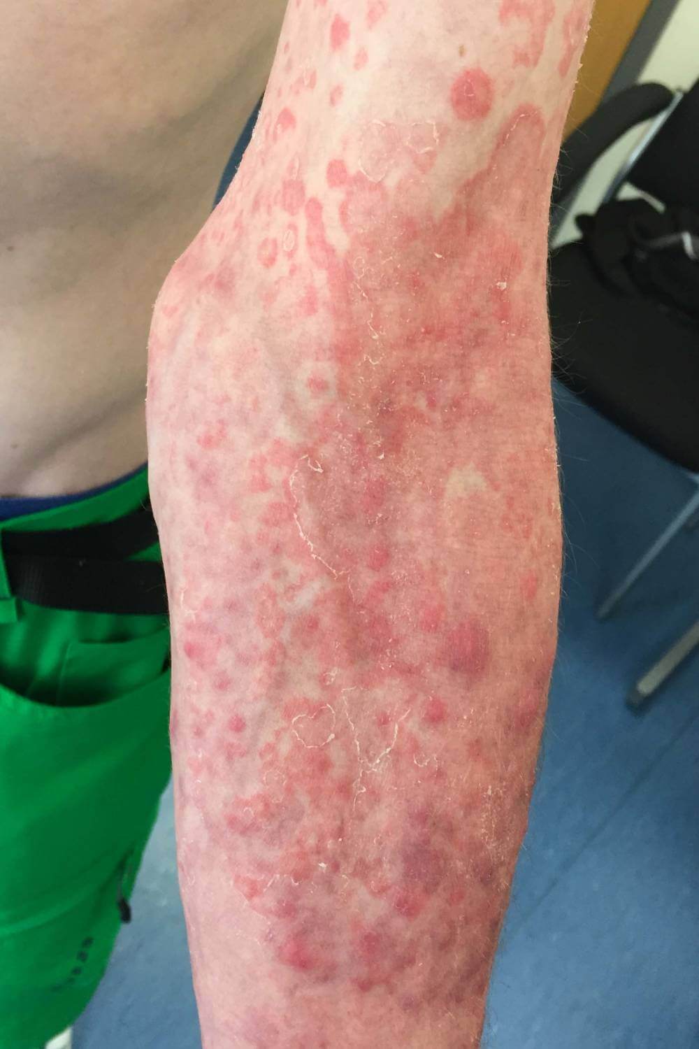
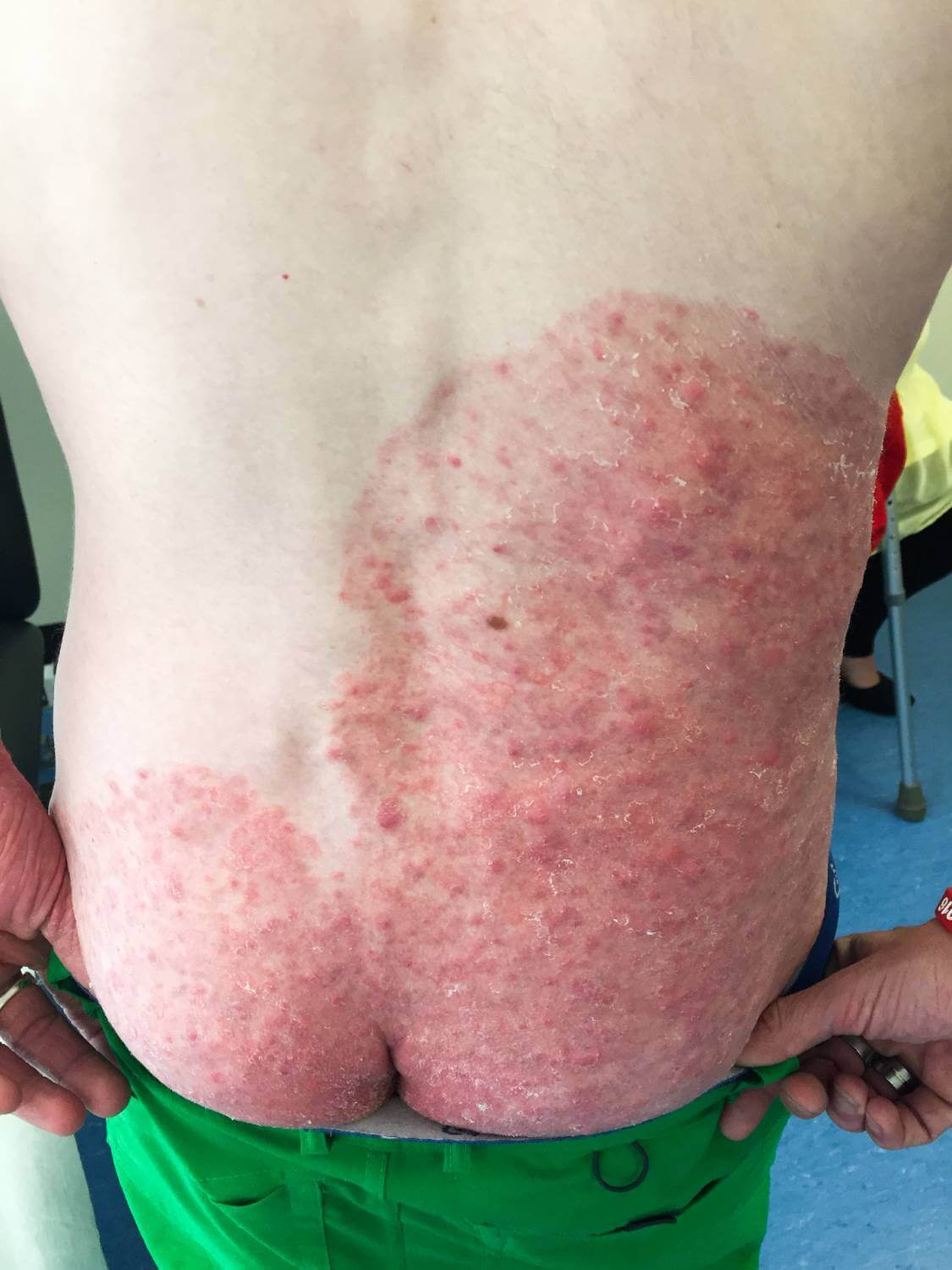
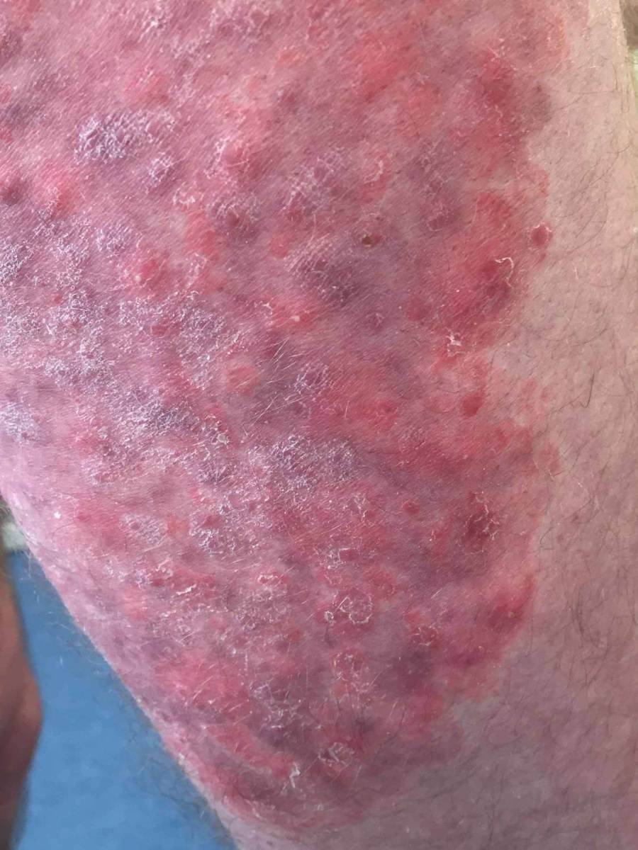
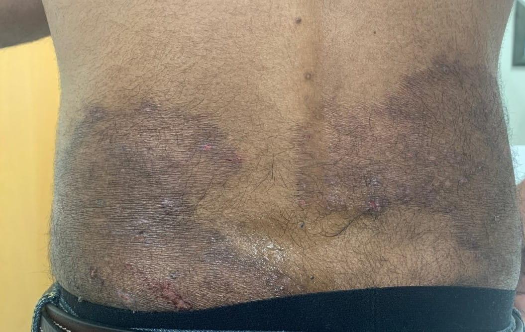
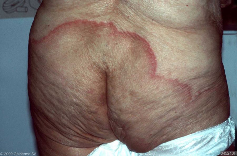
- Lesions are erythematous with scale and have an annular border. The erythema and scale tends to be most pronounced at the leading edge of the rash. Scale may be minimal or absent in flexural sites as a result of friction
- If the tinea is very inflammatory white / yellow pustules will be present within the affected patches
Tinea incognito
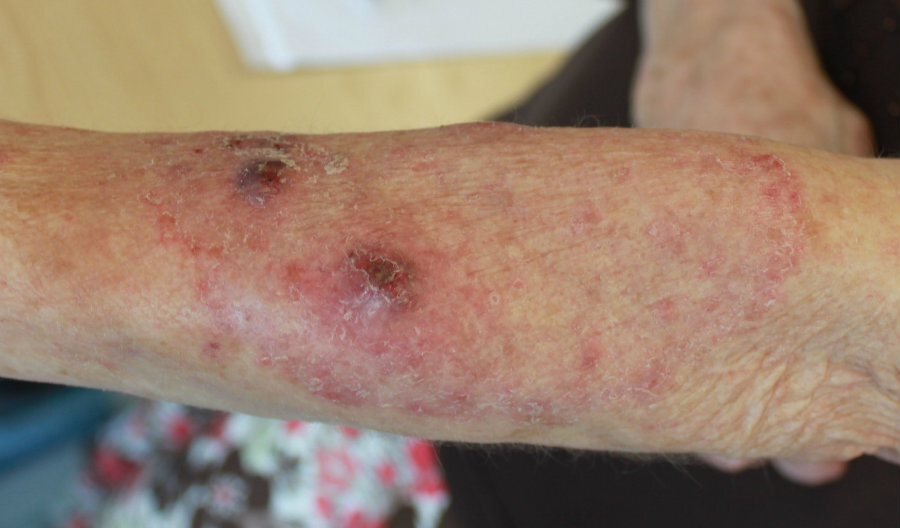
- Refers to tinea that has been misdiagnosed and treated inappropriately with topical steroids - the itch may settle a little with topical steroids giving a false sense of security, but the rash progresses
- Clinically there tends to be less scale and more pustules
Tinea imbricata
- A chronic superficial mycosis caused mainly by Trichophyton Concentricum
- Mainly found in isolated areas in developing countries. It is rare in developed countries
- The typical initial lesions consist of multiple, brownish red, scaly, pruritic papules. The papules then spread centrifugally to form annular and/or concentric rings that can extend to form serpinginous or polycyclic plaques with or without erythema
- With time, multiple overlapping lesions develop, and the plaques become lamellar with abundant thick scales adhering to the interior of the lesion, giving rise to the appearance of overlapping roof tiles, lace, or fish scales. Lamellar detachment of the scales is common
Clinical Images
Please refer to notes on image rights at bottom of the page with regards to individual image
ownership.
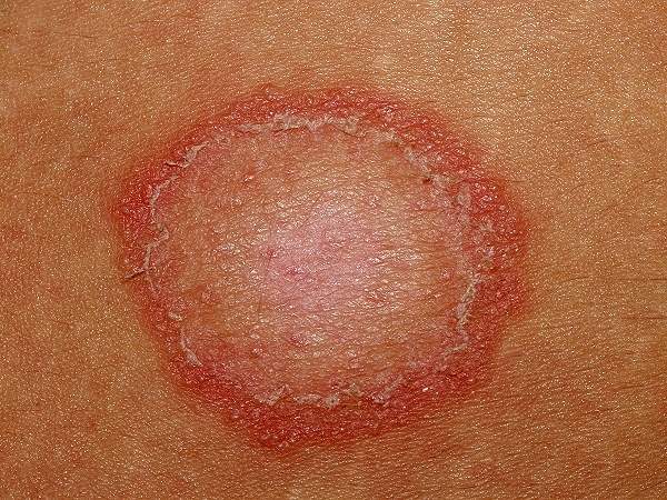
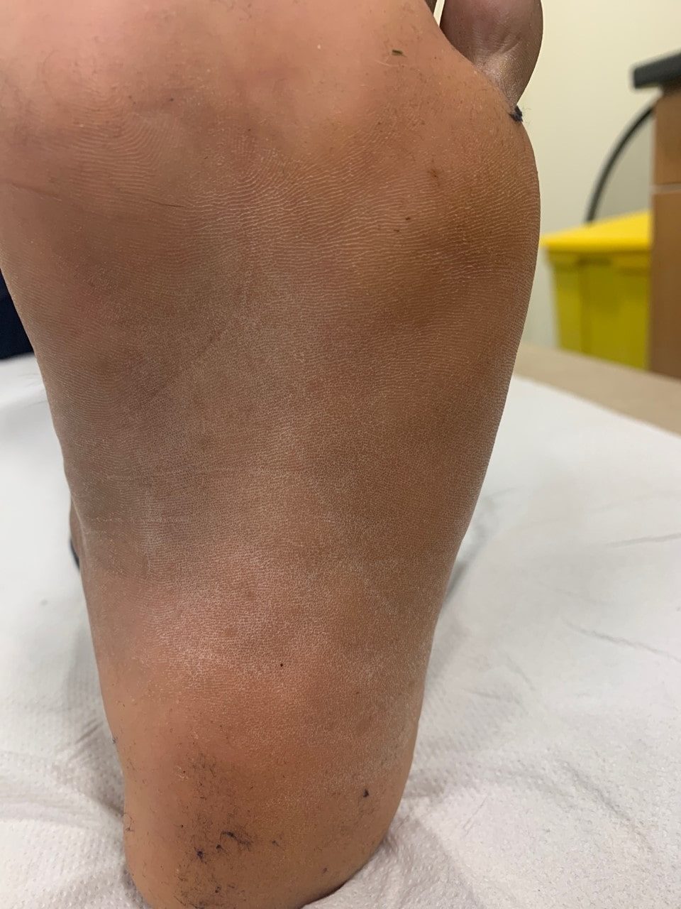
1
2
3
4
5
6
7
8
9
10
11
12
13
14
15
16
Investigations
-
Adequate scrapings should be taken with the back of a scalpel blade from the advancing edge of the lesion. The scrapings should be sent to microbiology either in a commercial sample pack or in folded black paper held by a paper clip. Adhesive tape (eg sellotape) stripping may be useful if scale is limited
Management
-
For milder cases treatment is normally with a topical antifungal agent eg terbinafine (Lamisil ®) cream BD for 2 weeks, or one of the imidazole creams eg miconazole (Daktarin ®) BD for 2-4 weeks. Terbinafine is more expensive but slightly more effective
-
If the rash is very extensive or inflammatory (pustules present) treat systemically eg terbinafine for two to four weeks depending on the extent
- Oral terbinafine is the drug of choice for the treatment of tinea imbricata
Disclaimer - the author PCDS cannot accept responsibility for any misleading or incorrect statements, and the management of individual patients remains the direct responsibility of the individual doctor. We do however hope that visitors to this site can contact us regarding comments that are considered misleading or incorrect so that we can continue to improve the site.
Image Rights - The PCDS would like to thank Dermatoweb, DermQuest (Galderma), and others who have contributed images. All named individuals and organisations maintain copyright for the relevant images.