Carcinoma cuniculatum (syn. epithelioma cuniculatum, verrucous carcinoma)
LAST UPDATED: Aug 02, 2021
Introduction
Carcinoma cuniculatum (CC), first described in 1954, is a locally aggressive, clinically exophytic, slow-growing, well-differentiated squamous cell carcinoma, primarily affecting the plantar surface of the foot. CC is regarded as low-grade with minimal metastatic potential.
This chapter is set out as follows:
Aetiology
-
There is contention regarding the role of human papillomavirus (HPV) types 1–4, 6, 11, 16 and 18 in the pathogenesis of verrucous carcinoma. Despite some specimens displaying oncogene expression or altered p53 activity, some authors have discovered no evidence of HPV
-
Trauma and long-term irritation have been implicated as weight-bearing areas have been found to be more frequently affected than non-weight-bearing areas
History
-
CC is more common in men with a mean age of presentation of 52-60 years
-
There is usually a delay in diagnosis due to the resemblance of the lesion to a wart or corn, which grows slowly and progressively despite topical treatment. The median time to diagnosis was 13 years in one study and 16 years in another case series
Clinical findings
Distribution
- Lesions are usually found on the sole of the foot (53%), toes (21%) and heel (16%)
- CC arising from other sites are very uncommon
Morphology
- Mainly occurs as a single mass or plaque, but there are rare reports of multiple lesions on the feet and ankles
- Lesions grow slowly into a large, hyperkeratotic, exophytic (ie growing outward beyond the surface epithelium from which it originates) mass
- Described in one account as squashy ‘with the consistency of an overripe orange’
- Lesions may become ulcerated and develop numerous sinuses from which a foul-smelling purulent keratinous debris can be expressed
Clinical Images
Please refer to notes on image rights at bottom of the page with regards to individual image
ownership.
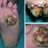
1
Investigations
-
Superficial / small biopsies are unsatisfactory and can provide false negative results. A good-sized incisional biopsy is required
Management
-
Patients should be referred urgently to Secondary Care (two-week wait)
-
Whilst CC is considered to have a very low incidence of metastases, tumours can invade the underlying structures such as bone, cartilage and tendons, accordingly surgical excision is regarded as first-line treatment. Recurrence rates of 19% have been reported. Attempts in electrodessication, cryotherapy and laser ablation results in higher excision rates. Moh's technique, under which microscopically controlled dissection allows total tumour removal with maximum preservation of normal tissue structure, has been reported to be successful in patients with CC
-
Amputations are necessary when the tumour is too extensive or recurs after multiple attempts of local excision. According to the literature, amputations were more likely to be performed when the tumour infiltrates the soft tissue between metatarsal heads or there is evidence of possible bony involvement on imaging. In these cases, it becomes impractical to perform local excision with good clearance margins
Disclaimer - the author PCDS cannot accept responsibility for any misleading or incorrect statements, and the management of individual patients remains the direct responsibility of the individual doctor. We do however hope that visitors to this site can contact us regarding comments that are considered misleading or incorrect so that we can continue to improve the site.
Image Rights - The PCDS would like to thank Dermatoweb, DermQuest (Galderma), and others who have contributed images. All named individuals and organisations maintain copyright for the relevant images.









