Xanthomata

LAST UPDATED: Aug 02, 2021

Introduction

The term xanthoma is derived from the Greek 'xanthos' meaning yellow. Xanthomata are skin lesions caused by the accumulation of fat in macrophages in the skin and more rarely the adipose tissue. Xanthomata are classified into the following, which are dependent on where they are found on the body and how they develop - xanthelasmata, tendon xanthomata, tuberous xanthomata, eruptive xanthomata, plane xanthomata and palmar xanthomata. Xanthoma disseminatum, a rare histiocytosis, is discussed in a related chapter.

Most types of xanthoma are indicative of disorders of lipid metabolism, and they may also be associated with other conditions such as diabetes mellitus or renal failure. Cerebrotendinous xanthomatosis (CTX) is a rare genetic metabolic disease that can present with tendon xanthomas but there is also a significant neurological component to the disease.  CTX patients often suffer from huge delays in diagnosis, yet it is very treatable if diagnosed early.

This chapter is set out as follows: 


Clinical findings

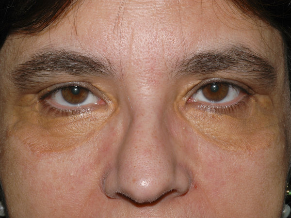
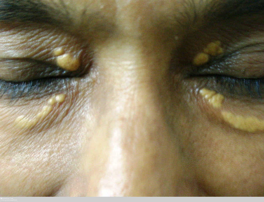
Xanthelasmata

  • Is the most common type of xanthomata
  • Distribution - lesions arise symmetrically on the upper and lower eyelids, and the medial canthus
  • Morphology - soft, velvety, yellow, flat-topped papules and plaques
  • May or may not be associated with hyperlipidaemia
  • Management
    • If LDL cholesterol levels are raised, treatment to reduce levels may improve the size of the lesions 
    • Physical treatments such as curettage, laser and chemical peels using trichloroacetic acid can all help, but lesions often recur

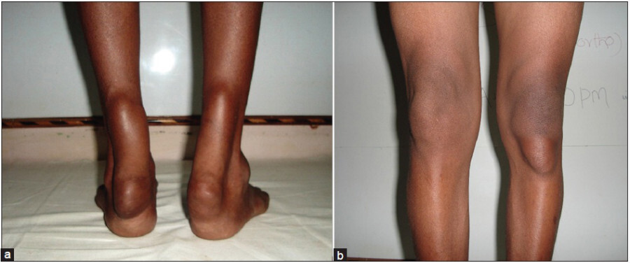
Tendon xanthomata

  • Distribution - most commonly found on dorsal aspects of the fingers, feet, and Achilles tendon
  • Morphology - slowly enlarging subcutaneous nodules related to the tendons or ligaments. As the lipid deposition is deep within the tendons, the skin is not yellow. Generally mobile, occasionally tethered
  • Associated with severe hypercholesterolaemia and elevated LDL levels 
  • CTX (cerebrotendinous xanthomatosis)
    • A rare genetic metabolic disease which, as the name suggests, can present with tendon xanthomas but there is also a significant neurological component to the disease. CTX patients often suffer from huge delays in diagnosis (a median of 26 years in one study) yet it is very treatable if diagnosed early
    • It is recommended to test for CTX when patients have two or more CTX symptoms, however, this is a little complicated as CTX is such a heterogeneous condition. The key early features are a history of prolonged cholestasis, chronic diarrhoea, neurological / psychiatric symptoms and presenile cataracts, along with tendon xanthomata
    • CTX is a progressive disorder with patients benefiting from early intervention -  patients suspected of having CTX should be referred to a metabolic specialist, although depending on the patient's specific issues they would likely also be managed by other disciplinaries eg genetics and neurology

Tuberous xanthomata

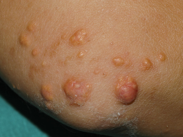
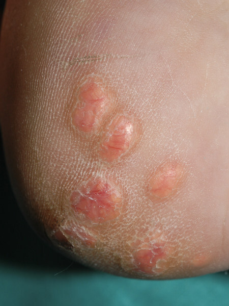
  • Distribution - develop around the pressure areas such as the knees, elbows, heels and buttocks
  • Morphology - firm, painless, red-yellow papules and nodules. The diameter can vary from 0.5 cm papules to 2.5 cm multilobulated nodules 
  • Associated with hypercholesterolaemia and elevated LDL levels

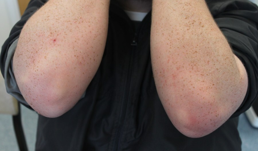
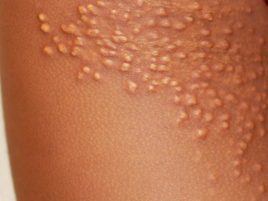
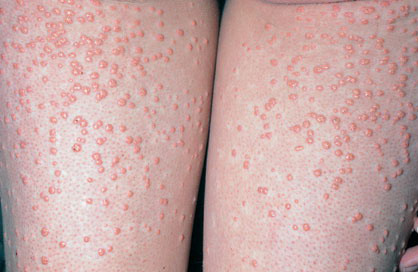
Eruptive xanthomata

  • Lesions may be tender and are usually itchy
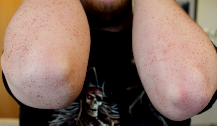
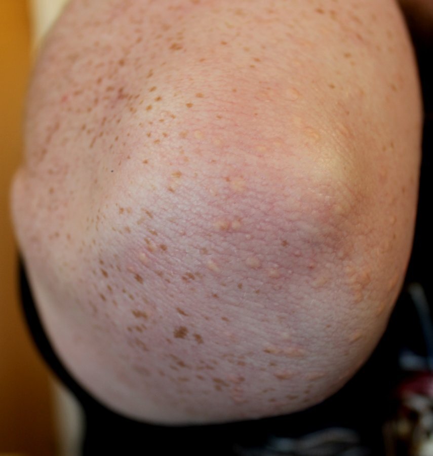
  • Distribution - most commonly arise over the buttocks, shoulders, arms and legs but may occur all over the body, although facial involvement is rare
  • Morphology - lesions typically erupt as crops of small, yellow papules 
  • Lesions may resolve spontaneously over a few weeks
  • Associated with hypertriglyceridaemia, which may or may not be secondary to diabetes mellitus
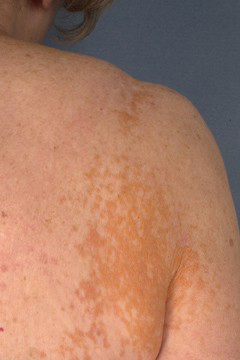
Plane xanthomata
  • Distribution - can occur anywhere on the body.
  • Morphology - multiple yellow / orange macules, or slightly elevated papules and plaques 
  • Associated with a wide range of lipid abnormalities, and a monoclonal gammopathy secondary to myeloma, lymphoma or macroglobulinaemia
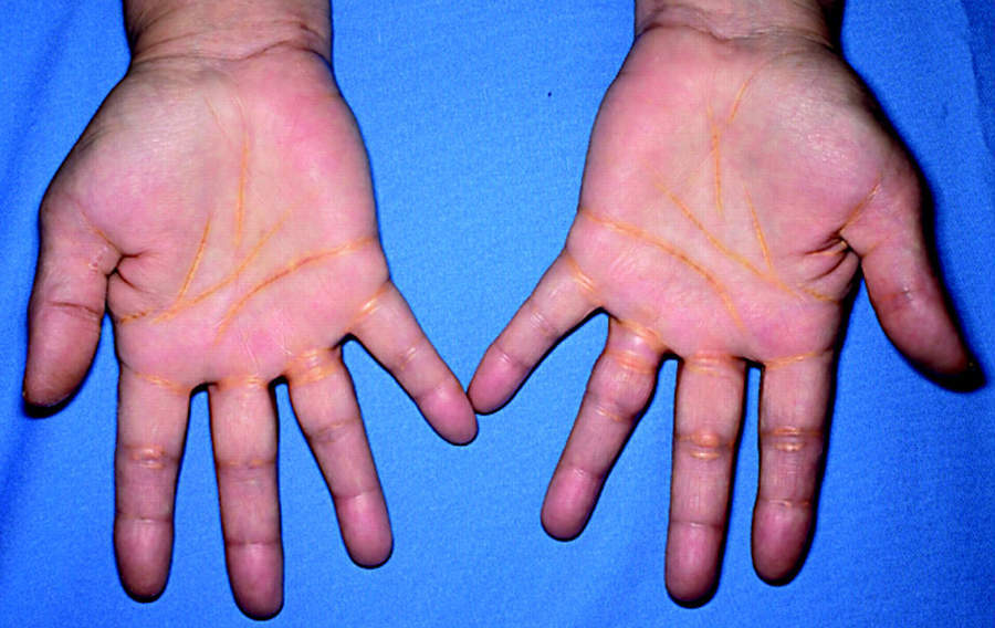
Palmar xanthomata
  • These have a similar appearance to plane xanthomata, but are found on the palmar creases 
  • Pathognomonic of type III / familial dysbetalipoproteinaemia

Clinical Images

Please refer to notes on image rights at bottom of the page with regards to individual image ownership.
Management

  • Many xanthomata improve with lipid lowering
  • For xanthelasmata refer to the section on clinical findings

Disclaimer - the author PCDS cannot accept responsibility for any misleading or incorrect statements, and the management of individual patients remains the direct responsibility of the individual doctor. We do however hope that visitors to this site can contact us regarding comments that are considered misleading or incorrect so that we can continue to improve the site.

Image Rights - The PCDS would like to thank Dermatoweb, DermQuest (Galderma), and others who have contributed images. All named individuals and organisations maintain copyright for the relevant images.

Quick Links

The following pharmaceutical companies have had no involvement in the content of this website or in our conference programmes


Almirall
Galderma
Glenmark
Johnson & Johnson
La Roche-Posay
LEO Pharma
Pierre Fabre
Schuco