Blaschko’s lines
LAST UPDATED: Aug 26, 2021
Introduction
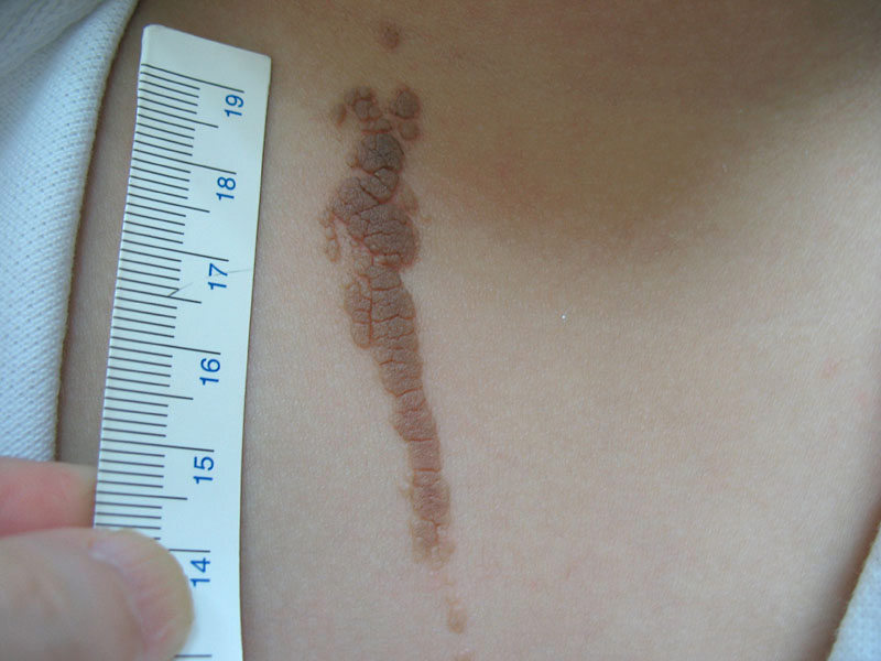
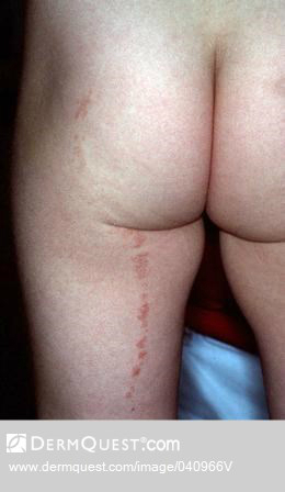
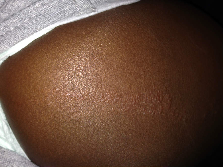
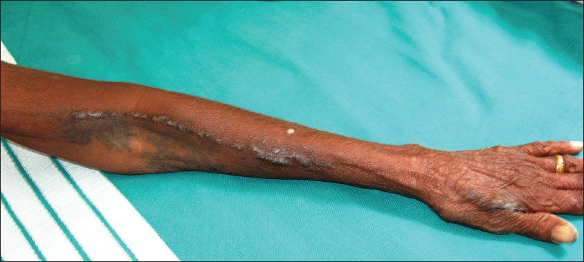
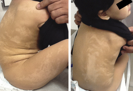
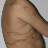
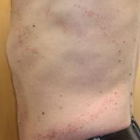
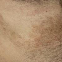
Blaschko's lines (BL) represent a classic pattern of cutaneous mosaicism (two or more genetically different populations of cells existing side by side), occuring in a wide variety of congenital and acquired skin conditions.
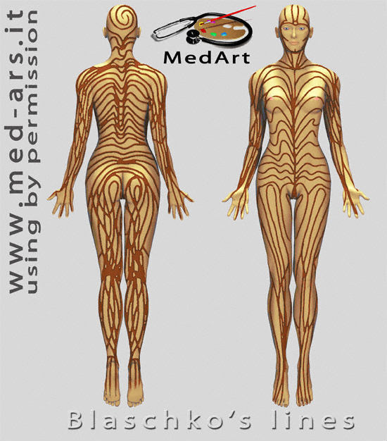
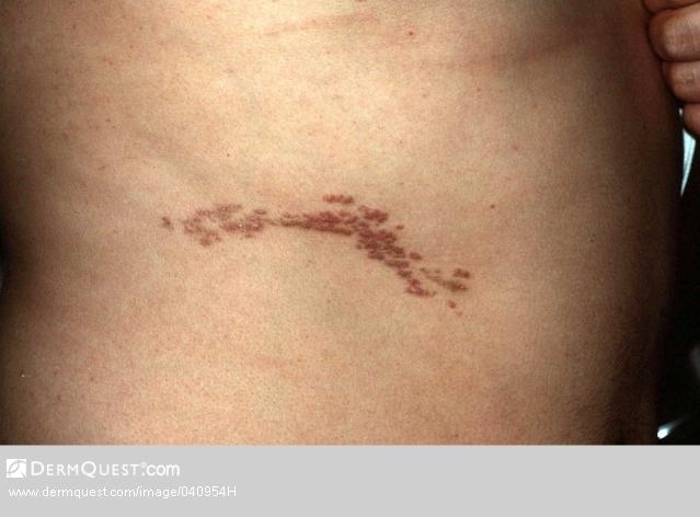
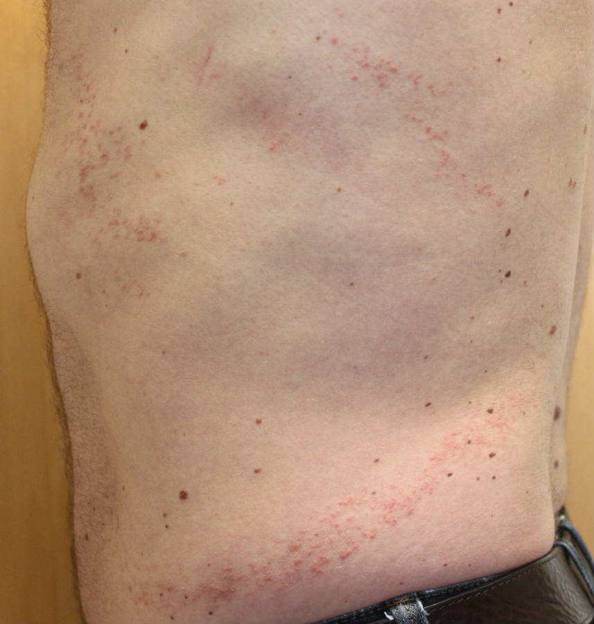
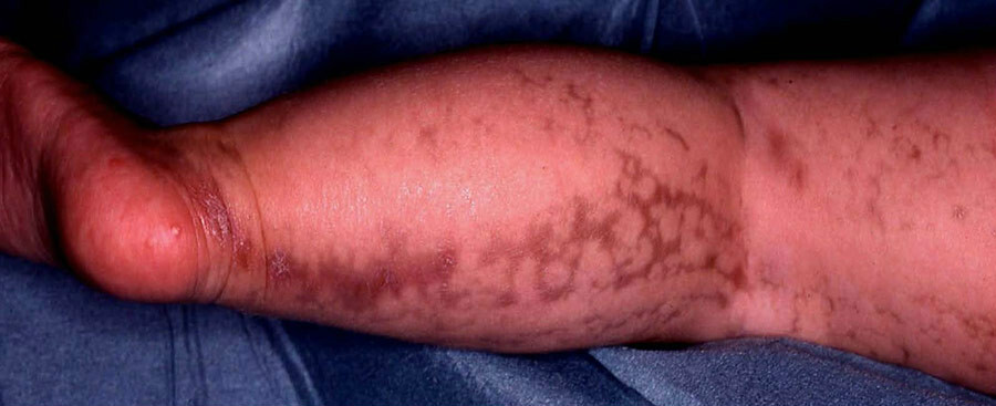
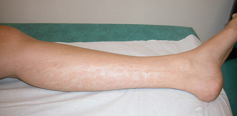
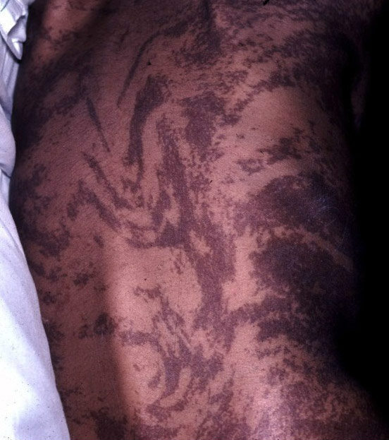
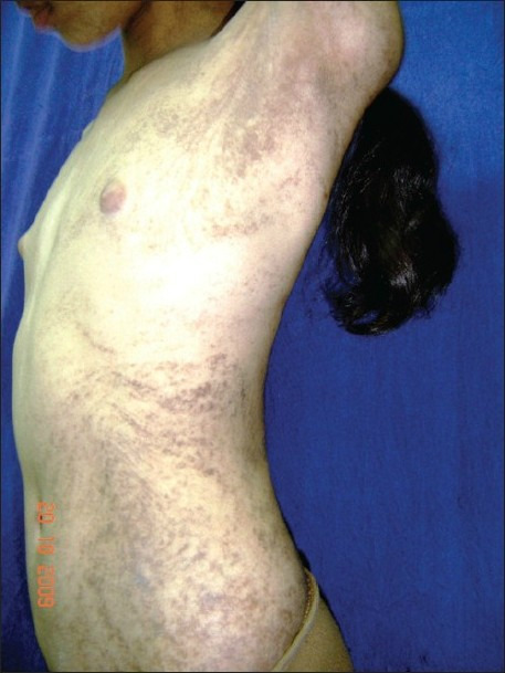
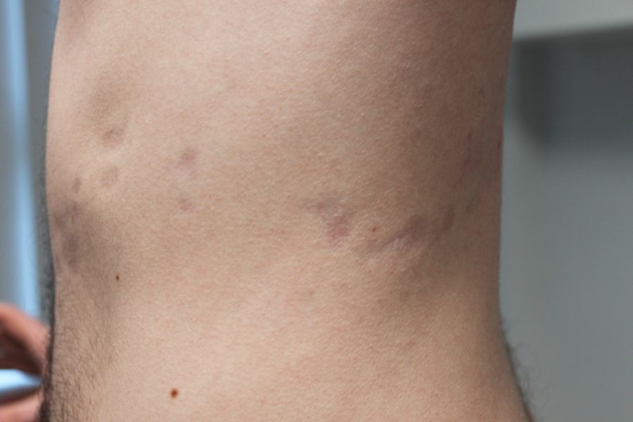
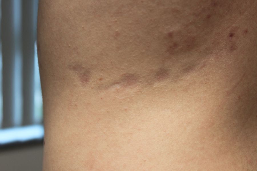
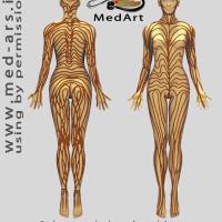
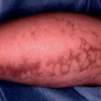
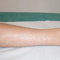
Blaschko's lines differ from dermatomes, and appear as single or multiple lines, whorls (swirls) and wave-like shapes in the skin. Cutaneous mosaicism will only arise as Blaschko's lines if the genetic mutations occur early enough in embryogenesis at a stage when cell migration from the neural crest is not yet complete.
This chapter is set out as follows:
History
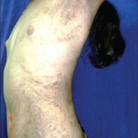
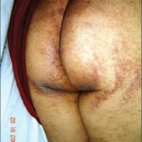
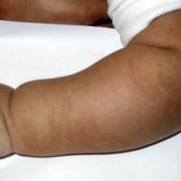
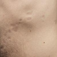
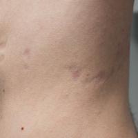
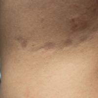
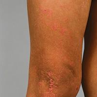
Numerous conditions can follow Blaschko's lines including:
- Most epidermal naevi
- Organoid naevi
- Linear presentations:
- Lichen striatus
- Linear lichen planus
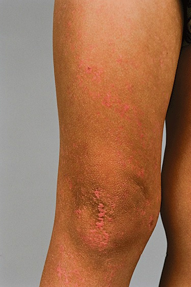
- Linear or naevoid psoriasis
- Linear cutaneous lupus erythematosus
- Linear Dariers disease
- Linear Hailey-Hailey disease
- Linear lichen nitidus
- Linear morphoea (some cases eg en coup de sabre follow Blaschko's lines, others do not)
- Linear and whorled naevoid hypermelanosis
- Linear atrophoderma of Moulin
- Linear porokeratosis
- Linear fixed drug eruptions
- Hypomelanosis of Ito
- Naevus depigmentosus (some cases are naevoid, others follow Blaschko's lines)
- Incontinentia pigmenti
- McCune-Albright syndrome (may have some Blaschkoid features)
- Focal dermal hypoplasia
- Blaschkitis (no apparent underlying cause)
Clinical findings
Blaschko's lines take on the following patterns:
-
Scalp - spiral
-
Face - vertical in the mid-face, laterally from the angles of mouth
-
Trunk - they differ from dermatomes in being more numerous and wave-like on the lateral trunk, and V-shaped in the middle of the back. Anteriorly they never cross the midline. Posteriorly the midline is often shifted from the anatomical midline
-
Arms and legs - linear or whorled (spiral), roughly parallel with long axis of the limbs
From a clinical perspective the different conditions can be grouped as follows:
Velvety / warty
- Incontinentia pigmenti
- Incontinentia pigmenti (IP) is a rare X‐linked dominant multisystemic ectodermal dysplasia that is usually lethal in males and that presents classically in females with skin lesions, teeth abnormalities, alopecia, nail dystrophy, and ocular and neurological findings
- The cutaneous findings are divided in to three stages
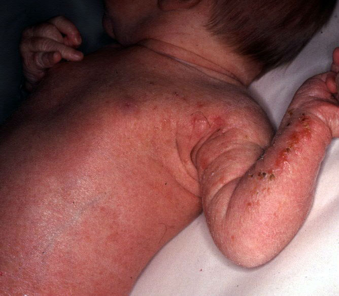
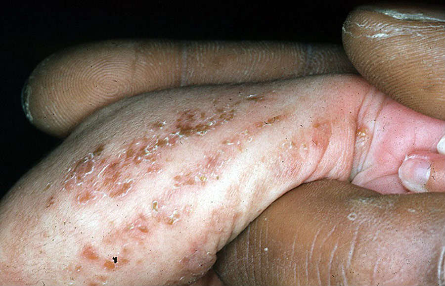
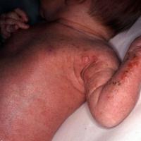
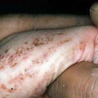
- Stage I typically present perinatally with an erythematous vesicular rash following Blaschko's lines
- Stage I evolves within a few months to a verrucous stage II, occurring mainly on the limbs
- Stage III consists of hyperpigmented streaks and whorls along Blaschko's lines that begin within months and fade in adolescence
Inflammatory
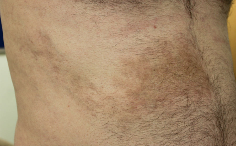
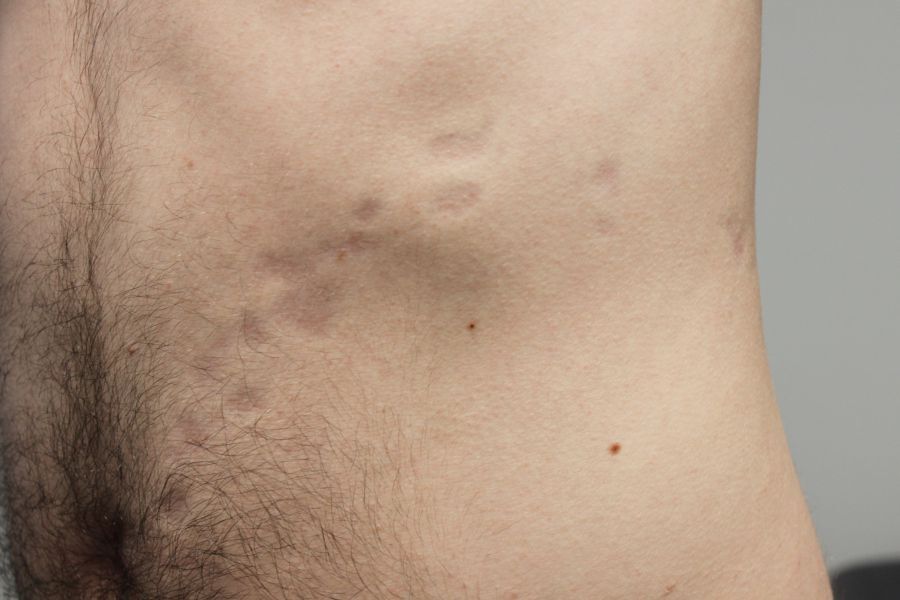
In the inflammatory phase the lesions will be red, but in the post-inflammatory phase the skin may become hyperpigmented.
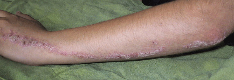
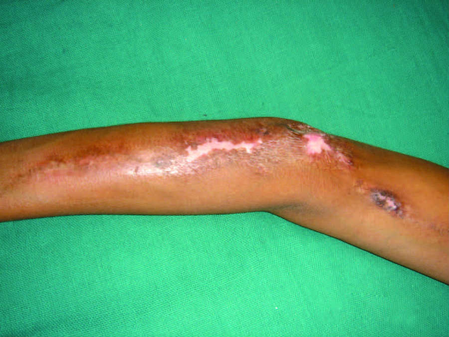
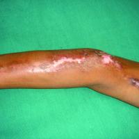
- Lichen striatus - mainly children. Most commonly proximal limb or neck. A sudden eruption of small papules that join together to form one or more dull-red, or hypopigmented, slightly scaly, irregular, linear bands. Self-limiting, spontaneously regressing within 3-12 months
- Inflammatory linear verrucous epidermal naevus syn. ILVEN - 75% of lesions arise during the first five years of life with a relatively gradual onset. Lesions are itchy and often persistent with an eczematous or psoriasiform appearance
- Blaschkitis - this term is used when no cause is found
Generally hyperpigmented (occasionally hypopigmented)
- A number of dermatoses that are in their post-inflammatory phase, eg lichen planus, and occasionally morphoea
- Incontinentia pigmenti: please refer to the section above 'velvety /warty'
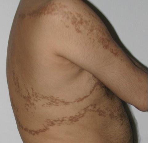
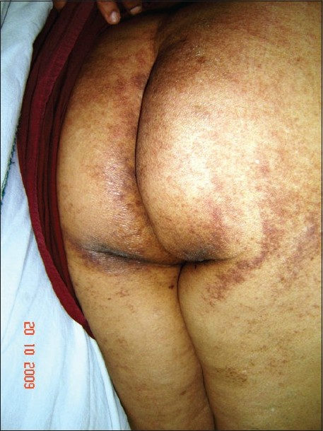
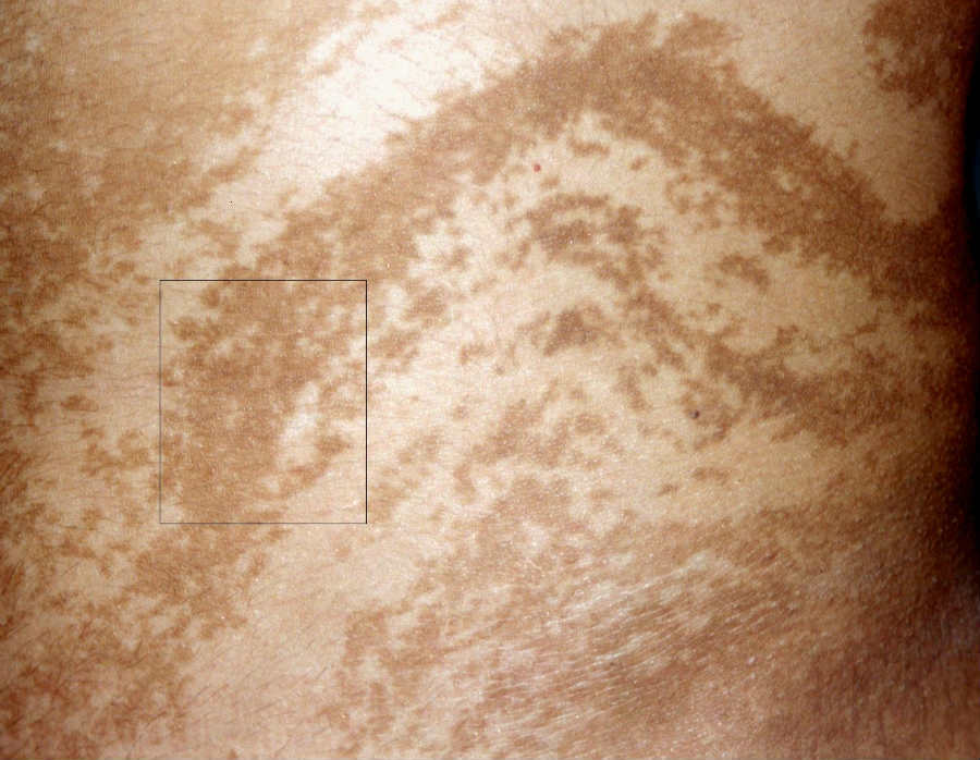
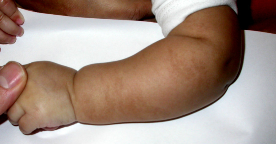
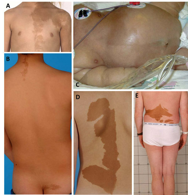
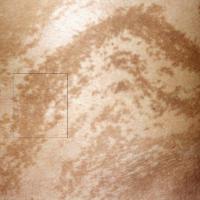
- Linear and whorled naevoid hypermelanosis (LWH)
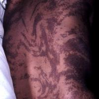
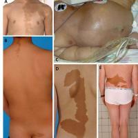
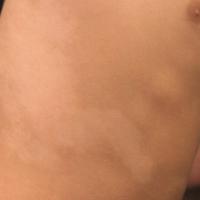
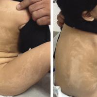
- Is characterised by the onset in infancy of hyperpigmented regions that follow the lines of Blaschko on the trunk and limbs. The striking colour changes are sometimes referred to as 'zebra-like'. There is no preceding inflammation or verruciform change
- The hyperpigmentation usually appears within a few weeks of birth, and progresses for 1 to 2 years before stabilisation. The changes usually persist indefinitely
- Striking similarities exist between LWH and hypomelanosis of Ito. In addition, several patients have been reported who display bands of both hypopigmentation and hyperpigmentation, making the distinction between LWH and hypomelanosis of Ito somewhat blurred, although in LWH accompanying non-cutaneous features appear to be very uncommon
- Linear atrophoderma of Moulin
- Linear atrophoderma of Moulin (LAM) is a rare linear dermatosis. The condition usually presents in childhood or early adolescence as asymptomatic, unilateral, hyperpigmented, atrophic, linear bands along Blaschko's lines without prior inflammation or sclerotic appearance
- The lesions usually progress over a few months, then stabilise and persist
- Focal dermal hypoplasia ‘Goltz syndrome’
- Focal dermal hypoplasia (FDH) belongs to the large group of ectodermal dysplasias. It is a rare genodermatosis that affects tissues of ectodermal and mesodermal origin. Clinically the syndrome is characterised by cutaneous, skeletal, dental, ocular and soft tissue defects
- A blaschkoid linear distribution is typical, lesions are prominent on the legs (especially the thighs), forearms, and cheeks (where lines radiate from the angles of the mouth). In mild cases, FDH involves only limited, sometimes unilateral, areas of skin. In severe cases, all areas of the body are involved
- The most prominent cutaneous features are reddish or red‐yellow cribriform atrophic skin lesions. Areas of hyperpigmentation or hypopigmentation, along with telangiectases, are common in atrophic sites. Other cutaneous abnormalities include lipomatous nodules, and a striking abnormality is the appearance of raspberry-like papillomas, these papillomas are multiple, often arising at junctions between the mucosa and the skin (ie, perioral, perivulvar, perianal, periocular junctions)
- Linear enamel hypoplasia is an important key feature. Numerous other oral alterations have been described including hypodontia, jaw cysts, clefting, hemihypoglossia of the tongue and papillomatosis
- McCune-Albright syndrome
- The classic triad that is McCune–Albright syndrome is the association of café‐au‐lait macules, polyostotic fibrous dysplasia and autonomously hyperfunctioning endocrinopathies. The clinical diagnosis can be made in the presence of two of these three features, and the genetic diagnosis can be made in the presence of one of these three with a detected mutation
- Typically the café‐au‐lait macules present at birth or appear early in the first decade, are large with irregular edges, and arranged in a segmental distribution. Lesions mostly affect the side overlying the bone defects, and rarely extend over the midline. Lesions may follow Blaschko's lines
- The commonest endocrinological abnormality is gonadotrophin‐independent precocious puberty
- If the condition is suspected then plain radiographs of the skull, mandible, pelvis and long bones should be performed to look for polyostotic fibrous dysplasia
If a distinct diagnosis cannot be made the terminology Blaschkoid hyperpigmentation is sometimes used. Current evidence suggests that additional cutaneous features of hyperkeratosis (thickened skin) or vascular anomalies are more likely to indicate other organ involvement.
Generally hypopigmented (occasionally hyperpigmented)
- Lichen striatus - see above under 'inflammatory'. Although many lesions are dull-red, they can be hypopigmented
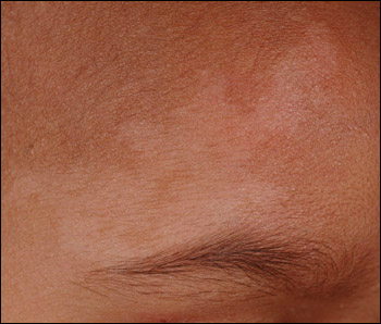
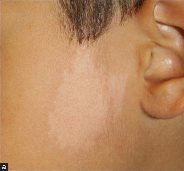
- Naevus depigmentosus (syn. achromic naevus)
- An uncommon naevus
- The lesion can be naevoid in appearance or follow Blaschko's lines
- It most commonly presents at birth or before three years of age
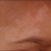
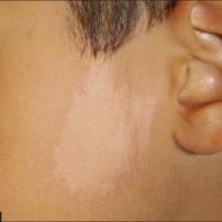
- The cutaneous appearance is usually that of a single, well‐marginated lesion on the trunk, although other sites can be affected. Often, smaller hypopigmented macules arise around the edges
- The name of the lesion is not quite right as it is hypomelanotic (reduced pigmentation) as opposed to amelanotic (completely white), as seen in congenital vitiligo
- The naevus remains stable over time
- Hypomelanosis of Ito
- A rare neuroectodermal disorder often associated with mental retardation and epilepsy
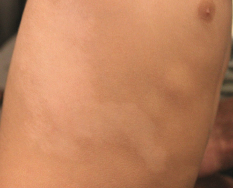
- Lesions usually present around birth as small 0.5-1 cm hypopigmented or white macules. Over time lesions become more confluent forming unilateral, or asymmetrical bilateral, hypopigmented whorls, streaks and patches
If a distinct diagnosis cannot be made the terminology Blaschkoid hypopigmentation is sometimes used. Current evidence suggests that additional cutaneous features of hyperkeratosis (thickened skin) or vascular anomalies are more likely to indicate other organ involvement.
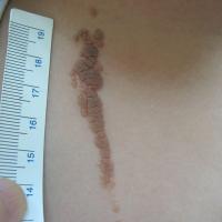
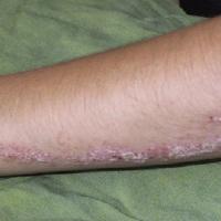
Clinical Images
Please refer to notes on image rights at bottom of the page with regards to individual image
ownership.
1
2
3
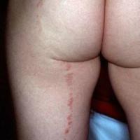
4
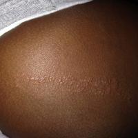
5
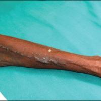
6
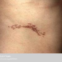
7
8
9
10
11
12
13
14
15
16
17
18
19
20
21
22
23
24
25
26
27
28
29
Management
-
The importance of conditions occuring in Blaschko's lines relates to:
-
How much the cutaneous features affect the patient
-
Whether they are associated with extra-cutaneous features eg McCune-Albright syndrome, focal dermal hypoplasia
-
If they are hereditary and can be passed onto other family members
-
For further information refer to the linked chapters
Disclaimer - the author PCDS cannot accept responsibility for any misleading or incorrect statements, and the management of individual patients remains the direct responsibility of the individual doctor. We do however hope that visitors to this site can contact us regarding comments that are considered misleading or incorrect so that we can continue to improve the site.
Image Rights - The PCDS would like to thank Dermatoweb, DermQuest (Galderma), and others who have contributed images. All named individuals and organisations maintain copyright for the relevant images.