Morphoea (syn. scleroderma)
LAST UPDATED: Oct 25, 2024
Introduction
Morphoea (syn. scleroderma) is a group of related conditions that share a common pathophysiology of increased collagen deposition in an autoimmune setting. Morphoea presents with varying degrees of sclerosis, fibrosis and atrophy in the skin and subcutaneous tissues sometimes extending into the fascia, muscle, bone, eye, and brain.
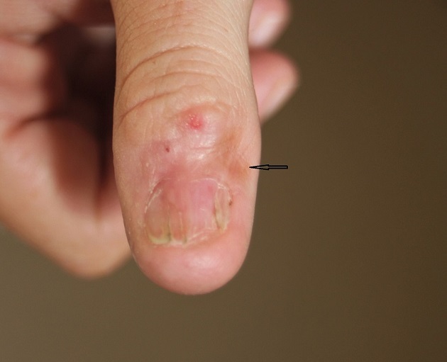
Morphoea is best considered as a separate entity to systemic sclerosis, which unlike morphoea results in Raynaud's phenomenon, sclerodactyly (spindled fingers), nailfold capillary changes, and systemic involvement.
This chapter is set out as follows:
Aetiology
- Although morphoea is of unknown aetiology, it is believed that both genetic and environmental factors have a role to play
- External environmental factors include injections (eg bleomycin), repeated friction, surgery, radiotherapy, and penetrating wounds
- Morphoea has also been associated with:
- Drugs eg carbidopa, penicillamine
- Chemicals eg polyvinyl chloride, solvents used in dry cleaning, and pesticides
- Graft-versus-host disease following bone marrow transplantation
History
-
Although any age can be affected, the peak incidence is 20-40 years of age, and 15% of cases occur before the age of 10 years
-
The female to male ratio is 3:1
-
It is less common in people with black skin
-
The onset of scleroderma is generally insidious, and although asymptomatic, affected skin often ceases to sweat
- While morphoea is not associated with systemic disease, up to one-third of cases (usually more severe cutaneous disease) have extracutaneous non-specific symptoms such as fatigue, arthralgia, muscle pain, and ocular symptoms
Clinical findings
Morphoea described by distribution
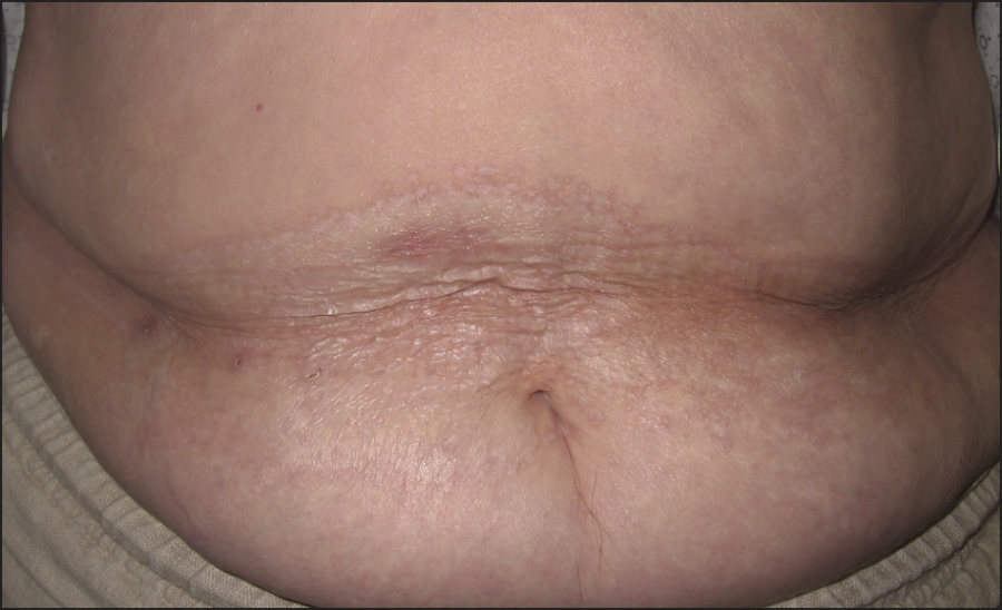
Although morphoea is primarily described by its distribution, the depth of tissue involvement is also important. Mixed morphoea is a term sometimes used when more than one pattern of morphoea is seen.
Limited plaque morphoea
-
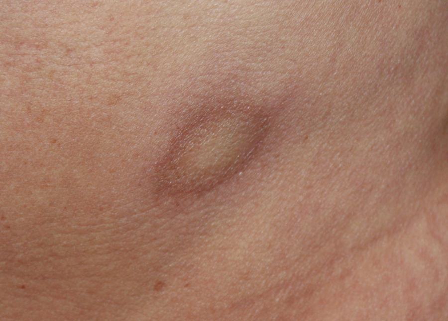
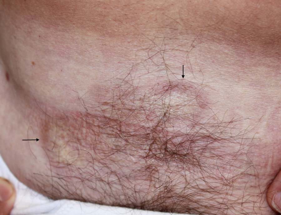
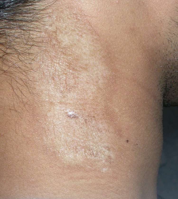
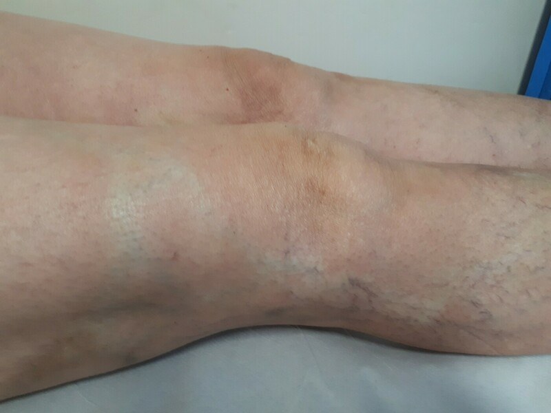
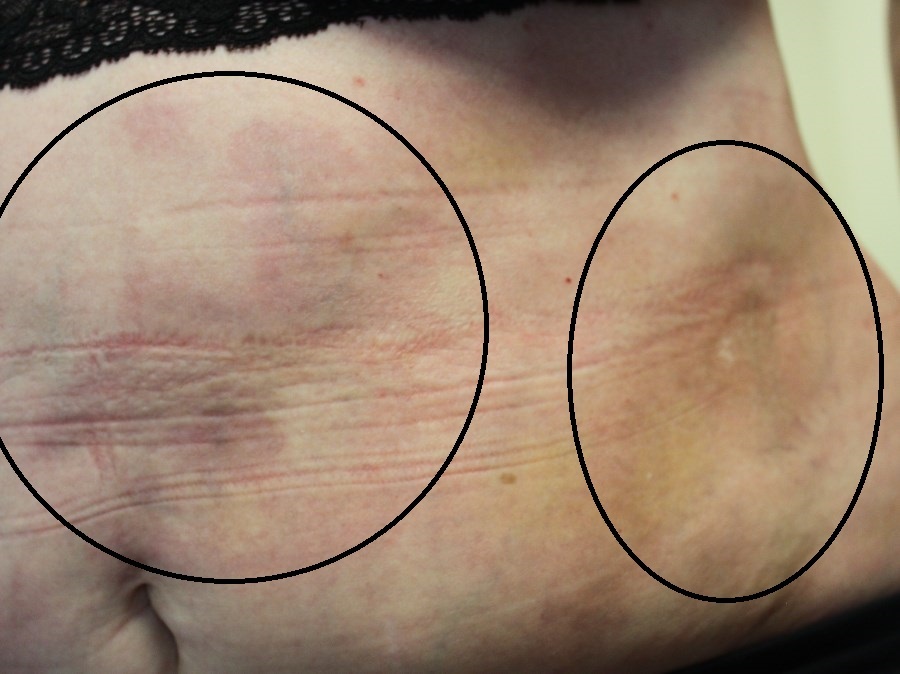
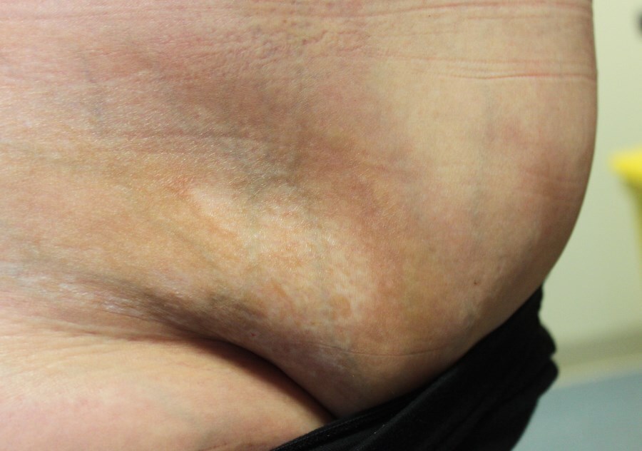
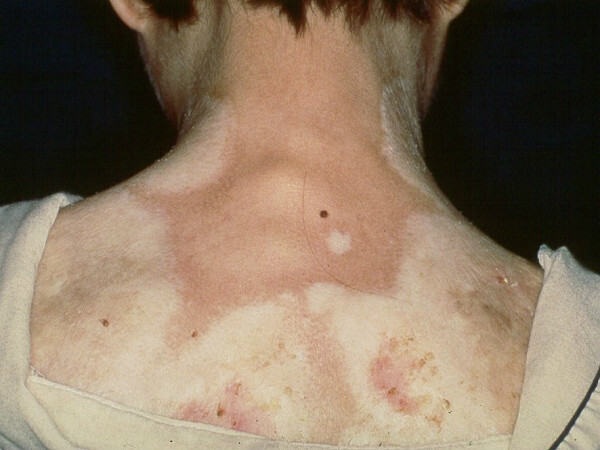
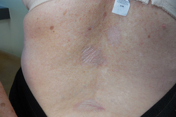
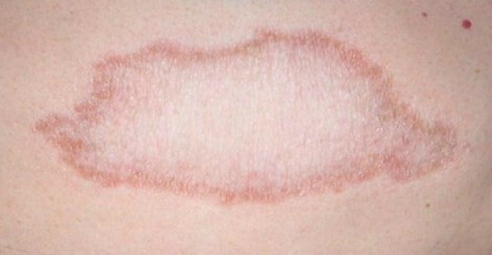
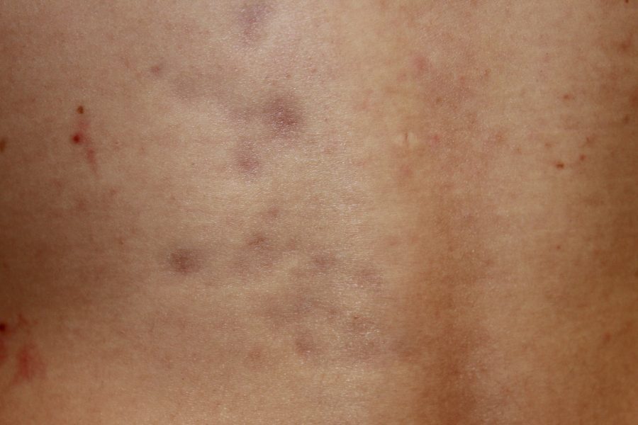
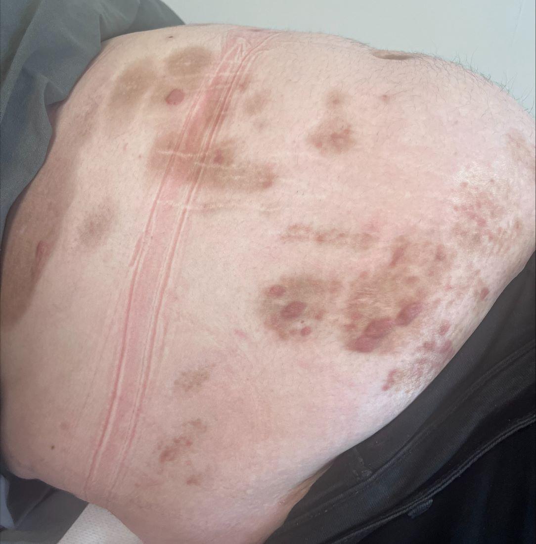
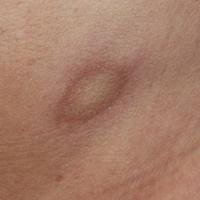
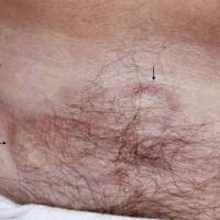
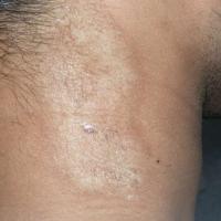
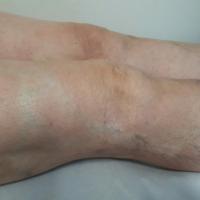
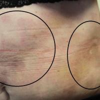
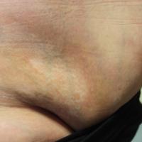
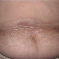
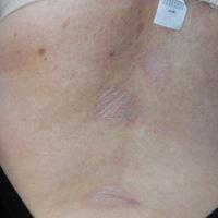
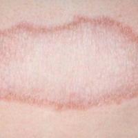
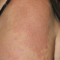
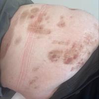
Indurated round or oval patches of skin that may enlarge to become several cm in diameter. Lesions are usually multiple and asymmetrical, involving one or two anatomical sites. The trunk and hips the most commonly affected areas
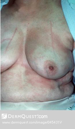
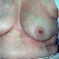
- An isomorphic distribution refers to morphoea arising at sites of pressures, eg under the waistband or bra
- Lesions are initially faintly purplish or mauve. Over several weeks-months the lesions develop a waxy, ivory white appearance in the centre with a lilac-coloured edge
-
Telangiectases may be seen, hairs are absent, and occasionally lesions blister
-
Deep involvement of underlying structures is uncommon
-
Old lesions become hyperpigmented
-
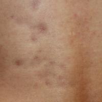
Occasionally lesions present in a ''guttate'' morphology, which are smaller in size and greater in number that typical plaque morphoea
-
The differential diagnosis includes:
Generalised plaque morphoea
-
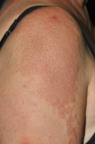
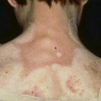
Is a rare condition in which idiopathic sclerosis of the skin occurs in a widespread fashion
-
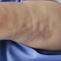
Lesions normally arise on the trunk and initially resemble plaque-type morphoea, but over time become much larger. Other sites can be affected. Refer below for pansclerotic morphoea
-
Contractures can arise as a result of limb involvement - occasionally, marked chest wall constriction can become life-threatening
-
Although generalised plaque morphoea does not have systemic involvement, it is associated with rheumatoid arthritis
-
The differential diagnosis includes systemic sclerosis:
-
Generalised plaque morphoea - Raynaud's phenomenon is uncommon, the trunk is the predominant site, the condition usually improves over time
-
Systemic sclerosis - Raynaud's phenomenon affects nearly all patients, the face and hands are the predominant sites, the condition is progressive
Linear morphoea
-
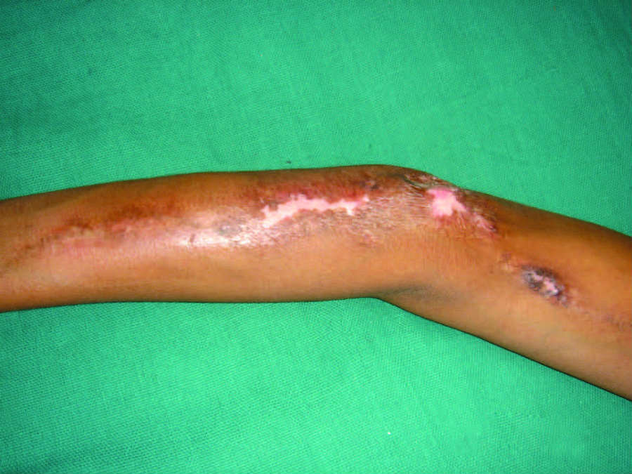
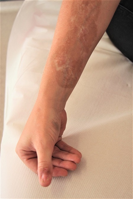
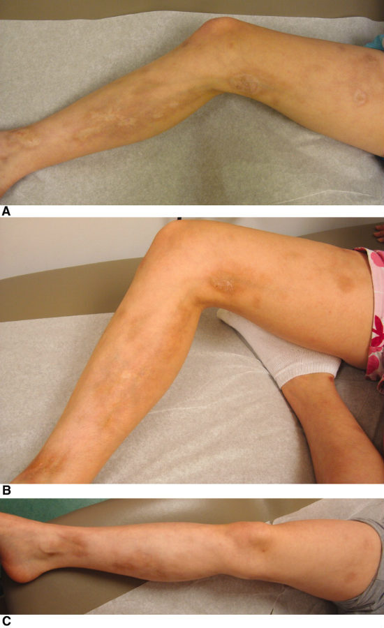
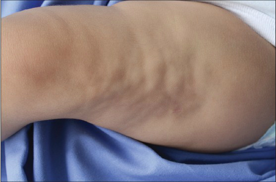
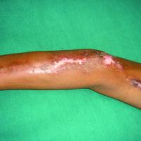
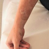
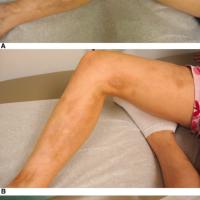
The most common presentation in children representing up to 65% of childhood cases of morphoea
- Preceded by trauma in up to 25% of cases
- Usually solitary and unilateral, but occasionally bilateral
- Most often presents on the limbs, or the head and neck, commonly following the lines of Blaschko
-
Lesions take on a similar appearance to plaque morphoea, but the lilac rings are less obvious
- Deeper involvement of the muscle or bone cause a growth disturbance in 20% of affected cases, and/or a flexion deformity
-
The diagnosis is usually straightforward, although many other conditions can grow along Blaschko's lines
Craniofacial morphoea
- There are two groups presenting in this way:
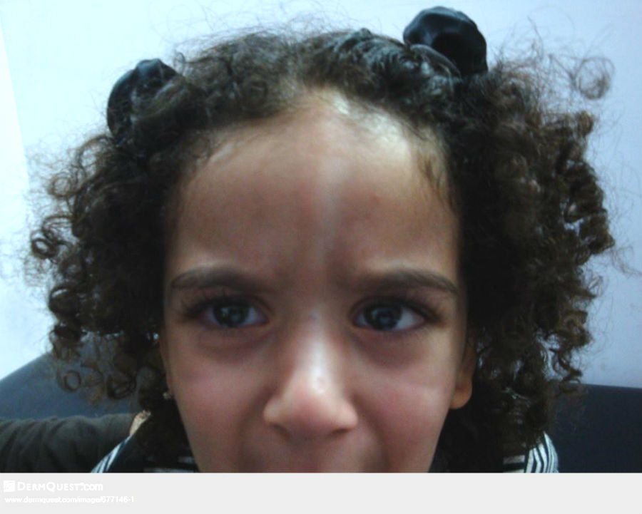
- En coup de sabre - a deep-seated form of linear scleroderma affecting the face and scalp, presenting with an ivory sclerotic plaque, which evolves into a linear depressed grove. The groove may extend from the scalp and forehead down to the cheek, nose, upper lip, and rarely the chin, neck, jaw, and tongue. Alopecia occurs at sites of scalp involvement
- Progressive hemifacial atrophy (syn. Parry-Romberg Syndrome) - is a slow and progressive atrophy affecting one side of the face, and sometimes the neck. The atrophy involves the skin, muscles, and skull causing a change in the shape of the face. 10-35% of cases have neurologic disturbances, which include focal epileptiform seizures and trigeminal neuralgia. Oral manifestations include atrophy of half of the lip and tongue, shortening of the body of the mandible and/or ramus of the mandible, and malformed tooth roots
Keloidal morphoea
- A very rare variant of morphoea. Clinically, it presents with asymptomatic firm keloid-like nodules or plaques, which are erythematous and irregularly shaped. Lesions have a predilection for the trunk and proximal extremities. The condition most commonly affects middle-aged women
Morphoea (scleroderma) described by depth
- The term subcutaneous morphoea refers to involvement of deeper tissues, and can occur in any of the groups described above, although it is infrequent in limited plaque morphoea
- Occasionally morphoea only arises in deeper tissues, which is referred to as morphoea profundus, although overlying skin may be hypo or hyperpigmented
- Osteoma cutis refers to bone formation within the skin, and differs from calcinosis cutis, which represents calcium salt deposits within the skin
- Involvement of muscles and other deep structures leads to changes in physical appearance, and can cause functional change
- Pansclerotic morphoea is characterised by near total body surface involvement, with circumferential lesions, sparing the fingers and toes, which usually extends into the subcutaneous tissue, and may affect the fascia, muscle, and bone. Raynaud's and systemic involvement are absent. Pansclerotic morphoea has a more insidious onset than eosinophilic fasciitis and post-infectious scleredema, symptoms progressively appearing over months
Clinical Images
Please refer to notes on image rights at bottom of the page with regards to individual image
ownership.
1
2
3
4
5
6
7
8
9
10
11
12
13
14
15
16
17
18

19
20
Investigations
- Blood tests
- CRP/ESR can be raised
- Autoantibodies - up to 50% of patients have elevated levels of ANA, anti-histone antibodies, or anti-ssDNA antibodies (single-stranded DNA)
- RF and anti-CCP can be raised with arthralgia
- For more information on tests including autoantibodies refer to the section on investigations
-
Radiology
- May be required if there is significant involvement of deeper tissues
- Histology
- In many cases a biopsy is not needed
- The histological findings of morphoea and systemic sclerosis are similar, with a fundamental process of thickening and homogenisation of collagen bundles
-
The epidermis is usually normal, but rete ridges may become flattened as the condition progresses
-
The dermis is initially oedematous with degeneration of collagen fibrils and a perivascular and interstitial variably dense infiltrate of lymphocytes. In the late sclerotic stage the inflammatory infiltrate typically disappears, and the dermis become thickened with dense collagen bundles in the reticular dermis and few fibroblasts
-
Depending on the subtype of morphoea, the histological changes may extend into the fascia and even underlying muscle, although in superficial plaque morphoea the thickened collagen bundles are restricted to the dermis
Management
Prognosis
-
Limited plaque morphoea tends to improve with time, often over 3-5 years, but some cases may last for many years with new lesions developing as old one's fade
-
Linear morphoea persists longer but most cases eventually resolve
-
Craniofacial lesions may improve, although facial hemiatrophy usually persists
-
Some improvement in generalised plaque morphoea is usually seen over a few years, although the condition may last for many years
Treatment
Step 1: general measures
-
Provide a patient information leaflet on morphoea
-
In uncomplicated cases, eg many cases of plaque morphoea, no treatment may be appropriate as the natural history is towards spontaneous resolution
Step 2: topical or intralesional treatments for localised morphoea
-
Corticosteroids - potent to super-potent topical steroids, and intralesional steroids may be helpful in some cases, but only during the inflammatory phase
-
Other topical treatments reported as sometimes beneficial include calcipotriol and tacrolimus
Step 3: linear morphoea
-
A combination of calcipotriol cream and phototherapy (UVA1 > PUVA) has found to be beneficial in some cases of linear morphoea
Step 4: systemic treatment for more progressive cases
-
Systemic treatment should be considered for progressive cases, eg linear morphoea affecting limb length or causing contractures, and generalised morphoea
- Options include methotrexate or mycophenolate mofetil, sometimes combined with pulse methylprednisolone
-
Other treatments sometimes used include hydroxychloroquine, ciclosporin, and penacillamine
Step 5: other treatments
- Include physiotherapy, surgery, and psychology
Disclaimer - the author PCDS cannot accept responsibility for any misleading or incorrect statements, and the management of individual patients remains the direct responsibility of the individual doctor. We do however hope that visitors to this site can contact us regarding comments that are considered misleading or incorrect so that we can continue to improve the site.
Image Rights - The PCDS would like to thank Dermatoweb, DermQuest (Galderma), and others who have contributed images. All named individuals and organisations maintain copyright for the relevant images.