Infantile haemangiomas (including strawberry naevi)
LAST UPDATED: Jun 15, 2022
Introduction
Infantile haemangiomas are rapidly growing, benign proliferations of endothelial cells, appearing within weeks of life, proliferating for approximately 9 months and then involuting, a process which is complete by 10-12 years of age.
This chapter is set out as follows:
Aetiology
-
Haemangiomas are the most common of the infantile tumours, most frequently seen in Caucasian female infants and in those born prematurely
-
The prevalence of these tumours is about 1%
-
They are presumed to be an abnormality of embryonic angioblastic development
-
The capillaries are the most commonly affected vessels
History
-
About 20% are present at birth, and 90% are seen within a month post-partum
-
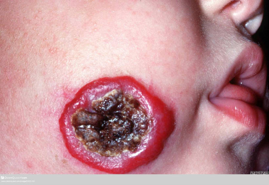
Lesions grow rapidly and can bleed or ulcerate
-
Apart from the occasional lesion that is fully developed at birth, 80% of growth occurs within the first 4 months, with lesions reaching a maximum size around 9 months of age. Deep lesions can grow for longer to become very large causing anatomical distortion
-
By year 4 there is approximately 80% regression. Ultimately up to 95% will completely / almost completely resolve
Clinical findings
Distribution
- Often single but multiple lesions can develop (more than 10 is uncommon)
- 60% of the lesions occur on the head and neck, other common sites include the buttocks and perianal area
Morphology
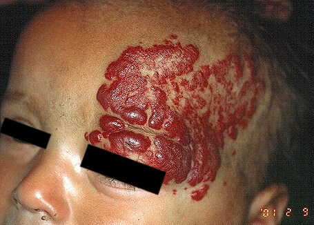
Tumours can be localised or segmental, and superficial or deep - although many have both superficial and deep components and are termed mixed infantile haemangiomas:
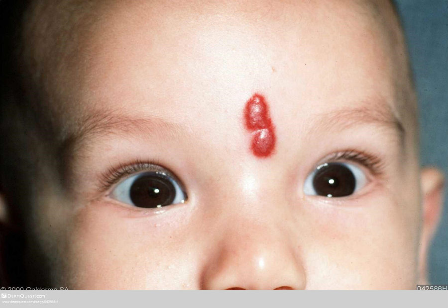
- The infantile superficial haemangioma is commonly known as a strawberry naevus (capillary haemangioma). Clinically they appear as red, relatively soft, and somewhat irregular in outline
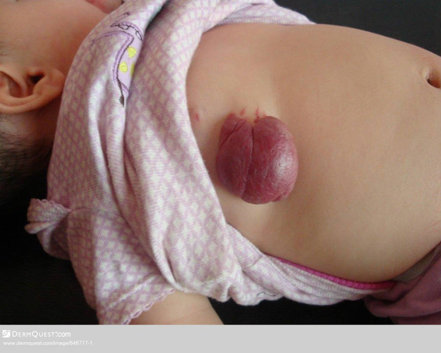
- Infantile deep haemangiomas are also called cavernous haemangiomas and are more deeply set in the dermis and subcutis. Clinically they appear as a bluish soft to firm swelling
- Both types of haemangiomas may occur together in mixed infantile haemangiomas when a strawberry naevus overlies a bluish swelling
Clinical Images
Please refer to notes on image rights at bottom of the page with regards to individual image
ownership.
Management
-
Most lesions need no treatment. Regular monitoring in Primary Care may reassure parents. Photographs at 6-12 month intervals are very useful
- Most of the problematic lesions are segmental proliferative haemangiomas as they occur at a younger age, grow up to ten times larger, and are more likely to have associated congenital anomalies
-
Urgent referral (same day) and intervention is required for the following lesions:
-
Lesions that are likely to grow into or within an important structure such as the eye, nose/mouth and ear leading to obstruction of the visual axis (amblyopia can occur within 10 days of lesions interfering with vision), airway or external auditory meatus
-
Interfering with feeding
- Lesions under the chin - can have laryngeal and other systemic involvement
- Lesions on the scalp can be associated with intracranial complications
- Lesions overlying the lumbosacral area - up to 50% of such cases are associated with significant problems such as tethering or compression of the spinal cord, and external genital abnormalities. MRI imaging should be performed in such patients
-
Timely referral is required for the following lesions:
- Lesions that have grown or are likely to grow into or within other important structure such as the genitalia
- Anywhere on the body if complicated by bleeding, ulceration or causing functional impairment
- Multiple haemangiomas (5 or more) can be associated with visceral haemangiomas including cardiac lesions
- Kasabach-Merritt syndrome (consumption coagulopathy) is an uncommon complication, mainly in larger lesions
-
Treatment
- If treatment is required, first-line therapy is a beta-blocker, usually systemic, although occasionally topically. To be effective treatment must be given early during the proliferative (growth) phase, which usually leads to a rapid and dramatic involution of the haemangioma. Treatment delay can lead to potentially serious outcomes including anatomical distortion
-
Other treatments sometimes used include laser, and systemic corticosteroids
Disclaimer - the author PCDS cannot accept responsibility for any misleading or incorrect statements, and the management of individual patients remains the direct responsibility of the individual doctor. We do however hope that visitors to this site can contact us regarding comments that are considered misleading or incorrect so that we can continue to improve the site.
Image Rights - The PCDS would like to thank Dermatoweb, DermQuest (Galderma), and others who have contributed images. All named individuals and organisations maintain copyright for the relevant images.