Melanocytic naevi (common type)
LAST UPDATED: Aug 06, 2026
Introduction
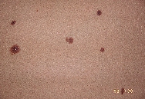
Benign melanocytic naevi, commonly referred to as 'moles', arise as a result of proliferation of melanocytes, the cells in the skin that produce pigment. This chapter focuses on common melanocytic naevi, which usually take on a familiar appearance. There are many variants of benign melanocytic naevi, which are detailed in other chapters.
This chapter is set out as follows:
Aetiology
- The cause of melanocytic naevi is unknown
- Melanocytic naevi have historically been classified by their histological appearance as being junctional (focal proliferation of melanocytes at the dermal-epidermal junction), compound (a combination of junctional activity and intradermal naevus cells) and intradermal (groups of mature naevus cells in the dermis only). The evolutionary theory was that naevi progress from junctional to compound and then intradermal, which is now thought not to be case for all such lesions
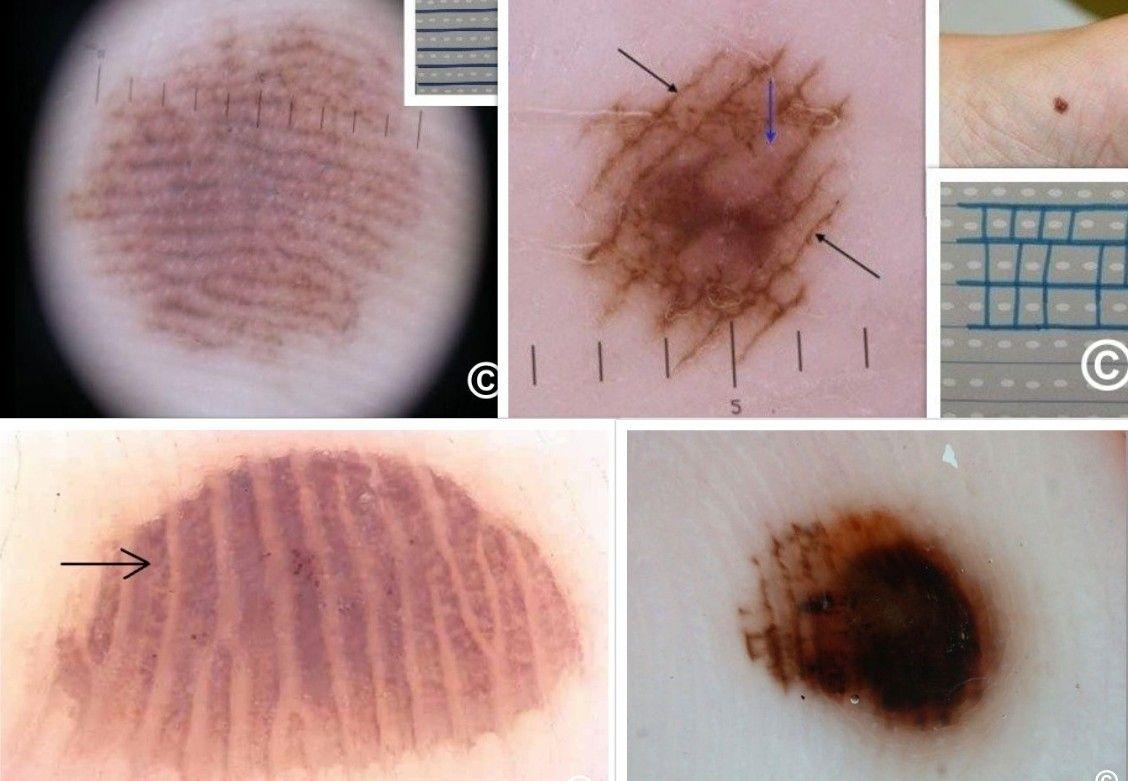
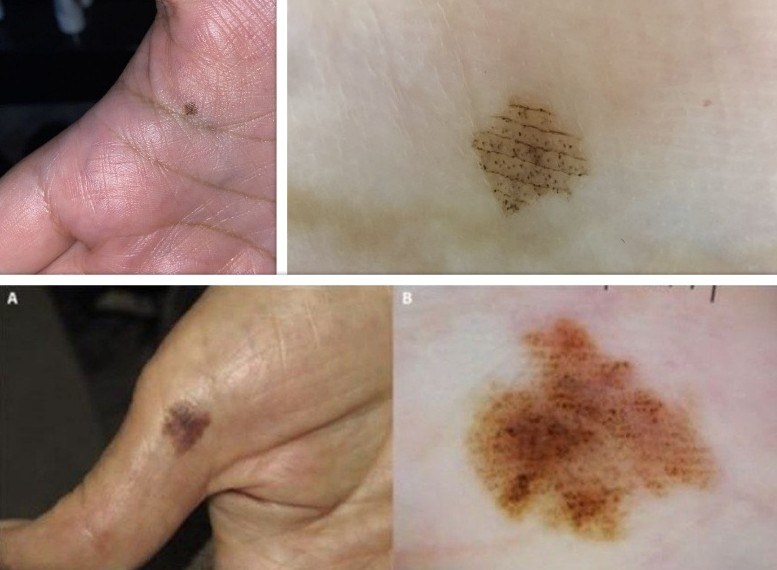
- Dermoscopy has given rise to a new classification of melanocytic naevi based on their pigment pattern, which in common moles can be:
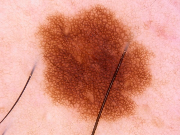
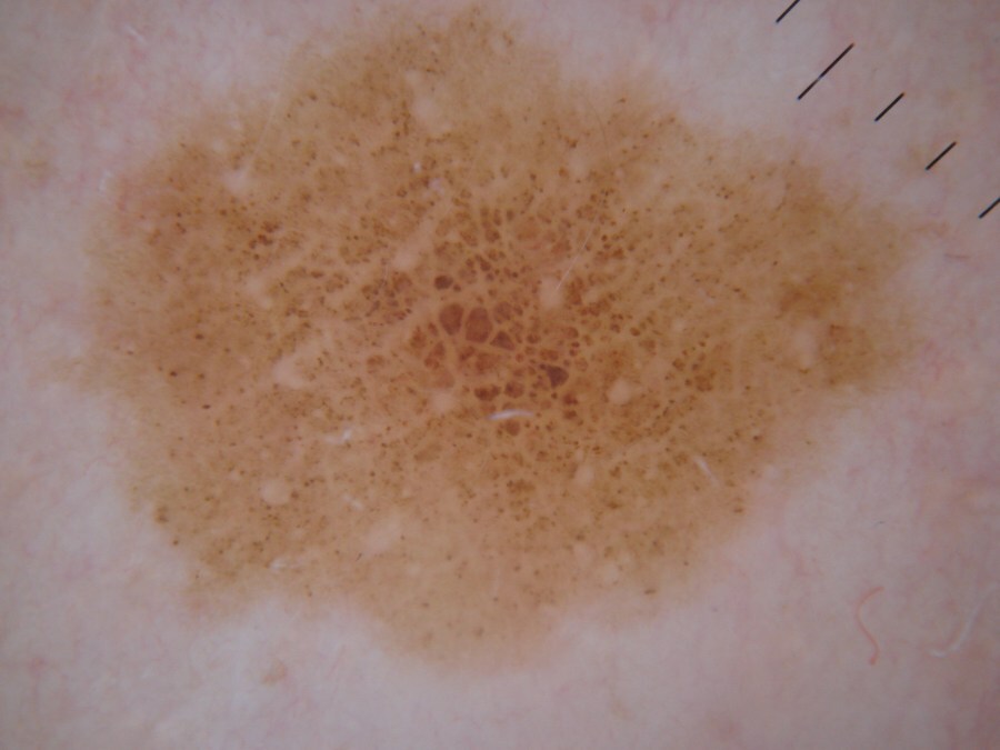
- Predominantly reticular (ie a pigment network) - these are acquired naevi, which have melanocytes at the dermoepidermal junction
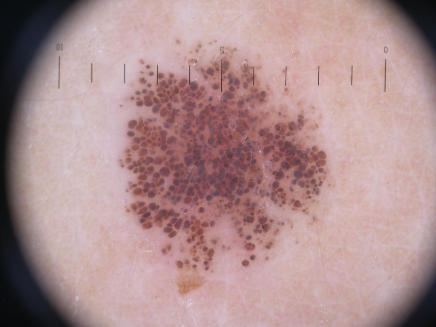
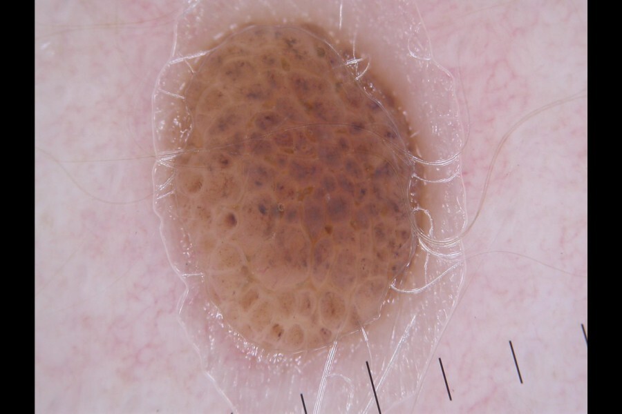
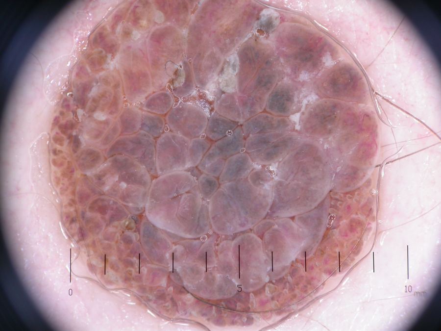
- Globular / cobblestone - nests of melanocytes found in the dermis (ie dermal naevi); probably congenital
History
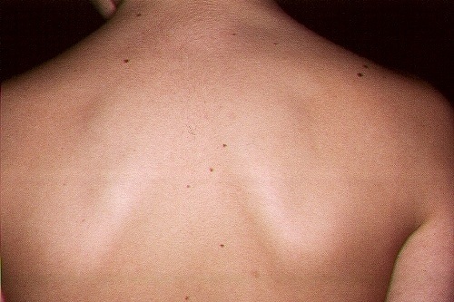
- Most melanocytic naevi become apparent during childhood or early adult life
- The number of melanocytic naevi that an individual has is related to genetic factors and exposure to UV radiation, greater exposure normally results in the development of a larger number of naevi
- In later life many melanocytic naevi slowly fade away
Clinical findings
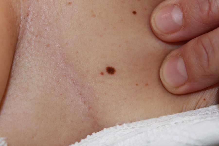
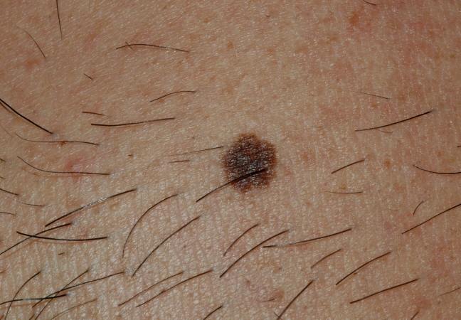
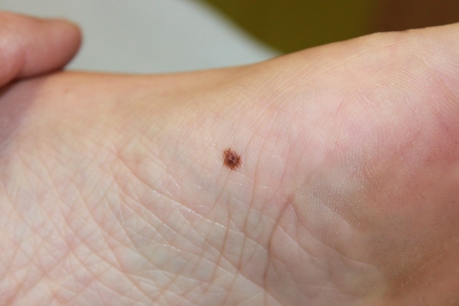
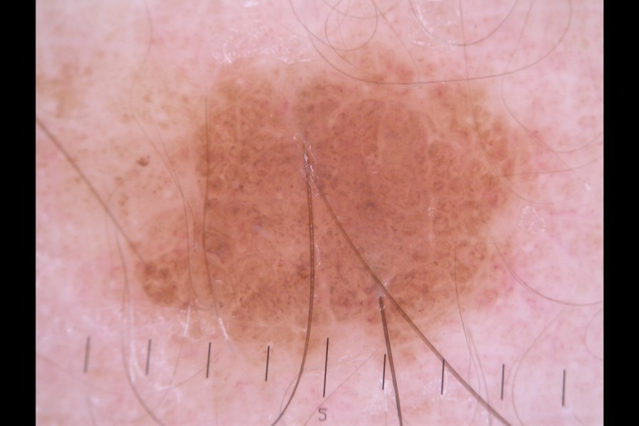
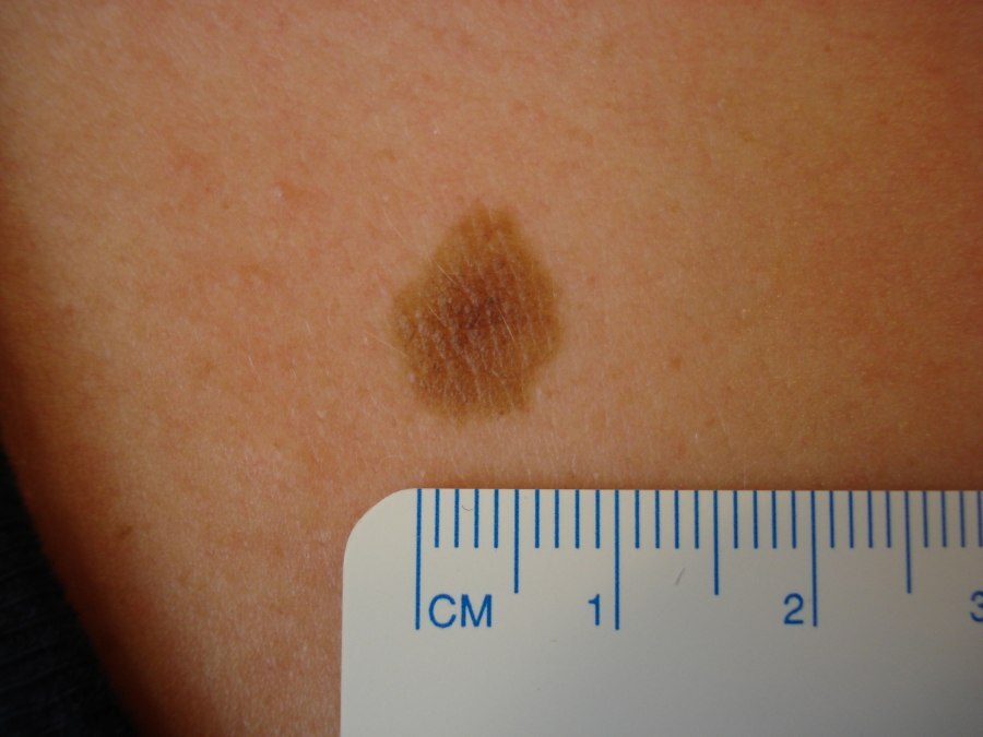
Acquired naevi - predominantly reticular
- Morphology
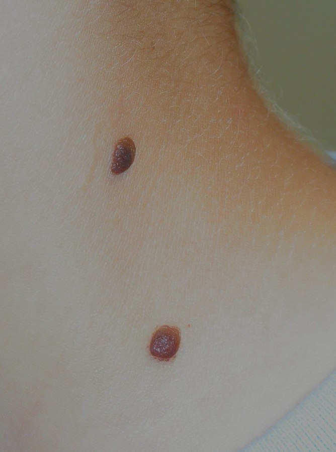
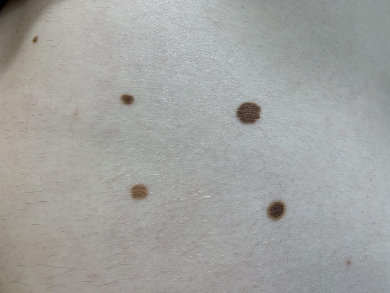
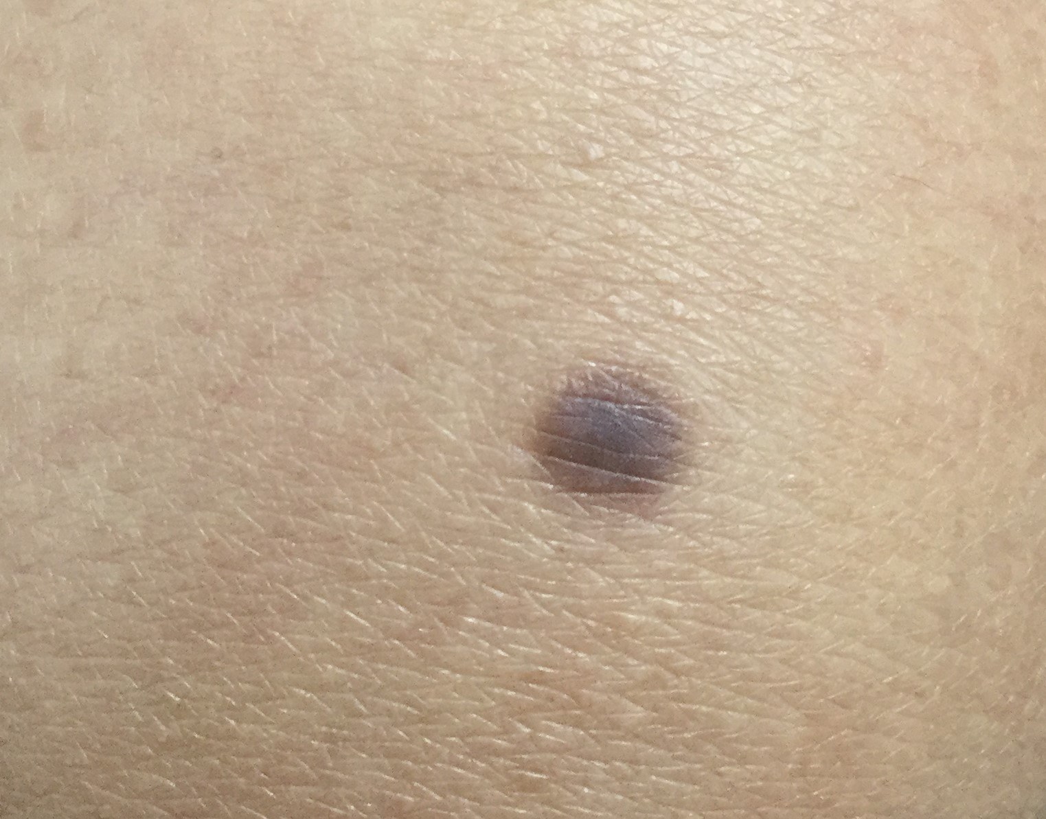
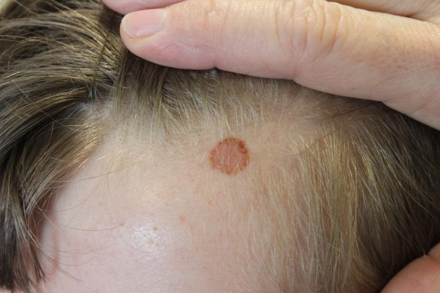
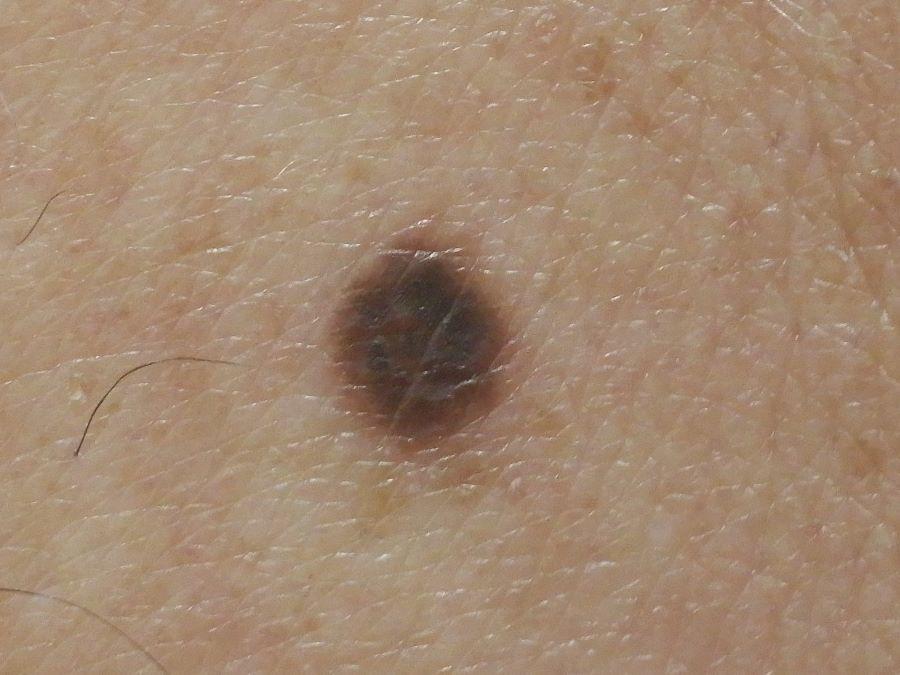
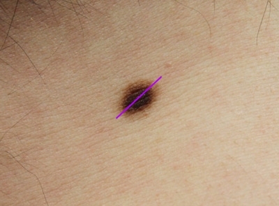
- Flat or slightly raised
- Generally symmetrical with a smooth border
- Colour - often brown but can be pink or blackish depending on skin type (darker lesions most common in skin types IV-VI). The colour tends to be evenly distributed throughout the lesion, although sometimes more pronounced in the centre and occasionally more pronounced at the edge. Lesions lighten with age
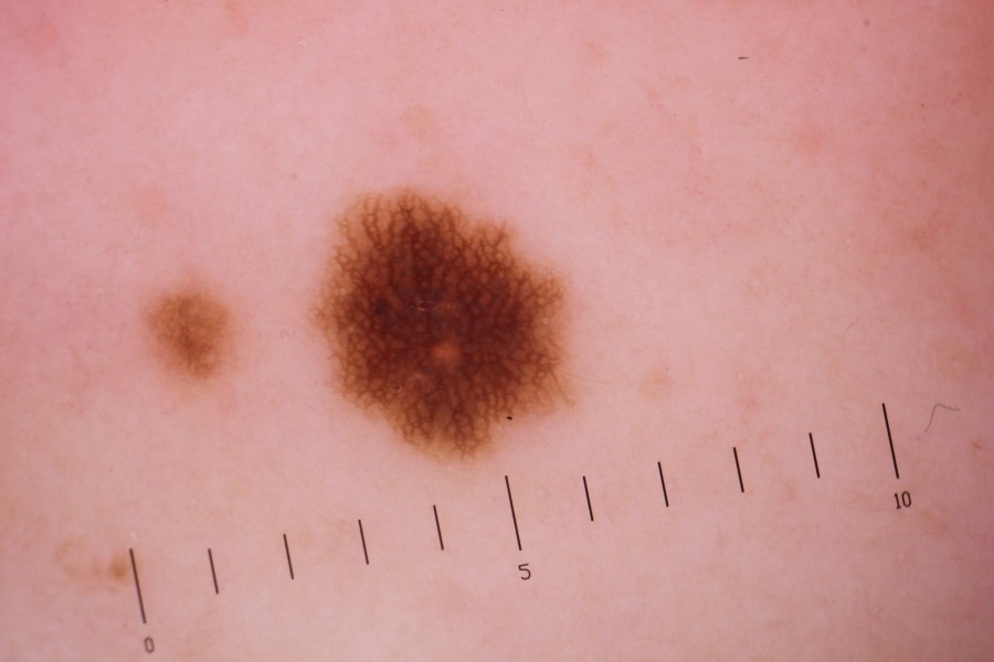
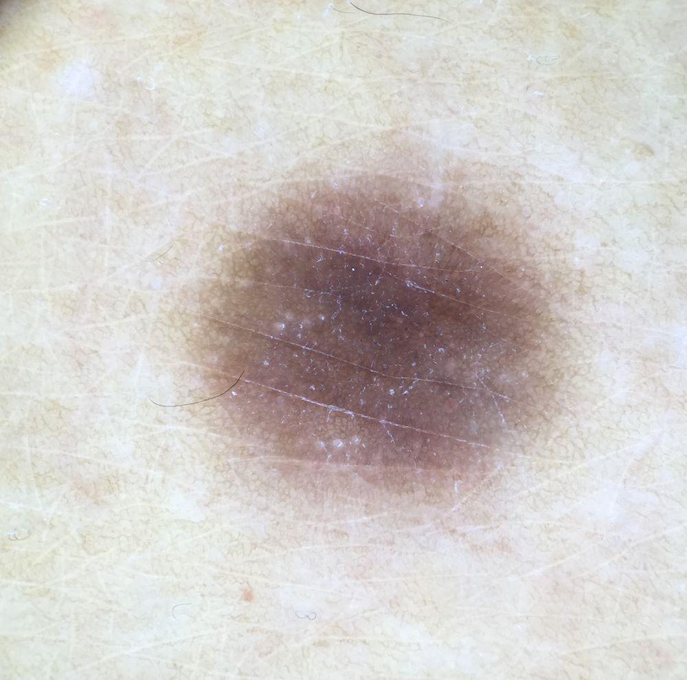
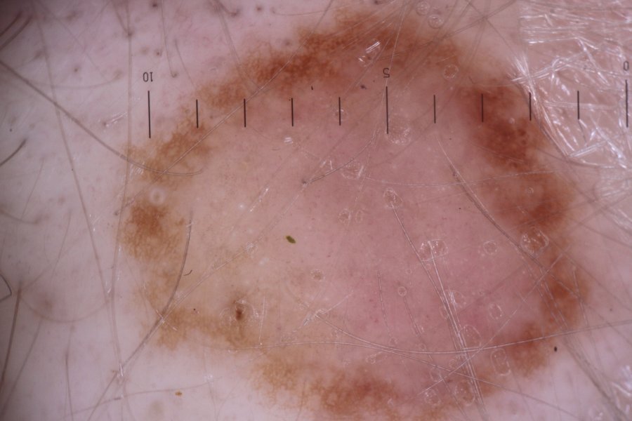
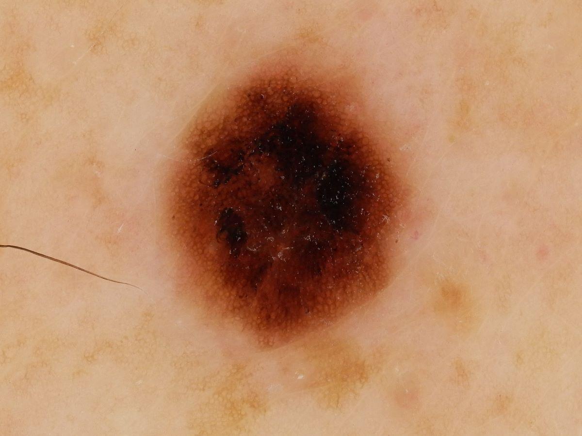
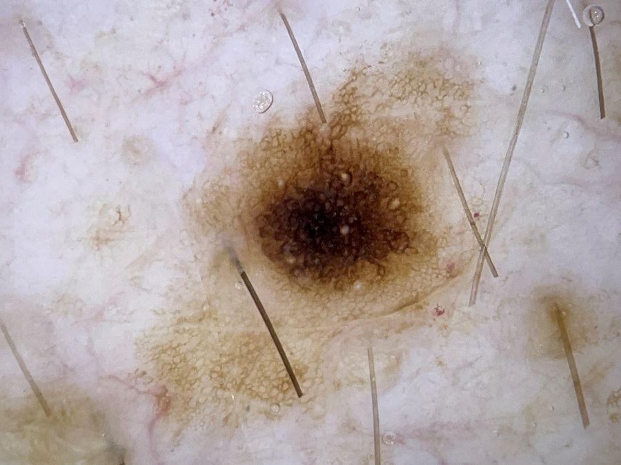
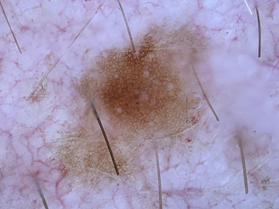
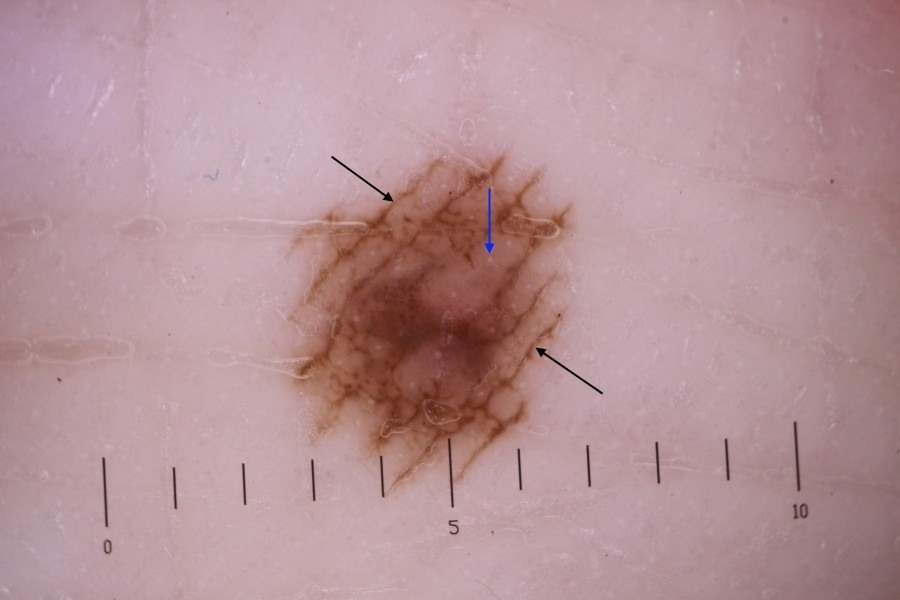
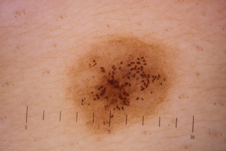
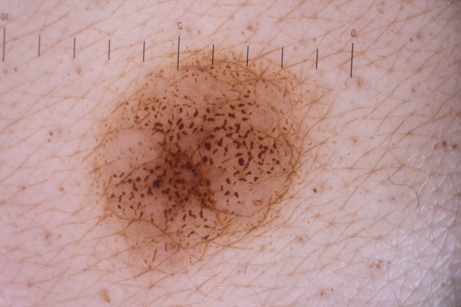
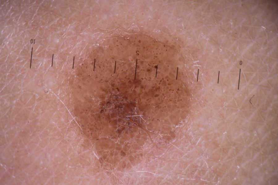
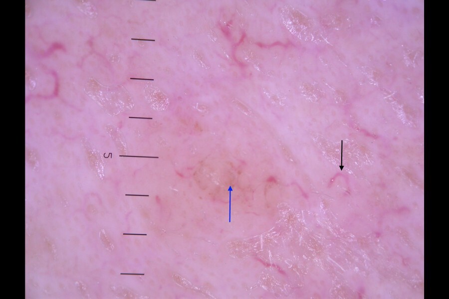
- Dermoscopic features of typical body sites - excluding head, palms and soles, mucosal surfaces (hover over terminologies for description)
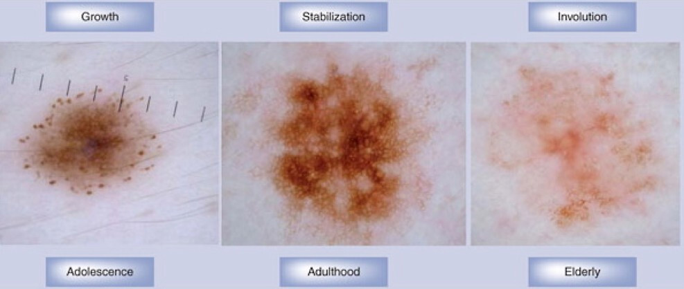
- The pattern tends to progress from having a peripheral rim of regular globules, to a predominantly reticular pigment network in adults, and then largely structureless in older people as the naevi involute
- In most patients the network is darker in the centre and fades to the edge. However, in some cases, eg skin type I-2 or lesions on the scalp, the network is more pronounced at the periphery. Regardless, with benign lesions the network should take on a symmetrical appearance
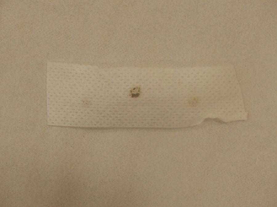
- Tape stripping - some benign melanocytic naevi are hypermelanotic; clinically black and sometimes suspicious for melanoma. These benign naevi are usually characterised by a large amount of melanin in the stratum corneum (black lamella), often most apparent centrally. The presence of a black lamella renders dermoscopic interpretation difficult. Tape stripping, in which an adhesive material such as Hypafix or Sellotape is applied to the lesion and then pulled off, has been shown to remove surface structures. If a lesion visualised after tape stripping is much lighter (with regular structures), this is likely to be reassuring
- As discussed in the chapter Melanoma - an overview the dermoscopic examination of melanocytic lesions requires considerable experience
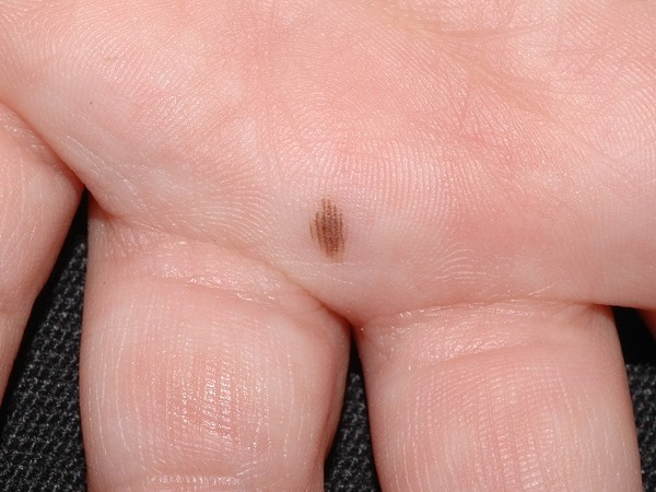
- In terms of benign and malignant lesions on the palms and soles refer to the chapter Melanoma: acral lentiginous melanoma. For the head and neck refer to the chapter Melanoma: lentigo maligna melanoma (including lentigo maligna)
- Pattern comparison
- It is important to examine all of a patient's moles - a melanoma / other skin cancer is likely to look different to the others
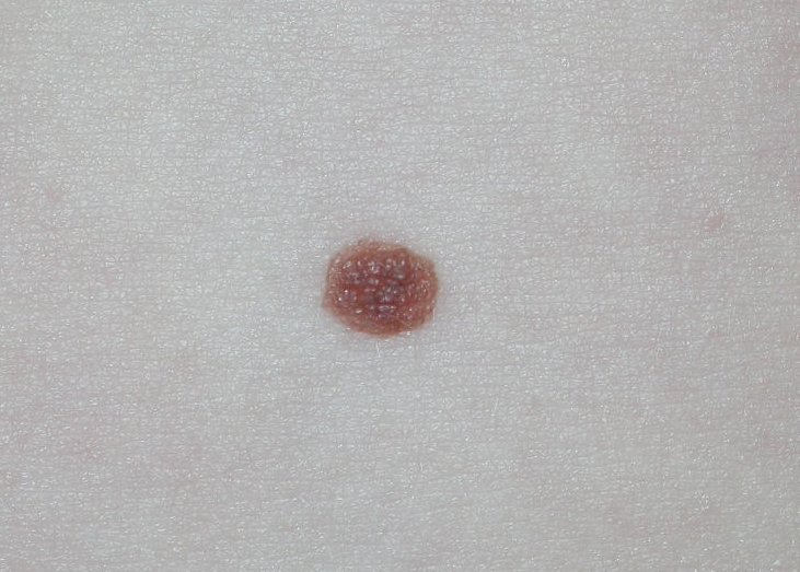
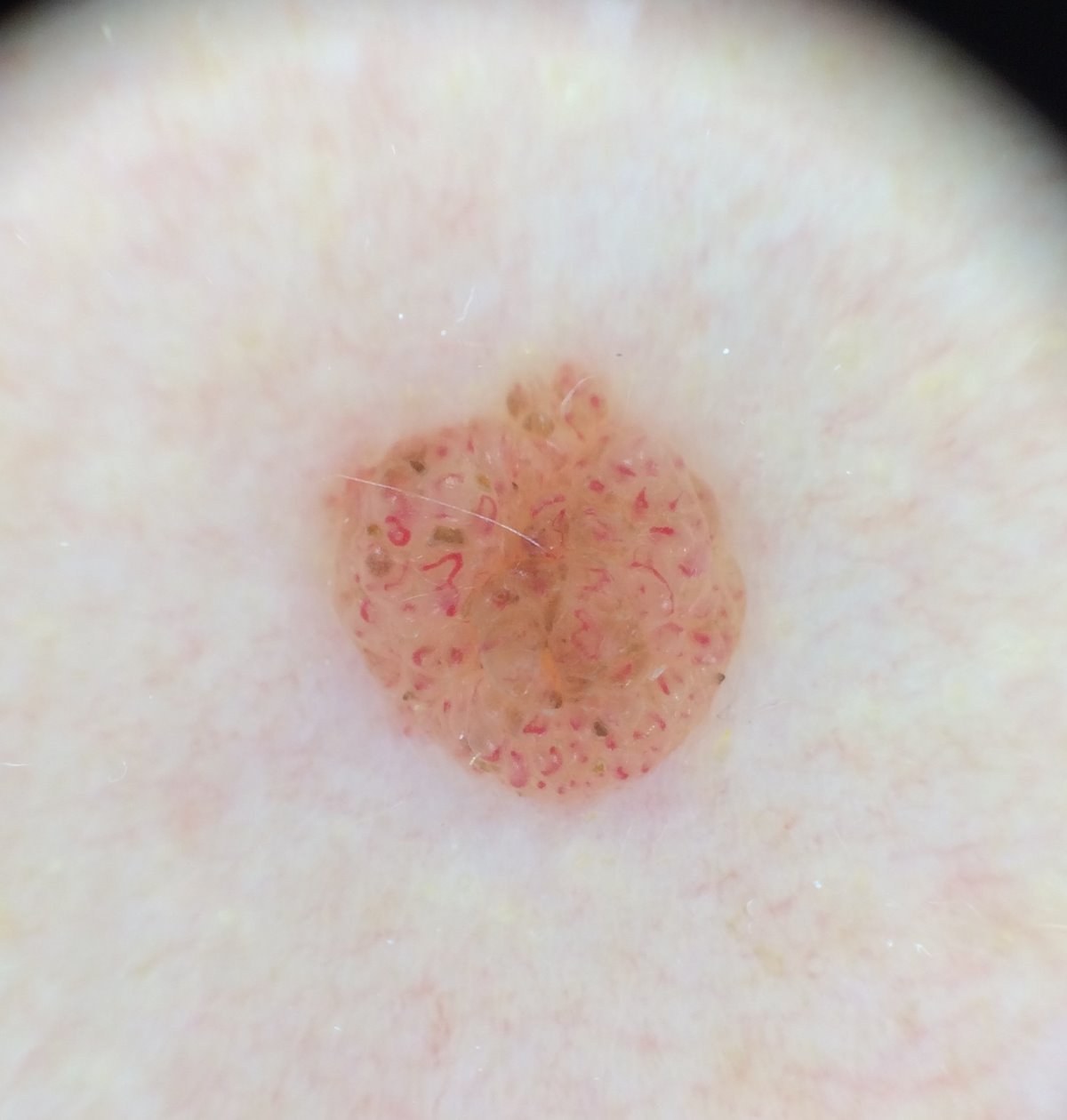
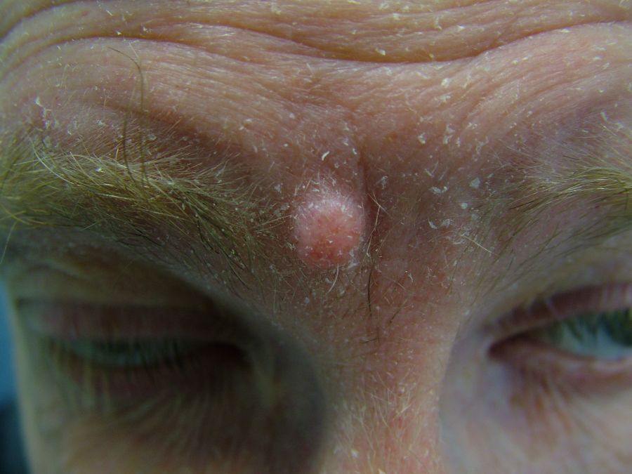
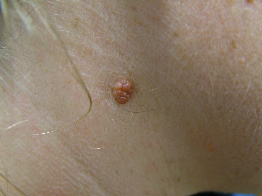
Dermal naevi - globular / cobblestone
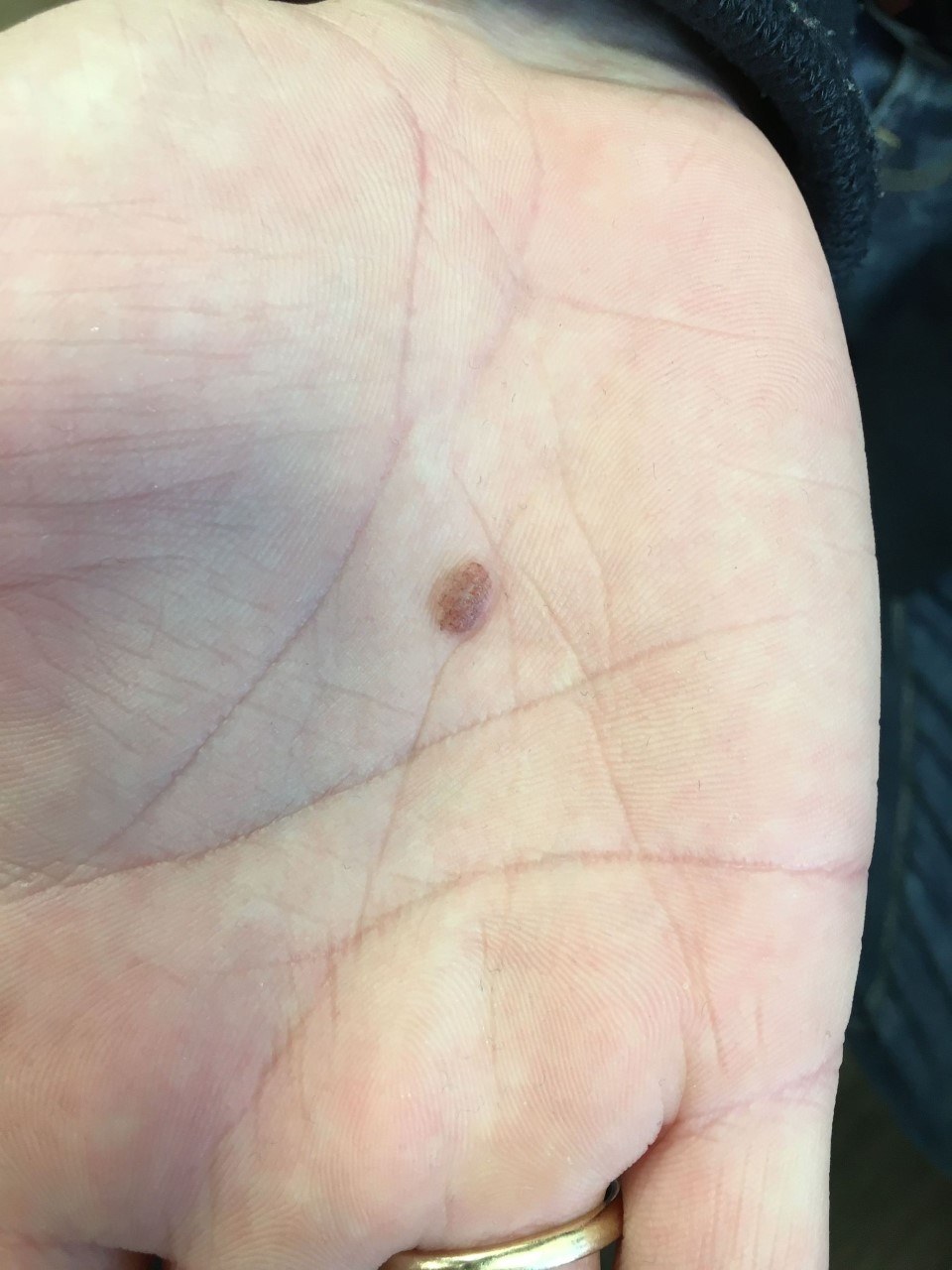
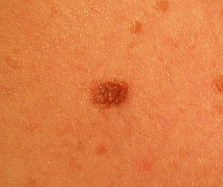
- Morphology
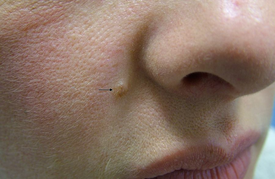
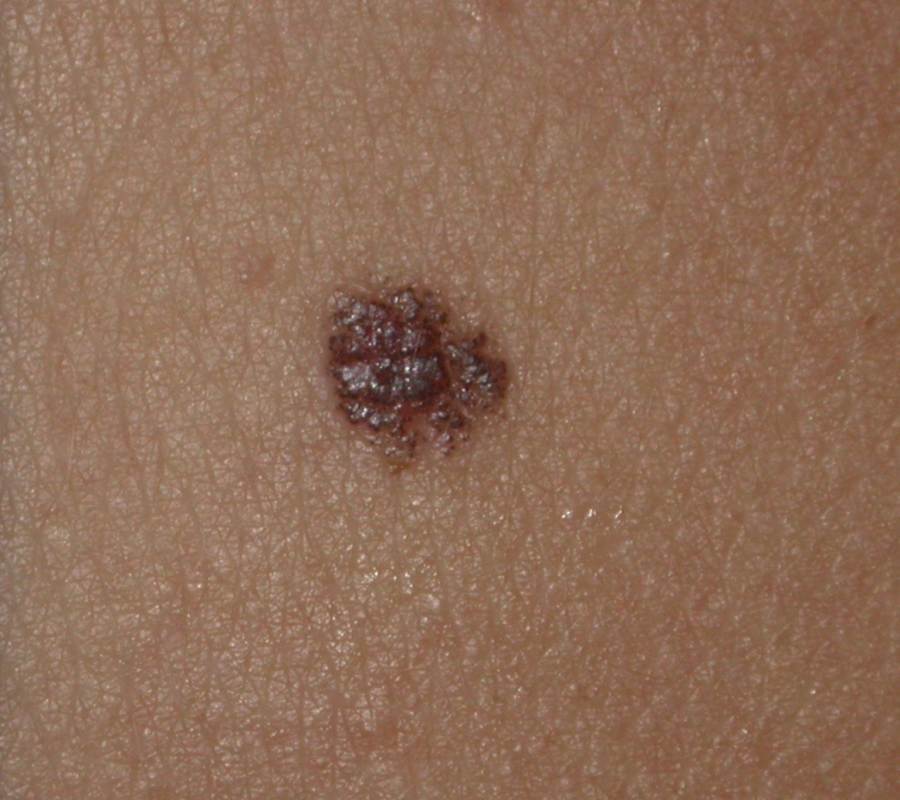
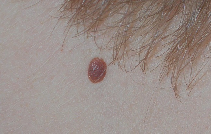
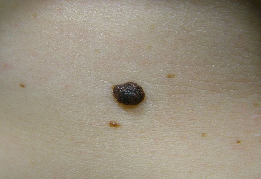
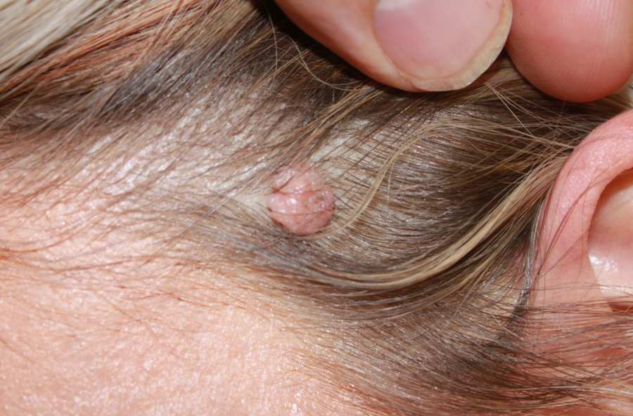
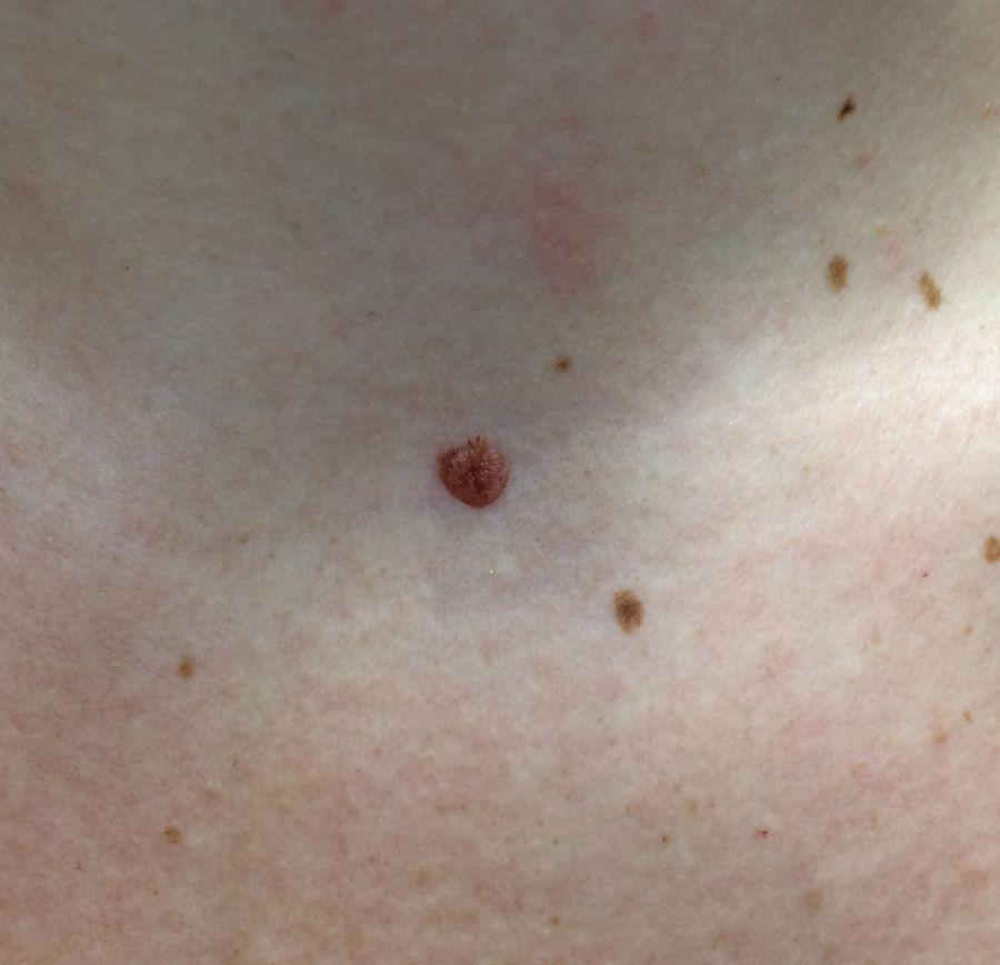
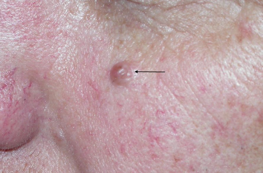
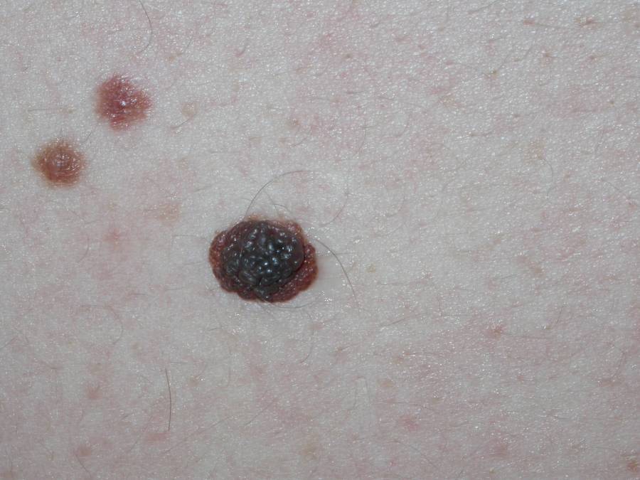
- Thickened and often protrude from the skin surface
- May become dome-shape or papillomatous and wobbly to palpate
- Colour - often brown. Lighten with age
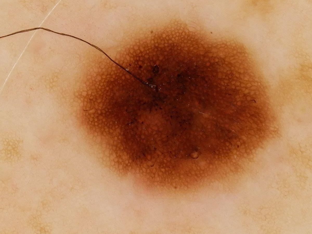
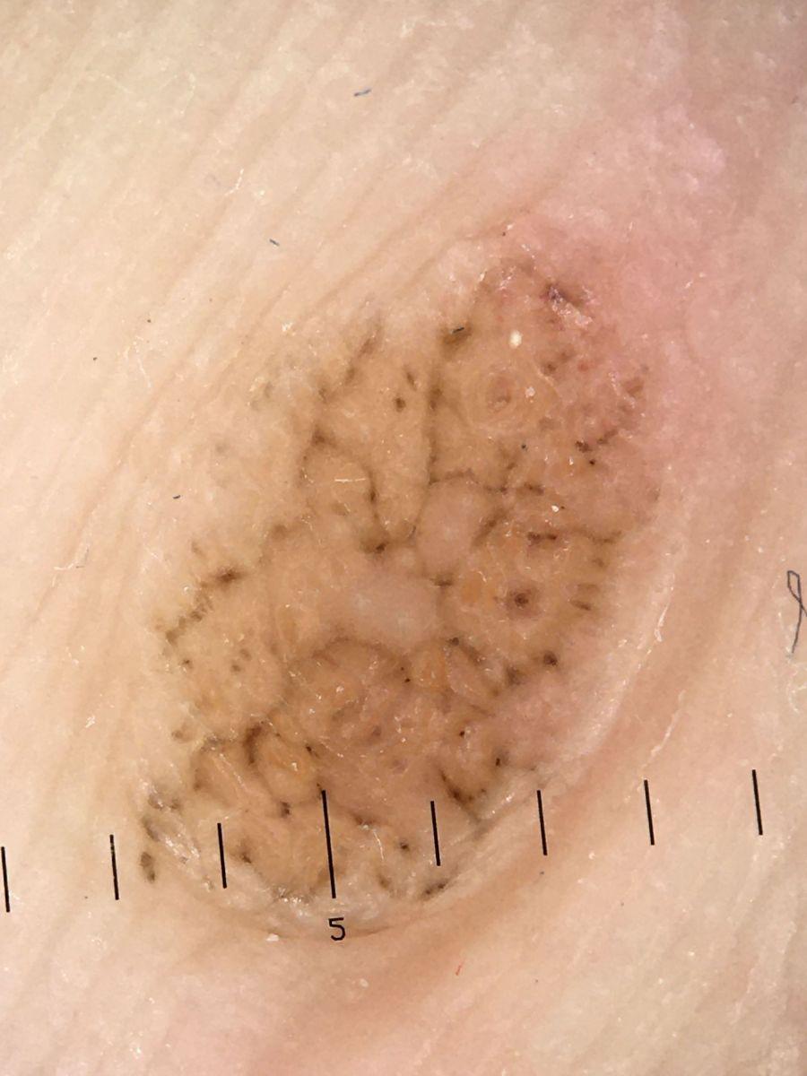
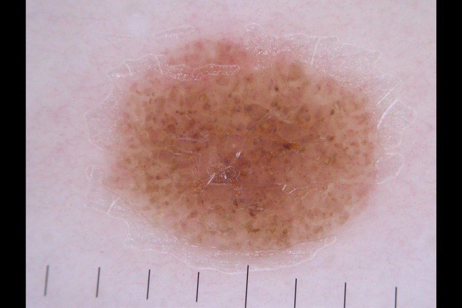
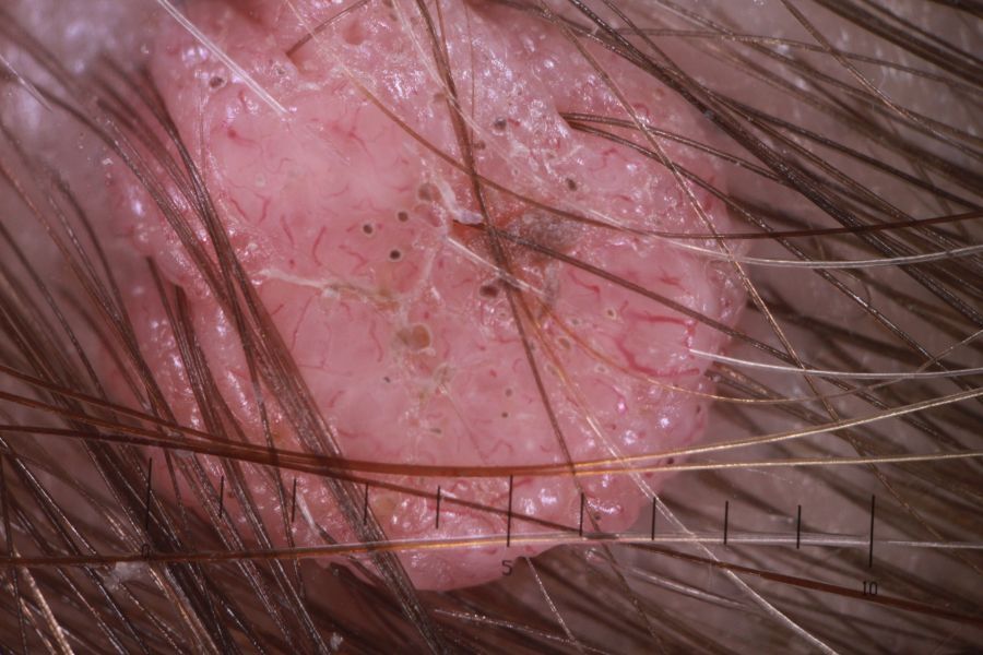
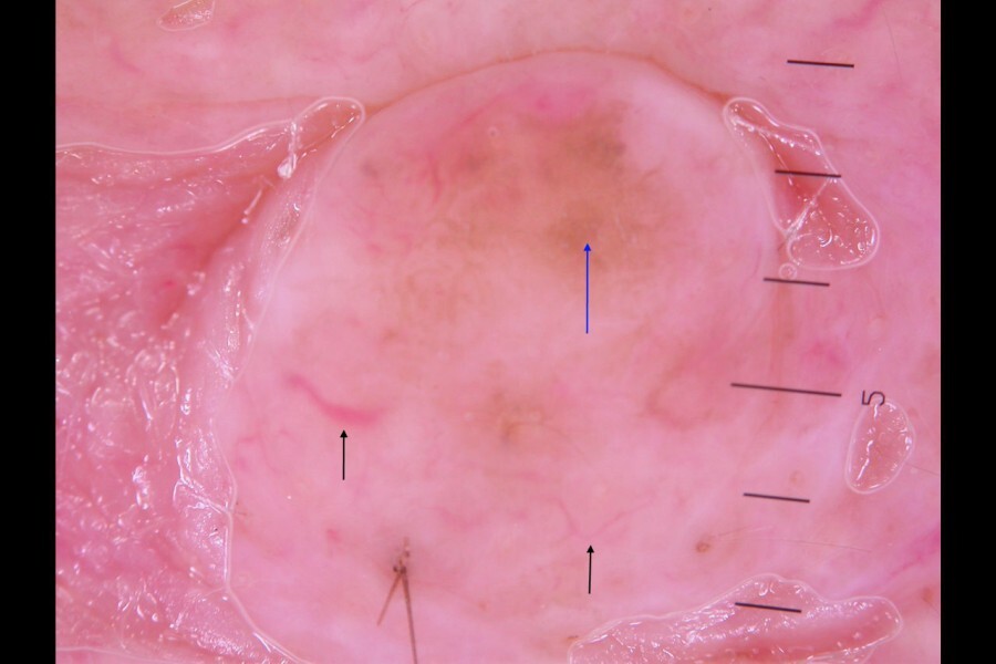
- Dermoscopic features (hover over terminologies for description)
- Tend to be uniform or darker in the centre
- Grouped smooth-looking globules, sometimes giving a cobblestone appearance
- Bloods vessels are ''comma-like'' in shape (click on the link for an overview of dermoscopic vessel patterns)
- In older people lesions may be relatively banal and show little remaining pigment
- As discussed in the chapter Melanoma - an overview the dermoscopic examination of melanocytic lesions requires considerable experience
Clinical Images
Please refer to notes on image rights at bottom of the page with regards to individual image
ownership.
1
2
3
4
5
6
7
8
9
10
11
12
13
14
15
16
17
18
19
20
21
22
23
24
25
26
27
28
29
30
31
32
33
34
35
36
37
38
39
40
41
42
43
44
45
46
47
48
49
50
51
52
Management
General advice - for all
- In general, the larger the number of naevi (typical or atypical), the greater the risk of melanoma - in patients with >100 common naevi on their body, the relative risk of developing cutaneous melanoma is approximately sevenfold higher compared to those with only 15 or less naevi. The arm appears to correlate most closely to total body naevus counts - 11 or more naevi on an arm predicts an increased risk of having >100 total body naevi
- Patients need to be taught on the self-examination of moles and informed to look for new lesions and change in existing lesions - approximately 20% of all melanomas arise from pre-existing naevi, with the remaining majority arising de novo
Surgical management of typical benign melanocytic naevi
- The chances of any one individual naevus becoming malignant is very low, as such benign naevi should not be excised as a preventative strategy
- Treatment for benign naevi should only be done if the lesion is causing problems eg bleeding from catching on clothing. Proud lesions are best removed by shave excision. All samples must be sent for histology
Larger numbers of moles with both typical and atypical melanocytic naevi
- Refer to the chapter Atypical melanocytic naevi
- Routine referral - patients with a mixture of typical and atypical melanocytic naevi require a thorough dermoscopic assessment and photography, from which the patient should receive a copy of their images to assist in self-monitoring
- Urgent skin cancer referral - if melanoma cannot be excluded
Disclaimer - the author PCDS cannot accept responsibility for any misleading or incorrect statements, and the management of individual patients remains the direct responsibility of the individual doctor. We do however hope that visitors to this site can contact us regarding comments that are considered misleading or incorrect so that we can continue to improve the site.
Image Rights - The PCDS would like to thank Dermatoweb, DermQuest (Galderma), and others who have contributed images. All named individuals and organisations maintain copyright for the relevant images.